Correspondence
Benign acute childhood myositis: an alarming condition with an
excellent prognosis!☆
To the Editor,
Benign acute childhood myositis (BACM) causes difficulty to walk due to severe bilateral calf pain. We report the clinical and laboratory features of children admitted with BACM to a level II hospital's pediatric department, from January 2001 to December 2012 (12 years). Data were collected retrospectively from patient's clinical records. The purpose was to characterize BACM's clinical picture, providing greater awareness to its benign course and helping to differentiate it from more serious disorders.
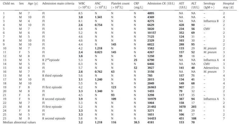
Twenty-eight admissions were identified (2.5/1000 admissions), corresponding to 25 children (Table). There was a 4.6:1 male:female ratio (21 boys, 84%). Median age was 7 years (range, 4-10 years), and 82% were older than 5 years. Most cases were seen from 2008 to 2010 (n = 19, 67.9%) and during winter and spring (n = 20, 71.4%) (Figs. 1 and 2). All occurred at the early convalescent phase of an upper respiratory infection. History of recent trauma or vigorous exercise and family history of neuromuscular disorders were negative. On physical examination at the pediatric emergency department, all patients were unable to walk/bear weight. Bilateral gastrocnemius-soleus muscles were tender to mild palpation without other inflammatory signs. Lower extremity sensation was intact, with normal strength, tone, and deep tendon reflexes. Children were admitted for clinical and laboratory follow-up because of serious functional impairment (n = 24, 85.7%) or previous history of BACM/recurrence (n = 4). The following laboratory abnormalities were detected (Table): leukopenia (n = 10), neutropenia (n = 7), thrombocytopenia (n = 6), and mildly elevated C-reactive protein (CRP) (n = 2). All children presented a markedly elevated serum creatine kinase (CK); median admission value was 4181 UI/L (range, 785-26863 UI/L). In the 22 available aminotransferase results, aspartate transaminase (AST) was elevated in all, with a less increased alanine transaminase (ALT) level in 12 cases. Blood urea nitrogen, creatinine, electrolytes, and urinalysis were normal. Serologic tests were performed in 20 cases (71.4%), with 9 positive results (Table): Mycoplasma pneumoniae(n = 3), cytomegalovirus and influenza A (n = 2), and influenza B and adenovirus (n = 1).
Median admission period was 3 days (range, 1-7 days), and all children revealed clinical and laboratory improvement with sup-portive therapy (bed rest, intravenous hydration, and oral ibupro-fen). No red to brown urine or renal/hydroelectrolytic abnormalities were seen. Children with recurrent episodes were reassessed at pediatric consultation 1 month after discharge, with full recovery and normal CK levels. The remaining patients were referred to their attending physician.
Benign acute childhood myositis is a rare disorder and continues to lead physicians into unnecessary workup when they are unfamiliar with its presentation [1-5]. Most commonly occur after influenza B and occasionally influenza A infection, but parainfluenza, adenovirus, herpes simplex, Epstein-Barr, Coxsackie, rotavirus, andM pneumoniaehave also been implicated [1-3,5-10], as found in our study. As evidenced, key elements in the diagnosis are a preceding upper respiratory infection followed by the acute onset of typical myositis clinicalfindings, predominantly affecting gastrocnemius-soleus muscles. Further clinical features include school-aged boys and late winter–early spring predomi-nance, elevated CK, and aminotransferase levels (especially AST) [1-10]. Transient hematological abnormalities (mild leukopenia, neutropenia, and thrombocytopenia) are also seen [1,3,8-10]. Erythrocyte sedimentation rate and CRP are usually normal but may be mildly elevated[1-3,5,6]. Patients with this typical clinical presentation can be treated as an outpatient[1,5]. However, as seen in our study, some children are admitted for hydration, observa-tion, and serial CK levels to rule out any progression to rhabdomy-olysis[1,6,8-10]. Recurrence is rare and has been demonstrated to be caused by different viruses or different influenza types[9]. In our study, 3 children had recurrent episodes with more than a year interval, and they totally recovered in between. If different etiologic agents were behind these episodes, it was not possible to assess. Treatment is supportive, and antivirals are unlikely to be beneficial [2-5]. The hallmark of BACM is its spontaneous and rapid clinical resolution within 1 week [1-5]. In this study, all children fully recovered within a week or less only with supportive measures. Higher CK levels were not associated with acute renal failure, progression to rhabdomyolysis, or other complications. Patients can be discharged if they have typical presentation, normal renal function, and follow-up to ensure complete resolution [2,10]. Further investigation is not indicated unless there is clinical concern for more serious disorders [1,6,8,10]. Findings not classically associated with BACM include myoglobinuria, recent trauma or vigorous exercise, family history of neuromuscular disorders, subacute or chronic progression, a new rash, frank muscle weakness, or abnormal neurologic findings. When such atypical features are present, other diagnosis as rhabdomyolysis, Guillain-Barré syndrome, primary inflammatory myositis, muscu-lar dystrophy, or metabolic disease must be excluded[2,6,8,10].
Benign acute childhood myositis should be part of the broad differential diagnosis of a child unable to walk/bear weight. Although potentially alarming, it is self-limited with an excellent prognosis. Recognition of this rare clinical entity by the emergency physician is essential to prevent unnecessary invasive testing and hospital admission.
American Journal of Emergency Medicine xxx (2014) xxx–xxx
☆ Conflict of interest: None.
0735-6757/© 2014 Elsevier Inc. All rights reserved.
Contents lists available atScienceDirect
American Journal of Emergency Medicine
j o u r n a l h o m e p a g e :w w w . e l s e v i e r . c o m / l o c a t e / a j e m
Joana Almeida Santos MD Pediatric Department, Hospital Dona Estefânia Centro Hospitalar Lisboa Central Lisbon, Portugal E:mail address:joanaasantos@gmail.com
Carolina Albuquerque MD David Lito MD Florbela Cunha MD Pediatric Department Hospital de Vila Franca de Xira Vila Franca de Xira, Portugal E:mail addresses:carolinalbuquerque@gmail.com(C. Albuquerque), dmlito@gmail.com(D. Lito),flor.cunha@gmail.com(F. Cunha)
http://dx.doi.org/10.1016/j.ajem.2014.08.022
References
[1]Hall G, Schranz CI. Benign acute childhood myositis—a rare cause of abnormal gait.
Am J Emerg Med 2014;32(2):193.e1–2.
[2]Heiner JD, Ball VL. A child with benign acute childhood myositis after influenza. J Emerg Med 2010;39(3):316–9.
[3]Koliou M, Hadjiloizou S, Ourani S, Demosthenous A, Hadjidemetriou A. A case of benign acute childhood myositis associated with influenza A (H1N1) virus infection. Clin Microbiol Infect 2010;16(2):193–5.
[4]Neocleous C, Spanou C, Mpampalis E, Xatzigeorgiou S, Pavlidou C, Poulos E, et al. Unnecessary diagnostic investigations in benign acute childhood myositis: a case series report. Scott Med J 2012;57(3):182.
[5]Rubín E, De la Rubia L, Pascual A, Domínguez J, Flores C. Benign acute myositis associated with H1N1 influenza A virus infection. Eur J Pediatr 2010;169(9):1159–61. [6]Agyeman P, Duppenthaler A, Heininger U, Aebi C. Influenza-associated myositis in
children. Infection 2004;32(4):199–203. Table
Clinical and laboratory features of 25 children admitted with BACM
Child no. Sex Age (y) Admission main criteria WBC
(×109/L)
ANC
(×109/L)
Platelet count
(×109/L)
CRP (mg/L)
Admission CK (UI/L) AST
(UI/L) ALT (UI/L)
Serology (IgM+)
Hospital stay (d)
1 M 7 FI 8.5 N N N 4095 NA NA – 2
2 M 10 FI 3.0 1.341 N N 4369 NA NA – 4
3 M 4 FI 8.1 N N N 4275 NA NA Influenza B 2
4 M 10 FI 2.6 0.754 N N 6629 428 90 – 2
5 M 7 FI 4.8 N N N 5020 234 56 CMV 3
6 M 6 FI 5.2 N N N 10 157 352 69 – 2
7 M 5 FI 4.6 N N N 7125 124 31 – 3
8 M 10 FI 4.6 N N N 2329 103 30 – 2
9 M 10 FI 4.4 N 145 N 6022 288 95 – 2
10 M 7 FI 4.2 1.218 N N 1582 133 29 M.pneum 2
11 F 5 FI 2.4 0.823 145 N 4267 157 52 M.pneum 3
12 M 8 FI 3.8 N N N 1250 59 18 – 2
13 M 5 R 2ndepisode 5.3 N N 25 6790 NA NA Influenza A 4
14 M 5 FI 6.3 N N N 6466 NA NA CMV 3
15 M 6 FI 5.7 N N 52 3927 141 40 Adenovírus 5
16 M 7 FI 2.6 0.762 63 N 3156 NA NA M.pneum 7
13 M 6 R third episode 5.6 N N N 785 157 71 – 4
17 M 10 FI 3.1 1.240 N N 2815 134 41 – 3
18 M 10 5.5 N N N 2049 149 68 – 4
19 F 8 FIfirst episode 4.2 N 123 N 26 863 907 21 – 2
20 M 8 FI 3.3 1.340 N N 1455 79 32 – 2
21 F 6 FI 4.5 N 93 N 3298 117 35 – 3
19 F 9 R second episode 3.9 N 109 N 10 979 387 94 Influenza A 5
22 M 7 FI 5.3 N N N 1064 158 17 – 2
23 M 8 FIfirst episode 5.2 N N N 21 492 1070 203 – 6
24 F 8 FI 3.3 N N N 3271 91 29 – 1
25 M 5 FI 3.3 N N N 1851 106 37 – 2
23 M 9 R second episode 5.8 N N N 14 453 451 108 – 7
Median abnormal values 3.2 1.218 116 38.5 4181 153 70
Child 13, 19, and 23 presented recurrent episodes (child 13 had a previous episode out of the study period). Bold numbers indicate abnormal values. Reference range: CRP, less than 5
mg/L; CK, 31 to 152 UI/L; AST, less than 31 UI/L; ALT, less than 39 UI/L. Leukopenia: white blood cell count less than 4 × 10⁹/L; neutropenia: absolute neutrophil count less than 1.5 ×
109/L; thrombocytopenia: platelet count less than 150 × 109/L. Viral and bacterial identification was based on serologic tests with a positive immunoglobulin M result. Abbreviations:
ANC, absolute neutrophil count;CMV, cytomegalovirus;F, female;FI, functional impairment;IgM, immunoglobulin M;M, male;M.pneum,Mycoplasma pneumoniae;n, absolute
number;N, normal value;NA, not available;R, recurrence;WBC, white blood cell count.
0 1 2 3 4 5 6 7 8 9
2001 2002 2003 2004 2005 2006 2007 2008 2009 2010 2011 2012
Number of admitted cases
Year
Fig. 1.Annual distribution of admitted cases with BACM. Most cases were seen from 2008 to 2010 (n = 19, 67.9%).
0 1 2 3 4 5 6 7 8 9 10
Jan Feb March April May June July Aug Sept Oct Nov Dec
Number of admitted cases
Month
Fig. 2.Monthly distribution of admitted cases with BACM. Most cases were seen during winter and spring.
2 Correspondence /American Journal of Emergency Medicine xxx (2014) xxx–xxx
[7]Hu JJ, Kao CL, Lee PI, Chen CM, Lee CY, Lu CY, et al. Clinical features of influenza A and B in children and association with myositis. J Microbiol Immunol Infect 2004;37(2):95–8.
[8]Jain S, Kolber MR. A stiff-legged gait: benign acute childhood myositis. CMAJ 2009; 181(10):711–3.
[9]King BA. Benign acute childhood myositis as a cause of failure to weight bear. J Paediatr Child Health 2003;39(5):378–80.
[10]Rennie LM, Hallam NF, Beattie TF. Benign acute childhood myositis in an accident and emergency setting. Emerg Med J 2005;22(10): 686–8.
3 Correspondence /American Journal of Emergency Medicine xxx (2014) xxx–xxx