Key words:
Biochemical Processes; Recurrence; Prostate; Prostate-Specific Antigen; Prostatectomy; Prostatic Neoplasms
Int Braz J Urol. 2013; 39: 779-92
__________________ Submitted for publication: April 12, 2013
__________________ Accepted after revision: July 04, 2013
Purpose: To analyze controversial clinicopathologic predictors of biochemical recurrence after surgery: age, race, tumor extent on surgical specimen, tumor extent on needle biop-sy, Gleason score 3 + 4 vs 4 + 3, and amount of extent of extraprostatic extension and positive surgical margins.
Materials and Methods: The needle biopsies and the correspondent surgical specimens were analyzed from 400 patients. Time to recurrence was analyzed with the Kaplan--Meier curves and risk of shorter time to recurrence using Cox univariate and multiva-riate analysis.
Results: Except for age, race, maximum percentage of cancer per core, and number of cores with cancer, all other variables studied were significantly predictive of time to biochemical recurrence using the Kaplan-Meier curves. In univariate analysis, except for focal extraprostatic extension, age, race, focal positive surgical margins, and maximum extent and percentage of cancer per core, all other variables were significantly predictive of shorter time to recurrence. On multivariate analysis, diffuse positive surgical margins and preoperative PSA were independent predictors.
Conclusions: Young patients and non-whites were not significantly associated with time to biochemical recurrence. The time consuming tumor extent evaluation in surgical spe-cimens seems not to add additional information to other well established predictive fin-dings. The higher predictive value of Gleason score 4 + 3 = 7 vs 3 + 4 = 7 discloses the importance of grade 4 as the predominant pattern. Extent and not simply presence or absent of extraprostatic extension should be informed. Most tumor extent evaluations on needle biopsies are predictive of time to biochemical recurrence, however, maximum percentage of cancer in all cores was the strongest predictor.
INTRODUCTION
While the overall survival of surgically trea-ted prostate cancer patients remains excellent, nearly
30% of patients treated definitively will have bioche-mical recurrence (BR), defined as prostate-specific antigen (PSA) ≥ 0.2 ng/ml according to recommen-dation of the American Urological Association (1).
Controversial predictors of biochemical recurrence after
radical prostatectomy: a study from a Latin American
(Brazilian) Institution
_______________________________________________
Marcelo R. Noronha, Maisa M. Q. Quintal, Luis A. Magna, Leonardo O. Reis, Athanase Billis, Luciana
R. Meirelles
Department of Pathology (MRN, MMQQ, AB, LRM); Department of Medical Genetics/Biostatistics (LAM) and Department of Urology (LOR), School of Medical Sciences, State University of Campinas (Unicamp), Campinas, Brazil
ABSTRACT
ARTICLE
INFO
There are well established clinical and pa-thological predictors of BR after radical prosta-tectomy (RP), including preoperative serum PSA, pathological tumor stage, Gleason histological grade, and positive surgical margins (2). However, other features are controversial predictors, which are the aim of our study.
The influence of age in the biological ag-gressiveness of prostate cancer is controversial (3-6). Some studies suggest that prostatic carcino-ma has worse outcome with increasing age while others suggest that young age is an adverse prog-nostic factor.
Prostate cancer may be biologically more aggressive among Black men though controversial (7-11). The prevalence of prostate cancer in Brazil is higher in Black men compared to White men (12-15). We studied the influence of race in regard to biochemical recurrence after radical prostatectomy.
Extent of tumor on needle biopsy may be useful to predict extent of cancer in RP, surgical margin status, pathological stage, and tumor re-currence. There are several ways to evaluate tu-mor extent on biopsy: number and percentage of positive cores, total linear length and percentage of carcinoma in all cores, length and percentage of carcinoma in a single core, and others. The con-troversy is related to which is the best predictor method (16-18).
One of the most controversial aspects of the pathological assessment of RP specimens is the measurement of tumor volume or extent. There is consensus that tumor volume or extent by itself correlates with adverse findings at RP (19). The cri-tical and controversial question concerns whether tumor volume or extent in RP is an independent prognostic parameter for BR after RP once other routinely variables are accounted for (20).
Tumors with a Gleason score of 7 have significantly worse prognosis than those with a Gleason score ≤ 6. Given the adverse prognosis as-sociated with Gleason pattern 4, one would expect whether a tumor Gleason score 3 + 4 = 7 or 4 + 3 = 7 would influence prognosis, however this issue is controversial (21-23).
Extraprostatic extension (EPE) and positi-ve surgical margins (PSM) hapositi-ve prognostic impor-tance in most studies (24-26) and may influence
therapeutic options after surgery. However, the amount of EPE and PSM seems to be more impor-tant than a simple report of presence or absence of these pathological findings. An additional concern is the absence of consensus among pathologists how to evaluate the amount of EPE and PSM (27,28).
MATERIALS AND METHODS
This retrospective study was based on nee-dle biopsies and correspondent surgical specimens of 400 consecutive patients submitted to retropubic RP from 1997 to 2011 at an institution located in the southeast region of Brazil. The clinical variables stu-died included age, race, and preoperative serum PSA; the pathological variables included tumor extent on needle biopsy; and, in surgical specimens tumor ex-tent, Gleason score, seminal vesicle invasion, extra-prostatic extension and positive surgical margins. According to race, patients were considered Whites and African-Brazilians. In Brazil African-Brazilians include Mulattos (White and African-Brazilian ad-mixture). Five patients of oriental ancestry were ex-cluded. Some other missing data are not included in the tables. Age was categorized as ≤ 55 years and > 55 years. All other continuous data were categorized by the median value.
Linear carcinoma extent in mm on needle prostatic biopsy was measured using a single Olym-pus (OlymOlym-pus Optical Co., Ltd., Tokyo, Japan) mi-crometer eyepiece with a linear array. In cases of discontinuous foci 1 mm apart, the tumor was con-sidered as continuous and the measure included 1 mm. In discontinuous foci more than 1 mm apart, the final extent was the sum of the measures. Tumor extent on biopsies was evaluated as: number and percentage of cores with carcinoma, total length and percentage of carcinoma in all cores, and maximum length and percentage of cancer per core.
recommendation (1). Patients without BR were censored at last follow-up. The study was approved by our institutional committee of ethics.
Surgical specimens were step sectioned at 3 to 5 mm intervals and embedded in paraffin. A mean of 32 paraffin blocks was processed. Sections (6 µm) of each block were stained with hemato-xylin and eosin. Each transverse section of the prostate was subdivided into 2 anterolateral and 2 posterolateral quadrants. Using the cone method, 8 sections from the bladder neck and 8 from the apex were obtained.
PSM was defined as cancer cells in contact with the inked specimen surface. PSM was con-sidered focal whenever present up to 1 quadrant and/or sections from the bladder neck or apex and diffuse in more than 1 quadrant or sections.
EPE was diagnosed when cancer was seen in adipose tissue, and in case of a desmoplastic response when a protuberance corresponding to tumor extension into periprostatic tissue was ob-served. EPE was considered focal whenever present up to 1 quadrant of the transverse sections and/or sections from the bladder neck or apex and diffuse in more than 1 quadrant or sections. Seminal vesi-cle invasion occurred when there was involvement of the muscular coat.
Tumor extent at RP was evaluated by a previously described semiquantitative point count method (29). Briefly, each quadrant of the trans-verse sections was drawn on paper and contained 8 equidistant points. During microscopic exami-nation of the slides, the tumor area was drawn on the correspondent quadrant on the paper. At the end of examination, the number of positive points represented an estimate of the tumor extent.
Histological tumor grading was performed according to the revised Gleason grading system (30). Gleason score was categorized as ≤ 6, 3 + 4 = 7, 4 + 3 = 7, ≥ 8. All cases were reviewed by a senior uropathologist (AB).
Statistical analysis
Time to biochemical recurrence (TBR) was analyzed with the Kaplan-Meier product-limit analysis using the log-rank test for comparison be-tween the groups. Univariate and multivariate Cox
stepwise logistic regression models were used to identify significant predictors of shorter TBR. Two--sided P value < 0.05 was considered statistically significant. All statistical analyses were performed using PASW® Statistics 18.0.
RESULTS
Table-1 shows the clinicopathologic cha-racteristics of 400 studied patients submitted to radical prostatectomy.
After RP 128 of the 400 men (32.0%) ex-perienced BR at a mean follow-up of 21 months (median 10, range 3 to 129). Of the remaining patients 253 (63.2%) who were censored remai-ned at risk at a mean follow-up of 53 months (median 48, range 3 to 155), while 19 (4.8%) had no serum PSA data available.
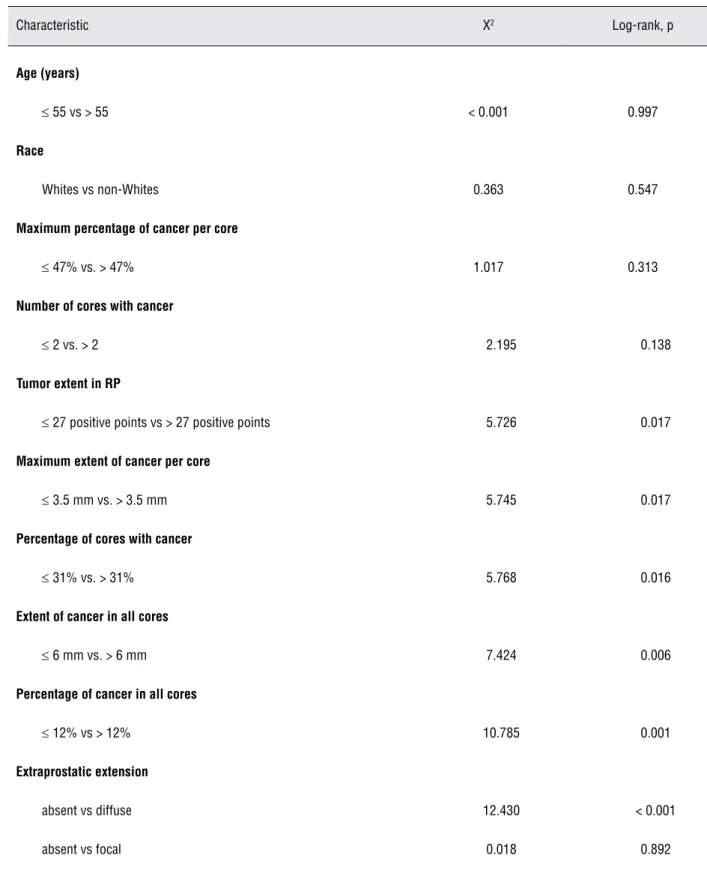
Table-2 shows the Kaplan-Meier product--limit analysis for TBR following RP according to several clinicopathologic characteristics. There was no statistical significant difference for age (log--rank, p = 0.997), race (log(log--rank, p = 0.547), ma-ximum percentage of cancer per core (log-rank, p = 0.313), number of cores with cancer (log-rank, p = 0.138), absent vs focal EPE (log-rank, p = 0.892), and absent vs focal PSM (log-rank, p = 0.069).
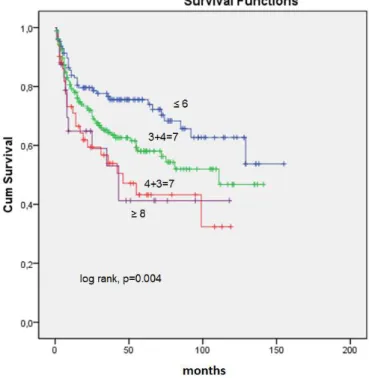
Percentage extent in mm of cancer in all cores was the strongest predictor of TBR (log-rank, p = 0.001) (Figure-1). Tumor extent of tumor in RP evaluated as positive points was significantly as-sociated with TBR (log-rank, p = 0.017) (Figure-2). Gleason score in RP categorized as ≤ 6, 3 + 4 = 7, 4 + 3 = 7, and ≥ 8 was significantly associated with TBR (Figure-3). There is almost an overlap comparing the curves of Gleason score 4 + 3 = 7 and Gleason score ≥ 8. Diffuse EPE (log-rank, p < 0.001) and diffuse PSM (log-rank, p < 0.001) were significantly associated with shorter TBR.
For multivariate analysis, we included only the statistically significant characteristics of uni-variate analysis using the backward stepwise lo-gistic regression method. For tumor extent evalu-ation on needle biopsy, we included the strongest predictor (percentage of cancer extent in all co-res). Only diffuse PSM (p < 0.001) and preopera-tive PSA (p = 0.034) were independent predictors of shorter TBR. Seminal vesicle invasion showed a trend toward significance (p = 0.060).
DISCUSSION
Age
The influence of age in the biological aggressiveness of prostate cancer is controver-sial. Carter et al. (3) and Herold et al. (4) suggest that prostatic carcinoma is higher grade and has worse outcome with increasing age. However, the data are conflicting on this issue. Bauer et al. (5) and Catalona and Smith (6) have not found age to be related to outcome. Parker et al. (31) in a meta-analysis of 34 studies which included a to-tal of 27551 patients concluded that evidence su-ggests that young age was an adverse prognostic factor in some series of radiation therapy before the advent of PSA assays, when men typically presented clinically with locally advanced dise-ase, but that age has no significant prognostic effect in contemporary series of localized prosta-te cancer. In our study, TBR was not statistically different comparing patients ≤ 55-year-old vs > 55-year-old.
Race
Prostate cancer may be biologically more aggressive among Black men though controversial (7-9). In a study by Cross et al. (10), even thou-gh African-American men presented at a younger age and with more advanced disease compared with White men with prostate cancer, PSA outco-me after RP when controlled for known clinical predictive factors was not statistically different. Freedland et al. (11) described no differences be-tween Black and White men in the preoperative clinical factors or the pathologic features of the RP specimens and race was not an independent predictor of biochemical recurrence.
The existence of racial differences in pros-tate carcinoma treatment outcomes remains con-troversial and although Latino patients/Hispanic race were included in one report (32), our study is unique dealing with this issue in a Latin American (Brazilian) country. The frequency of non-Whites in our study (19.7%) is similar to this population in the southeast region of Brazil. The results of our cohort of patients showed that race is not signifi-cantly associated with TBR following surgery.
In contrast to the bifurcated United States model, where Blacks and Whites are clearly se-parate groups, with Blacks defined as those with any African ancestry, racial classification in Brazil is far more complex, ambiguous, and fluid. Non--Whites in Brazil includes an intermediate Bro-wn (pardo) category along a white-to-black color continuum, often used as a proxy for mulattos or persons with White and Black admixture (14).
Tumor extent in radical prostatectomy
One of the most controversial aspects of the pathological assessment of radical prostatec-tomy specimens is the measurement of tumor vo-lume or extent. Some institutions have accurately calculated the tumor volume through computer--assisted image analysis systems (33). As this me-thod is not feasible for routine clinical practice, there are alternative simpler means for measuring tumor extent. We applied a practical method for estimating tumor extent in RP, which can be used by any general pathologist in the laboratory (29).
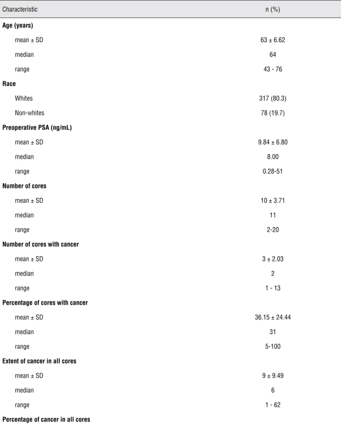
Table 1 - Clinicopathologic characteristics of 400 study patients submitted to radical prostatectomy for prostate cancer. SD, standard deviation.
Characteristic n (%)
Age (years)
mean ± SD 63 ± 6.62
median 64
range 43 - 76
Race
Whites 317 (80.3)
Non-whites 78 (19.7)
Preoperative PSA (ng/mL)
mean ± SD 9.84 ± 6.80
median 8.00
range 0.28-51
Number of cores
mean ± SD 10 ± 3.71
median 11
range 2-20
Number of cores with cancer
mean ±SD 3 ± 2.03
median 2
range 1 - 13
Percentage of cores with cancer
mean ± SD 36.15± 24.44
median 31
range 5-100
Extent of cancer in all cores
mean ± SD 9 ± 9.49
median 6
range 1 - 62
mean ± SD 18.5 ± 18.28
median 12
range 0.4 - 100
Maximum extent of cancer per core
mean ± SD 4 ± 3.31
median 3.5
range 1 - 20
Maximum percentage of cancer per core
mean ± SD 51.58± 30.14
median 47
range 1 - 100
Gleason score in RP
≤ 6 134 (33.8)
3 + 4 = 7 186 (46.9)
4 + 3 = 7 53 (13.4)
≥ 8 24 (6.0)
Tumor extent in RP (positive points)
mean ± SD 35.80
median 27
range 1 - 225
Seminal vesicle invasion
absent 353 (89.6)
present 41 (10.4)
Extraprostatic extension
absent 254 (64.0)
focal 40 (10.1)
diffuse 103 (25.9)
Positive surgical margins
absent 213 (53.5)
focal 49 (12.3)
diffuse 136 (34.2)
Table 2 - Kaplan-Meier product-limit analysis for time to biochemical recurrence following radical prostatectomy according to several clinicopathologic characteristics.
Characteristic X2 Log-rank, p
Age (years)
≤ 55 vs > 55 < 0.001 0.997
Race
Whites vs non-Whites 0.363 0.547
Maximum percentage of cancer per core
≤ 47% vs. > 47% 1.017 0.313
Number of cores with cancer
≤ 2 vs. > 2 2.195 0.138
Tumor extent in RP
≤ 27 positive points vs > 27 positive points 5.726 0.017
Maximum extent of cancer per core
≤ 3.5 mm vs. > 3.5 mm 5.745 0.017
Percentage of cores with cancer
≤ 31% vs. > 31% 5.768 0.016
Extent of cancer in all cores
≤ 6 mm vs. > 6 mm 7.424 0.006
Percentage of cancer in all cores
≤ 12% vs > 12% 10.785 0.001
Extraprostatic extension
absent vs diffuse 12.430 < 0.001
Figure 1 - Kaplan-Meier product-limit analysis shows time to PSA biochemical progression-free outcome by the median value of percentage of cancer extent in mm in all cores. Cum, cumulative.
Gleason score in RP
≤ 6 vs 3 + 4 = 7 vs 4 + 3 = 7 vs ≥ 8 13.477 0.004
Preoperative PSA (ng/mL)
≤ 10 vs >10 25.800 < 0.001
Seminal vesicle invasion
absent vs present 26.071 < 0.001
Positive surgical margins
absent vs focal vs. diffuse 33.562 < 0.001
absent vs focal 5.350 0.069
Figure 3 - Kaplan-Meier product-limit analysis shows time to PSA biochemical progression-free outcome by Gleason score ≤ 6, 3+4=7, 4+3=7, and ≥ 8 in surgical specimens. Cum, cumulative.
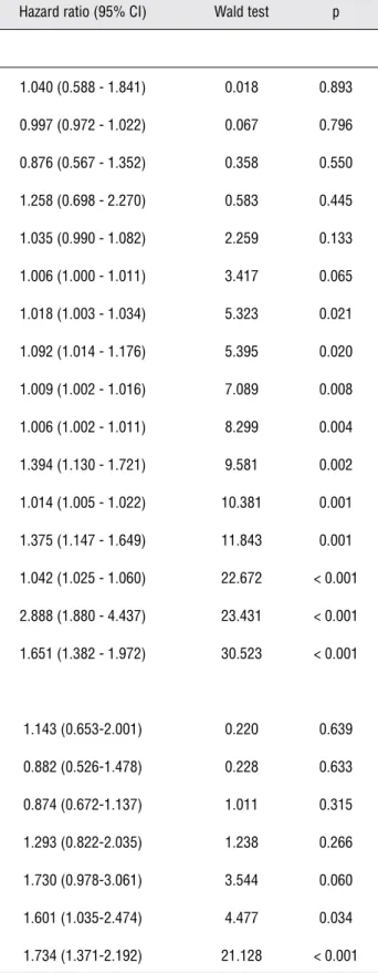
Table 3 - Cox univariate and multivariate proportional hazard analysis of several clinicopathologic characteristics predicting shorter time to biochemical recurrence following radical prostatectomy. CI, confidence interval.
Characteristic Hazard ratio (95% CI) Wald test p
Univariate analysis
Focal EPE 1.040 (0.588 - 1.841) 0.018 0.893
Age 0.997 (0.972 - 1.022) 0.067 0.796
Race 0.876 (0.567 - 1.352) 0.358 0.550
Focal PSM 1.258 (0.698 - 2.270) 0.583 0.445
Maximum extent of cancer per core 1.035 (0.990 - 1.082) 2.259 0.133
Maximum percentage of cancer per core 1.006 (1.000 - 1.011) 3.417 0.065
Extent of cancer in all cores 1.018 (1.003 - 1.034) 5.323 0.021
Number of cores with cancer 1.092 (1.014 - 1.176) 5.395 0.020
Percentage of cores with cancer 1.009 (1.002 - 1.016) 7.089 0.008
Tumor extent in RP 1.006 (1.002 - 1.011) 8.299 0.004
Gleason score in RP 1.394 (1.130 - 1.721) 9.581 0.002
Percentage of cancer in all cores 1.014 (1.005 - 1.022) 10.381 0.001
Diffuse EPE 1.375 (1.147 - 1.649) 11.843 0.001
Preoperative PSA 1.042 (1.025 - 1.060) 22.672 < 0.001
Seminal vesicle invasion 2.888 (1.880 - 4.437) 23.431 < 0.001
Diffuse PSM 1.651 (1.382 - 1.972) 30.523 < 0.001
Multivariate analysis
Gleason score in RP 1.143 (0.653-2.001) 0.220 0.639
Tumor extent in RP 0.882 (0.526-1.478) 0.228 0.633
Diffuse EPE 0.874 (0.672-1.137) 1.011 0.315
Percentage of cancer in all cores 1.293 (0.822-2.035) 1.238 0.266
Seminal vesicle invasion 1.730 (0.978-3.061) 3.544 0.060
Preoperative PSA 1.601 (1.035-2.474) 4.477 0.034
Gleason score in radical prostatectomy
Tumors with a Gleason score of 7 have significantly worse prognosis than those with a Gleason score ≤ 6. Given the adverse prognosis as-sociated with Gleason pattern 4, one would expect whether a tumor Gleason score 3 + 4 = 7 or 4 + 3 = 7 would influence prognosis. No significant sur-vival advantage was reported for Gleason score 3 + 4 = 7 over 4 + 3 = 7 by Oefelein et al. (21). Other investigators have shown that Gleason score 4 + 3 = 7 has a worse prognosis than Gleason score 3 + 4 = 7 (18,19). Due to this controversy, the prog-nostic Gleason grouping according to score may be either ≤ 6, 7, ≥ 8 or ≤ 6, 3 + 4 = 7, 4 + 3 = 7, ≥ 8. In our cohort of patients the Kaplan-Meier curve showed that the Gleason score 4 + 3 = 7 behaved similarly to score ≥ 8 and different to score 3 + 4 = 7. This result discloses the importance of grade 4 as the predominant pattern and favors a 4 score prognostic grouping: ≤ 6, 3 + 4 = 7, 4 + 3 = 7, and
≥ 8. Gleason score in our study was significantly predictive of TBR in univariate analysis but not in multivariate analysis probably due to fewer cases in the prognostic groups 4 + 3 = 7 and ≥ 8. Ano-ther limitation could be the relatively short mean follow-up time.
Extraprostatic extension and positive surgical margins
Extraprostatic extension and positive sur-gical margins have prognostic importance and may influence therapeutic options after surgery. However, the amount of EPE and PSM seems to be more important than a simple report of presence or absence of these pathological findings (24-26).
During the International Society of Uro-logical Pathology (ISUP) consensus conference on handling and staging of radical prostatectomy spe-cimens held in Boston during the 98th meeting of the United States and Canadian Academy of Pa-thology (USCAP), no consensus could be reached as to how evaluate extent of EPE and PSM (27,28). In this study we evaluated the influence of focal and diffuse EPE and PSM on TBR using a simple method for EPE and PSM extent quantitation that can be used in the daily practice of all pathologists who step section and totally process the surgical specimen. Considering that in the 2009 version of
the TNM Classification of Malignant Tumors mi-croscopic involvement of the bladder neck is pT3a, in our study this finding was included as EPE.
Our study is in accordance with authors that consider extent of EPE or PSM an important information in the pathology report. Diffuse EPE or PSM were significantly associated with shorter time of TBR in univariate analysis, however, only diffuse PSM was significantly associated with TBR in univariate (p < 0.001) and multivariate analysis (p < 0.001). Diffuse PSM was the strongest and an independent predictor of TBR in our cohort of patients. The incidence of PSM in the literature ranges from 16% to 50% (34). In our study the incidence was 46.5%. One of the largest potential sources of discrepancy for incidence of PSM is the different methods used to process the radical pros-tatectomy specimens. It is also worth mention that positive surgical margins result of surgical factors more than biology of disease.
Tumor extent on needle prostatic biopsies
Recent efforts have focused on incorpo-rating prostate needle biopsy measurements as an adjunct to improve pretreatment risk stratification (35-38). However, it is controversial which is the best predictor method (16-18). Bismar et al. (18) studied the prediction of pathologic stage in a scre-ening population of multiple measures of carcino-ma on prostate needle biopsy tissue. In univariate analysis all measures were significantly associa-ted with pathologic stage > T2 but in multivariate analysis the percentage of positive cores was the strongest predictor.
In our study, except for maximum per-centage of cancer per core and number of cores with cancer all other methods were significantly predictive of TBR using the curves of Kaplan--Meier. Using the Cox univariate analysis, all measures were statistically predictive of risk to TBR except maximum extent and percentage of cancer per core. The strongest predictor was per-centage of cancer in all cores, however was not an independent predictor of shorter time of TBR in multivariate analysis.
All methods for measurement are sim-ple and may be done in the daily practice of the surgical pathologist. However, they are not equi-valent in application because measuring with an ocular micrometer foci of carcinoma can be time consuming. Total number and percentage of po-sitive cores are the easiest way of evaluation ac-cessible to all pathologists, and in our study were statistically significant in univariate analysis.
In summary, age and race were not sig-nificantly associated with TBR after surgery, ho-wever other more powered studies could show significance. Tumor extent evaluation in the surgical specimen was predictive of TBR in uni-variate analysis but not in multiuni-variate analysis favoring that this time consuming report does not add additional information to other well es-tablished predictive findings. The higher predic-tive value of Gleason score 4 + 3 = 7 vs 3 + 4 = 7 disclosed the importance of grade 4 as the pre-dominant pattern and favors a 4 score prognostic grouping: ≤ 6, 3 + 4 = 7, 4 + 3 = 7, and ≥ 8. Ex-tent of EPE or PSM is an important information in the pathology report. Diffuse EPE or PSM were significantly associated with TBR in univariate analysis and diffuse PSM was independent and the strongest predictor of shorter time to BR in multivariate analysis. Most tumor extent evalua-tions on needle biopsies were predictive of TBR, however, maximum percentage of cancer in all cores was the strongest predictor. While shedding light to important and controversial predictors of biochemical recurrence after radical prostatec-tomy among Latin Americans, limitations of this study are the relatively short follow-up and the retrospective design, even in a prospectively col-lected database.
ABBREVIATIONS
PSA = prostate specific antigen;
SD = standard deviation;
CI = confidence interval;
RP = radical prostatectomy;
BR = biochemical recurrence;
TBR = time to biochemical recurrence;
EPE = extraprostatic extension;
PSM = positive surgical margin
CONFLICT OF INTEREST
None declared.
REFERENCES
1. Cookson MS, Aus G, Burnett AL, Canby-Hagino ED, D’Amico AV, Dmochowski RR, et al.: Variation in the definition of bio-chemical recurrence in patients treated for localized prostate cancer: the AmericanUrological Association Prostate Guide-lines for Localized Prostate Cancer Update Panel report and recommendations for astandard in the reporting of surgical outcomes. J Urol. 2007; 177: 540-5.
2. Epstein JI, Amin M, Boccon-Gibod L, Egevad L, Humphrey PA, Mikuz G, et al.: Prognostic factors and reporting of prostate carcinoma in radical prostatectomy and pelvic lymphadenec-tomy specimens. Scand J Urol Nephrol Suppl. 2005; 34-63. 3. Carter HB, Epstein JI, Partin AW: Influence of age and
prostate-specific antigen on the chance of curable prostate cancer among men with nonpalpabledisease. Urology. 1999; 53: 126-30. 4. Herold DM, Hanlon AL, Movsas B, Hanks GE: Age-related
prostate cancer metastases. Urology. 1998; 51: 985-90. 5. Bauer JJ, Connelly RR, Seterhenn IA, Deausen J, Srivastava
S, McLeod DG, et al.: Biostatistical modeling using traditional preoperative and pathological prognostic variables in the se-lection of men at highrisk for disease recurrence after radical prostatectomy for prostate cancer. J Urol. 1998; 159: 929-33. 6. Catalona WJ, Smith DS: Cancer recurrence and survival rates
after anatomic radical retropubic prostatectomy for prostate cancer: intermediate-term results. J Urol. 1998; 160: 2428-34. 7. Evans S, Metcalfe C, Ibrahim F, Persad R, Ben-Shlomo Y: In-vestigating Black-White differences in prostate cancer prog-nosis: A systematic review and meta-analysis. Int J Cancer. 2008; 123: 430-5.
9. Powell IJ, Bock CH, Ruterbusch JJ, Sakr W: Evidence supports a faster growth rate and/or earlier transformation to clinically significant prostate cancer in black than inwhite American men, and influences racial progression and mortality disparity. J Urol. 2010; 183: 1792-6.
10. Cross CK, Shultz D, Malkowicz SB, Huang WC, Whittington R, Tomaszewski JE, et al.: Impact of race on prostate-specific antigen outcome after radical prostatectomy for clinically local-ized adenocarcinoma of the prostate. J Clin Oncol. 2002; 20: 2863-8.
11. Freedland SJ, Jalkut M, Dorey F, Sutter ME, Aronson WJ: Race is not an independent predictor of biochemical recurrence after radical prostatectomy in an equal access medical center.Urol-ogy. 2000; 56: 87-91.
12. Paschoalin EL, Martins AC, Pastorello M, Sândis KA, Maciel LM, Silva WA Jr, et al.: Racial influence on the prevalence of prostate carcinoma in Brazilian volunteers. Int Braz J Urol. 2003; 29: 300-5.
13. Barros MS, Silva VR, Santos GB, Hughes A, Silveira MA: Prevalence of prostate adenocarcinoma according to race in an university hospital. Int Braz J Urol. 2003; 29: 306-11; discus-sion 312.
14. Romero FR, Romero AW, Almeida RM, Tambara Filho R: The prevalence of prostate cancer in Brazil is higher in Black men than in White men: systematic review and meta-analysis. Int Braz J Urol. 2012; 38: 440-7.
15. Romero FR, Romero AW, de Almeida RM, de Oliveira FC Jr, Tambara Filho R: The significance of biological, environmental, and social risk factors for prostate cancer in a cohort study in Brazil. Int Braz J Urol. 2012; 38: 769-78.
16. Brimo F, Vollmer RT, Corcos J, Kotar K, Bégin LR, Humphrey PA, et al.: Prognostic value of various morphometric measure-ments of tumour extent in prostate needle core tissue. Histo-pathology. 2008; 53: 177-83.
17. Epstein JI, Potter SR: The pathological interpretation and sig-nificance of prostate needle biopsy findings: implications and current controversies. J Urol. 2001; 166: 402-10.
18. Bismar TA, Lewis JS Jr, Vollmer RT, Humphrey PA: Multiple measures of carcinoma extent versus perineural invasion in prostate needle biopsy tissue in prediction ofpathologic stage in a screening population. Am J Surg Pathol. 2003; 27: 432-40. 19. Humphrey PA, Vollmer RT: Intraglandular tumor extent and
prognosis in prostatic carcinoma: application of a grid method to prostatectomy specimens. Hum Pathol. 1990; 21: 799-804. 20. Salomon L, Levrel O, Anastasiadis AG, Irani J, De La Taille A,
Saint F, et al.: Prognostic significance of tumor volume after radical prostatectomy: a multivariate analysis of pathological prognosticfactors. Eur Urol. 2003; 43: 39-44.
21. Oefelein MG, Smith ND, Grayhack JT, Schaeffer AJ, McVary KT: Long-term results of radical retropubic prostatectomy in men with high grade carcinoma of the prostate. J Urol. 1997; 158: 1460-5.
22. Han M, Partin AW, Pound CR, Epstein JI, Walsh PC: Long-term biochemical disease-free and cancer-specific survival following anatomic radical retropubic prostatectomy. The 15-year Johns Hopkins experience. Urol Clin North Am. 2001; 28: 555-65.
23. Chan TY, Partin AW, Walsh PC, Epstein JI: Prognostic signifi-cance of Gleason score 3+4 versus Gleason score 4+3 tumor at radical prostatectomy. Urology. 2000; 56: 823-7.
24. Sung MT, Lin H, Koch MO, Davidson DD, Cheng L: Radial distance of extraprostatic extension measured by ocular micrometer is an independent predictor of prostate-specific antigen recurrence: A new proposal for the substaging of pT3a prostate cancer. Am J Surg Pathol. 2007; 31: 311-8. 25. Shikanov S, Song J, Royce C, Al-Ahmadie H, Zorn K,
Stein-berg G, et al.: Length of positive surgical margin after radical prostatectomy as a predictor of biochemical recurrence. J Urol. 2009; 182: 139-44.
26. van Oort IM, Bruins HM, Kiemeney LA, Knipscheer BC, Witjes JA, Hulsbergen-van de Kaa CA: The length of positive surgical margins correlates with biochemical recurrence af-ter radical prostatectomy. Histopathology. 2010; 56: 464-71. 27. Magi-Galluzzi C, Evans AJ, Delahunt B, Epstein JI, Griffiths
DF, van der Kwast TH, et al.: International Society of Urologi-cal Pathology (ISUP) Consensus Conference on Handling and Staging of RadicalProstatectomy Specimens. Working group 3: extraprostatic extension, lymphovascular invasion and locally advanceddisease. Mod Pathol. 2011; 24: 26-38. 28. Tan PH, Cheng L, Srigley JR, Griffiths D, Humphrey PA,
van der Kwast TH, et al.: International Society of Urologi-cal Pathology (ISUP) Consensus Conference on Handling and Staging of RadicalProstatectomy Specimens. Working group 5: surgical margins. Mod Pathol. 2011; 24: 48-57. 29. Billis A, Magna LA, Ferreira U: Correlation between tumor
extent in radical prostatectomies and preoperative PSA, his-tological grade, surgical margins, andextraprostatic exten-sion: application of a new practical method for tumor extent evaluation.Int Braz J Urol. 2003; 29: 113-9; discussion 120. 30. Epstein JI, Allsbrook WC Jr, Amin MB, Egevad LL; ISUP
Grading Committee: The 2005 International Society of Uro-logical Pathology (ISUP) Consensus Conference on Gleason Grading of ProstaticCarcinoma. Am J Surg Pathol. 2005; 29: 1228-42.
31. Parker CC, Gospodarowicz M, Warde P: Does age influence the behaviour of localized prostate cancer? BJU Int. 2001; 87: 629-37.
32. Latini DM, Elkin EP, Cooperberg MR, Sadetsky N, Duch-ane J, Carroll PR: Differences in clinical characteristics and disease-free survival for Latino, African American, and non-Latino white men withlocalized prostate cancer: data from CaPSURE. Cancer. 2006; 106: 789-95.
34. Watson RB, Civantos F, Soloway MS: Positive surgical mar-gins with radical prostatectomy: detailed pathological analy-sis and prognoanaly-sis. Urology. 1996; 48: 80-90.
35. D’Amico AV, Whittington R, Malkowicz SB, Schultz D, Fon-durulia J, Chen MH, et al.: Clinical utility of the percentage of positive prostate biopsies in defining biochemical outcome after radical prostatectomyfor patients with clinically local-ized prostate cancer. J Clin Oncol. 2000; 18: 1164-72. 36. Grossfeld GD, Latini DM, Lubeck DP, Broering JM, Li YP,
Mehta SS, et al.: Predicting disease recurrence in intermedi-ate and high-risk patients undergoing radical prostintermedi-atectomy using percentpositive biopsies: results from CaPSURE. Urol-ogy. 2002; 59: 560-5.
37. Presti JC Jr, Shinohara K, Bacchetti P, Tigrani V, Bhargava V: Positive fraction of systematic biopsies predicts risk of relapse after radical prostatectomy. Urology. 1998; 52: 1079-84.
38. Freedland SJ, Csathy GS, Dorey F, Aronson WJ: Percent prostate needle biopsy tissue with cancer is more predic-tive of biochemical failure or adverse pathology after radi-cal prostatectomy than prostate specific antigen or Gleason score. J Urol. 2002; 167: 516-20.
_____________________
Correspondence address: Athanase Billis, MD, PhD Depatment of Anatomic Pathology School of Medical Sciences State University of Campinas (Unicamp)