DOI: 10.1590/0004-282X20150215
ARTICLE
Surgical clipping is still a good choice for the
treatment of paraclinoid aneurysms
A clipagem cirúrgica é uma boa opção para o tratamento de aneurismas paraclinóideos
Felix Hendrik Pahl1,2,3, Matheus Fernandes de Oliveira1,2,3, Roger Schmidt Brock2,3,JoséErasmo Dal Col
Lucio 2,3, José Marcus Rotta1
Paraclinoid aneurysms are lesions located adjacent to the clinoid and ophthalmic segments of the internal carotid ar-tery, distal to the proximal dural ring and proximal to the
pos-terior communicating artery origin. hey are usually divided
into clinoid segment aneurysms and ophthalmic-hypophyseal segment aneurysms. Frequently, part of an ophthalmic seg-ment aneurysm occupies the clinoid segseg-ment. Paraclinoid aneurysms are an uncommon cause of aneurysmal subarach-noid hemorrhage, and, in large series, account for approxi-mately 1.4–9.1% of all patients with ruptured aneurysms1,2,3,4,5.
Because of their location close to the skull base, paracli-noid aneurysms can be challenging to repair surgically, due to proximity with the optic apparatus, bone structures, and
cavernous sinus; surgery often requires extensive drilling of
the roof of the optic canal, anterior clinoid process (ACP) and
optic strut to obtain proximal control and expose the aneu-rysm neck in its entirety6,7,8,9,10. he diiculty of proximal con
-trol and the narrow operative ield might lead to a higher
frequency of failed clipping procedures, as well as to higher surgical morbidity and mortality. Because of these challenges,
paraclinoid aneurysms have been one of the most common indications for endovascular treatment. Nevertheless, despite availability of adjunctive techniques, such as balloon-assisted
and stent-assisted coiling, coil embolization continues to be associated with a high rate of residual/recurrent
aneurysm illing11,12,13,14,15.
In recent years, low diverter stents (FDS) have been intro
-duced as an alternative and more efective endovascular tech -nique than coil embolization, and in April 2011, the Pipeline
Embolization Device (Chestnut Medical Technologies, Menlo
1IAMSPE, Hospital do Servidor Público Estadual de São Paulo, Departamento de Neurocirurgia, Sao Paulo SP, Brazil;
2Hospital Sirio Libanês, Departamento de Neurocirurgia, Sao Paulo SP, Brazil;
3DFV Neuro, São Paulo SP, Brazil.
Correspondence: Felix Hendrik Pahl; Alameda Franca, 432 / apt. 31; 01422-002 São Paulo SP, Brasil; E-mail: fpahl@globo.com Conflict of interest: There is no conlict of interest to declare.
Received 17 August 2015; Received in inal form 27 October 2015; Accepted 26 November 2015.
ABSTRACT
Paraclinoid aneurysms are lesions located adjacent to the clinoid and ophthalmic segments of the internal carotid artery. In recent years, low diverter stents have been introduced as a better endovascular technique for treatment of these aneurysms. Method: From 2009 to 2014, a total of 43 paraclinoid aneurysms in 43 patients were surgically clipped. We retrospectively reviewed the records of these patients to analyze clinical outcomes. Results: Twenty-six aneurysms (60.5%) were ophthalmic artery aneurysms, while 17 were superior hypophyseal artery aneurysms (39.5%). The extradural approach to the clinoid process was used to clip these aneurysms. One hundred percent of aneurysms were clipped (complete exclusion in 100% on follow-up angiography). The length of follow-up ranged from 1 to 60 months (mean, 29.82 months). Conclusion: Surgical clipping continues to be a good option for the treatment of paraclinoid aneurysms.
Keywords: intracranial aneurysm; endovascular treatment; surgery.
RESUMO
Aneurismas paraclinóideos são lesões localizadas adjacentes aos segmentos clinóideos e oftálmicos da artéria carótia interna. Os stents desviadores de luxo tem sido crescentemente aplicados com sucesso. Métodos: De 2009 a 2014, um total de 43 aneurismas paraclinóideos foram clipados em 43 pacientes. Analisamos retrospectivamente os dados dos pacientes e desfechos clínicos. Resultados: Vinte seis aneurismas (60,5%) foram de artéria oftálmica e 17 de artéria hipoisária superior (39,5%). O acesso extradural à clinóide foi utilizado para todos aneurismas. Cem por cento dos aneurismas foram clipados com oclusão de 100% na angiograia controle. O tempo de follow-up oscilou de 1 a 60 meses, com media de 29 meses. Conclusão: A clipagem cirúrgica é uma opção boa e segura para o tratamento de aneurismas paraclinóideos.
Park, CA) was approved by the FDA for treatment of large or
giant wide-neck intracranial aneurysms in the proximal in-tracranial ICA, including the ophthalmic segment1,13,16,17.
Good results have been published in the literature with the use of FDS, with occlusion rates of up to 90% and compli
-cations lower than 5%, however surgical clipping remains an acceptable option to achieve high occlusion rates with aver -age complications1,2,3,4,5,18,19,20.
he purpose of this study is to expose our surgical results and match them with those of the FDS era.
METHOD
his paper describes the surgical results of a senior vas -cular neurosurgeon (Pahl, FH). Indications for neurosurgi-cal treatment of paraclinoid aneurysms were unruptured
aneurysms with 5mm or above, symptomatic aneurysms (visual deicits) and/or associated subarachnoid hemor
-rhage (SAH).
From 2009 to 2014 (5 years), a total of 43 paraclinoid
an-eurysms in 43 patients were surgically clipped. We retrospec
-tively reviewed the records of these patients to analyze clini
-cal outcomes, which are expressed as modiied Rankin s-cale (mRs). Additionally, we divided aneurysms in two groups ac
-cording to size (< 10 mm and 10 mm or above) to compare
surgical results in both groups.
Data distribution was evaluated with Kolmogorov-Smirnov test when applicable. Statistical analysis was per
-formed using Chi-Square test and multiple variate analysis.
Sample data (Table 1)
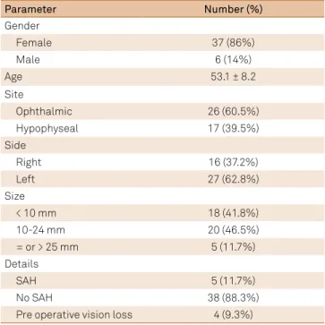
Of the 43 patients, 37 (86%) were women and 6 (13%)
were men. Overall, ages ranged between 34 and 74 years old, with the mean age of 53.18 years and a standard deviation of 8.2 years. he mean age was 53.3 years among men and 53.1
years among women.
Twenty-six aneurysms (60.5%) were ophthalmic artery
aneurysms, while 17 were superior hypophyseal artery aneu-rysms (39.5%). Among men, four aneuaneu-rysms were superior
hypophyseal and two were ophthalmic; among women, 13 were superior hypophyseal and 24 were ophthalmic. Sixteen
aneurysms were located on the right (37.2%) and 27 (62.8%) on the left.
Aneurysm size ranged from 2 to 25 mm (mean, 12.3 mm). Eighteen (41.8%) were smaller than 10 mm, 20 (46.5%) were in the 10 to 24 mm range, and 5 (11.7%) were 25 mm or
larg-er. Five (11.7%) of the 37 patients sufered SAH, including the case with a 2mm aneurysm. he other 38 patients had un -ruptured aneurysms.
Four patients (9.3%) had preoperative vision loss: three
ipsilateral to the aneurysm and one with bitemporal
hemi-anopia. hree patients recovered vision after surgical decom -pression of the optic apparatus.
RESULTS
Surgical technique
he extradural approach to the ACP was used to clip
these aneurysms (Figure 1). In ophthalmic segment aneu-rysms, the last part of the ACP was taken out intradurally, facing the aneurysm itself. In superior hypophyseal aneu-rysms, the ACP was taken out in completely extradural fashion, because of the low risk of damaging the aneurysm during drilling.
Proximal control of the neck was used in aneurysms with clinoid segment extension only, because of the high
risk of drilling with the aneurysm under the ACP; other
-wise, proximal control was achieved at the clinoid segment
of the ICA.
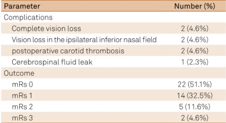
Complications
There were seven complications in seven patients (Table 2). Complete vision loss occurred in two patients, and vision loss in the ipsilateral inferior nasal field oc
-curred in two others. There were two cases of postopera
-tive carotid thrombosis. The first occurred at the site of
proximal control in the neck, with contralateral hemipare-sis and aphasia due to intimal dissection, and the second was due to accidental injury of the right carotid with trap-ping, which resulted in contralateral left hemiparesis. In
the latter case, the patient also developed a cerebrospinal fluid (CSF) leak, which was treated successfully with lum -bar drainage for 1 week.
Table 1. Summarized demographic of patients submitted to microneurosurgical treatment of paraclinoid aneurysms.
Parameter Number (%)
Gender
Female 37 (86%)
Male 6 (14%)
Age 53.1 ± 8.2
Site
Ophthalmic 26 (60.5%)
Hypophyseal 17 (39.5%)
Side
Right 16 (37.2%)
Left 27 (62.8%)
Size
< 10 mm 18 (41.8%)
10-24 mm 20 (46.5%)
= or > 25 mm 5 (11.7%)
Details
SAH 5 (11.7%)
No SAH 38 (88.3%)
Pre operative vision loss 4 (9.3%)
Outcome (Table 2)
he length of follow-up ranged from 1 to 60 months
(mean, 29.82 months). One hundred percent of aneurysms were clipped (complete exclusion in 100% on follow-up
angiography – Figure 2). Twenty-two (51.1%) patients were discharged with a mRs of 0, and 14 patients (32.5%) with a mRs score of 1. In ive patients (11.6%), the mRs was 2, and in two patients (4.6%), the mRs was 3.
Two patients with no previous visual deicits lost vision in the ipsilateral eye, and two others had partial visual loss in the nasal ield. Conversely, three patients with preoperative visual loss recovered their sight postoperatively.
here were no deaths in this case series, despite the com -plexity of the lesions.
Small aneurysms (< 10mm) versus large aneurysms (10mm or above) (Table 3)
In the small aneurysm group, from the eighteen patients,
ive were men (27.7%) and thirteen were women (72.3%). Mean age was 49.2 years old and aneurysm were of ophthal -mic artery in 12 patients (66.6%) and hypophyseal in 6
pa-tients (33.4%). here were four complications in four papa-tients. Eight (44.4%) patients were discharged with a mRs of 0, 5 pa
-tients (27.7%) with a mRs score of 1. In three pa-tients (16.6%), the mRs was 2, and in two patients (11.1%), the mRs was 3. SAH happened in three patients (1 male and 2 females), being
one hypophiseal aneurysm and two ophthalmic.
In the large aneurysm group, from the twenty-ive pa -tients, twenty-four were women (96%) and just one man
(4%). Mean age was 55.8 years old and aneurysm were of
ophtalmic artery in 14 patients (56%) and hypophyseal in 11
patients (44%). here were three complications in three pa
-tients. All patients three patients with post operative vision recovery were from this group. Fourteen (56%) patients were discharged with a mRs of 0, and nine patients (36%) with a mRs score of 1. In two patients (2%), the mRs was 2. SAH hap -pened in two female patients, being one hypophiseal aneu-rysm and one ophthalmic.
Table 2. Surgical data of patients (complications and outcomes).
Parameter Number (%)
Complications
Complete vision loss 2 (4.6%)
Vision loss in the ipsilateral inferior nasal ield 2 (4.6%) postoperative carotid thrombosis 2 (4.6%)
Cerebrospinal luid leak 1 (2.3%)
Outcome
mRs 0 22 (51.1%)
mRs 1 14 (32.5%)
mRs 2 5 (11.6%)
mRs 3 2 (4.6%)
mRs: modiied Rankin scale.
Table 3. Comparison between small and large aneurysms.
Parameter Small aneurysms
(< 10 mm)
Large aneurysms (10 mm or >)
Number of patients 18 25
Gender*
Male 5 1
Female 13 24
Mean age (years)* 49.2 55.8
Aneurysmal site
Ophthalmic 12 14
Hypophyseal 6 11
Number of SAH 3 2
Number of complications 4 3
Aneurysmal occlusion 100% 100%
Percentage of good outcome (mRs 0 or 1)
73% 92%
* = statistically signiicant difference (p < 0.05). SAH: subarachnoid hemorrhage; mRs: modiied Rankin scale.
Figure 1. Standard surgical approach to paraclinoid aneurysms. In (A) positioning and incision to pterional craniotomy. In (B) pterional craniotomy and drilling of orbital roof. In (C) Clinoidal space after ressection of anterior clinoidal process. In (D) opening of duramater. In (E) Ophthalmic artery aneurysm revealing proximal and distal neck. In (F) aneurysm after clipping.
C
D
E
F
A
B
Figure 2. Typical subject from the sample. In (A) pre operative angiography revealing large aneurysm. In (B) post operative angiography, with complete exclusion of aneurysm.
After performing multivariate analysis, there was no sta
-tistically signiicant diference between both groups when evaluating aneurismal site, complications, SAH and outcome in mRs (p > 0.05). here was statistical association of age and size of aneurysm and gender and size of aneurysm (p < 0.05).
Large aneurysms were more frequent in women than in men and patients with large aneurysms were older than those with small aneurysms.
DISCUSSION
SAH following intracranial aneurysmal rupture is a major
cause of morbidity and mortality23,24,25. Several factors may
interfere with the probability of rupture, such as smoking, use of alcohol, size, shape, location of the aneurysm, presence of
intraluminal thrombus and even the gender of the patient. Korja et al.18 disclosed that even patients with small (< 7 mm)
unruptured aneurysms could have a lifelong risk for rupture
of up to 25%, depending on many factors.
Although being uncommon causes of SAH, paraclinoid aneurysms may promote varied symptomatology, espe
-cially due to compression of optic nerve and surrounding
structures24,25. heir treatment is challenging and demanding,
whether by surgical or endovascular approaches. Recent en
-dovascular advances, such as FDS, have been introduced as a promising treatment alternative, being more efective than
coil embolization. In experienced hands, surgical treatment of these lesions can be accomplished with quite high success
rates, but carry signiicant morbidity1,2,3,4,5.
In our sample of 43 patients, 100% of aneurysms were clipped (complete exclusion in 100% on follow-up angiogra-phy). ICA occlusion occurred in two cases (4.6%) and was
un-related to clipping itself. here were no cases of rebleeding during the follow-up period. A good outcome (mRs of 0 or 1) was achieved in 84% of patients, with no diference related to aneurysm size. here was no mortality. he rate of vision loss was 9% (4.5% total ipsilateral vision loss; 4.5% partial ip
-silateral vision loss), and the rate of vision recovery, 7%. he rate of vision recovery among patients with previous visual deicits was 75% (three out of four patients). here was no statistically signiicant diference between small and large aneurysms when evaluating aneurysmal site, complications, SAH and outcome in mRs (p > 0.05). Conversely, there was a
clear trend for more complications in small aneurysm group.
here is no apparent rationale for such inding, and we be
-lieve it is an aleatory inding, without statistical signiicance. Several series have reported the outcomes of patients un
-dergoing surgical or endovascular treatment. In endovascu -lar approaches with stent-assisted coiling, balloon-assisted
coiling, coiling without adjunctive techniques, and stent -ing alone, complication rates approach 15%, and total
aneu-rysm occlusion is achieved in up to 50-85% of patients. All above endovascular techniques appear equally successful.
Procedure-related complications may be observed in 10% of patients, visual complications in 8% and the recurrence rate
may be up to 20% during follow-up1,2,3,4,5,20,21,22,23,24,25,26. Some
authors propose endovascular treatment as an efective mean for small paraclinoid aneurysms (≤ 10 mm) with a low
rate of recurrence. In contrast, large paraclinoid aneurysms
(> 10 mm) may exhibit a high rate of recurrence27,28,29.
Increasing evidence on FDS in paraclinoid aneurysms have demonstrated successful application in up to 100% of
aneurysms, near 90% complete or near-complete
oblitera-tion on angiographic follow-up, preserved patency of the
ophthalmic artery, and minor procedure-related
complica-tions, clearly qualifying FDS as an endovascular approach with superior results compared to other endovascular treat -ments, without an increased rate of complications1,30.
he largest experience to our knowledge treated 107
patients with only one procedure related complication and 9% of recurrence15. On the other hand, some
com-plications are described but poorly understood, such as post-procedure aneurismal bleeding and occlusion of the ophthalmic artery at its origin, which may produce undesir-able outcomes1,19,20,21,22,23,24,25,26.
In a study by Zanaty et al.30, forty-one patients harboring
44 paraclinoid aneurysms were treated by low-diversion. At inal angiographic follow-up, 77.2% had complete oc -clusion, 6.8% had near-complete occlusion and 15.9% had incomplete occlusion30. Of the 22 symptomatic, complete
resolution or signiicant improvement was noted in 72.7%,
while worsening of symptoms occurred in 4.5%30. Five pa
-tients out of 22 (22.7%) had no signiicant changes in their symptoms. he complication rate was 2.2% and mortality
rate was 0%30.
Another study by Moon et al.1 evaluated 29 patients with
38 aneurysms submitted to FDS. It was successfully deployed
for all lesions, with 92.1% complete or near-complete oblit-eration rate at angiographic follow-up1. All but one patient
were found to have a patent ophthalmic artery at short-term follow-up and 100% of patients retained intact vision1. Five
patients had minor periprocedural hemorrhagic
complica-tions but no permanent morbidities. here were no intra
-cranial hemorrhages, thromboembolic phenomena, vessel
dissections, or mortalities1.
Despite sample heterogeneity, the results of surgical re
-pair of paraclinoid aneurysms are well documented. Surgical success can be high (over 90%), with acceptable complica
-tions, morbidity and mortality. Good outcomes are achieved in 70-90% of patients, with progressive improvement of im
-mediate postoperative deicits on late follow-up. he proxim -ity of paraclinoid lesions to the ophthalmic artery and optic
nerve is the main concern, with risk of visual loss. However, the possibility of direct decompression of the optic nerve after aneurysmal treatment may reverse visual deicits.
Restoration of eyesight in patients with preopera
-tive visual loss is a surgical advantage less reported in endovascular series, but can be achieved with clipping. In our series, three of four patients with preoperative visual loss recovered their sight1,2,4,7,23.
Although our results with surgical treatment were
ani-mating, they still disclose signiicant morbidity rates, like 4.6% of complete and 4.6% partial ipsilateral vision loss and are not strong enough to provide evidence of superiority of surgery over endovascular means.
Besides, even in expert hands, paraclinoid aneu
-rysms do have significant surgical morbidity. Lower endovascular morbidity and widespread availability of endovascular therapy make this the chosen alternative for many of these aneurysms, however lower complete
obliteration rate and individual anatomical anomalies
suggest the need for surgical options1,30.
Anyway, potential outcome predictors in paraclinoid
aneurysms include the presence of SAH, vasospasm, in -farcts, hydrocephalus and patient age. A multidisciplinary,
combined surgical and endovascular team can formulate in
-dividualized treatment strategies for patients.
CONCLUSIONS
In conclusion, surgical clipping is a good option for the treatment of paraclinoid aneurysms, especially in
experi-enced hands. Surgical clipping may facilitate improvements in vision by decompression of the visual apparatus.
References
1. Moon K, Albuquerque FC, Ducruet AF, Webster Crowley R, McDougall CG. Treatment of ophthalmic segment carotid aneurysms using the pipeline embolization device: clinical and angiographic follow-up. Neurol Res. 2014;36(4):344-50. doi:10.1179/1743132814Y.0000000322
2. Jeon JS, Ahn JH, Huh W, Son YJ, Bang JS, Kang HS et al. A retrospective analysis on the natural history of incidental small paraclinoid unruptured aneurysm. J Neurol Neurosurg Psychiatry. 2014;85(3):289-94. doi:10.1136/jnnp-2013-305019
3. Lai LT, Morgan MK. Outcomes for unruptured
ophthalmic segment aneurysm surgery. J Clin Neurosci. 2013;20(8):1127-33. doi:10.1016/j.jocn.2012.12.004
4. Colli BO, Carlotti CG Jr, Assirati JA Jr, Abud DG, Amato MC, Dezena RA. Results of microsurgical treatment of paraclinoid carotid aneurysms. Neurosurg Rev. 2013;36(1):99-114; discussion 114-5. doi:10.1007/s10143-012-0415-0
5. Mattingly T, Kole MK, Nicolle D, Boulton M, Pelz D, Lownie SP. Visual outcomes for surgical treatment of large and giant carotid ophthalmic segment aneurysms: a case series utilizing retrograde suction decompression (the “Dallas technique”). J Neurosurg. 2013;118(5):937-46. doi:10.3171/2013.2.JNS12735
6. Gross BA, Du R. Microsurgical treatment of ophthalmic segment aneurysms. J Clin Neurosci. 2013;20(8):1145-8.
doi:10.1016/j.jocn.2012.11.005
7. Yadla S, Campbell PG, Grobelny B, Jallo J, Gonzalez LF, Rosenwasser RH et al. Open and endovascular treatment of unruptured carotid-ophthalmic aneurysms: clinical and radiographic outcomes. Neurosurgery. 2011;68(5):1434-43. doi:10.1227/NEU.0b013e31820b4f85
8. Nanda A, Javalkar V. Microneurosurgical management of ophthalmic segment of the internal carotid artery aneurysms: single-surgeon operative experience from Louisiana State University, Shreveport. Neurosurgery. 2011;68(2):355-70. doi:10.1227/NEU.0b013e3182039819
9. Dehdashti AR, Le Roux A, Bacigaluppi S, Wallace MC. Long-term visual outcome and aneurysm obliteration rate for very large and giant ophthalmic segment aneurysms: assessment of surgical treatment. Acta Neurochir (Wien). 2012;154(1):43-52. doi:10.1007/s00701-011-1167-2
10. Malatesta E, Nuzzi NP, Divenuto I, Fossaceca R, Lombardi M, Cerini P et al. Endovascular treatment of intracranial aneurysms with low-diverter stents: preliminary single-centre experience. Radiol Med (Torino). 2013;118(6):971-83. doi:10.1007/s11547-013-0944-9
11. Fang S, Lanzino G. Paraclinoid aneurysms: is there a new endovascular standard? Neurol Res. 2014;36(4):314-22. doi:10.1179/1743132814Y.0000000326
12. Wang Y, Li Y, Jiang C, Jiang F, Meng H, Siddiqui AH et al. Endovascular treatment of paraclinoid aneurysms:
142 aneurysms in one centre. J Neurointerv Surg. 2013;5(6):552-6. doi:10.1136/neurintsurg-2012-010494
13. Chen Z, Yang Y, Miao H, Li F, Zhang J, Feng H et al. Experiences and complications in endovascular treatment of paraclinoid aneurysms. J Clin Neurosci. 2013;20(9):1259-63. doi:10.1016/j.jocn.2012.09.043
14. Sorimachi T, Ito Y, Morita K, Jimbo Y, Nishino K, Sasaki O et al. Long-term follow-up of intra-aneurysmal coil embolization for unruptured paraclinoid aneurysms. Neurol Res. 2012;34(9):864-70. doi:10.1179/1743132812Y.000000008
15. D’Urso PI, Karadeli HH, Kallmes DF, Cloft HJ, Lanzino G. Coiling for paraclinoid aneurysms: time to make way for low diverters?. AJNR Am J Neuroradiol. 2012;33(8):1470-4. doi:10.3174/ajnr.A3009
16. Loumiotis I, D’Urso PI, Tawk R, Cloft HJ, Kallmes DF, Kairouz V et al. Endovascular treatment of ruptured paraclinoid aneurysms: results, complications, and follow-up. AJNR Am J Neuroradiol. 2012;33(4):632-7. doi:10.3174/ajnr.A2825
17. Puffer RC, Kallmes DF, Cloft HJ, Lanzino G. Patency of the ophthalmic artery after low diversion treatment of paraclinoid aneurysms. J Neurosurg. 2012;116(4):892-6. doi:10.3171/2011.11.JNS111612
18. Korja M, Lehto H, Juvela S. Lifelong rupture risk of intracranial aneurysms depends on risk factors: a prospective Finnish cohort study. Stroke. 2014; 45(7):1958-63. doi:10.1161/STROKEAHA.114.005318
19. Lanzino G, Brown RD Jr. Natural history of unruptured intracranial aneurysms. J Neurosurg. 2012;117(1):50-1. doi:10.3171/2012.1.JNS129
20. Loumiotis I, Brown RD Jr, Vine R, Cloft HJ, Kallmes DF, Lanzino G. Small (< 10-mm) incidentally found intracranial aneurysms, Part 2: treatment recommendations, natural history, complications, and short-term outcome in 212 consecutive patients. Neurosurg Focus. 2011;31(6):E4. doi:10.3171/2011.9.FOCUS11237
21. Heller RS, Lawlor CM, Hedges TR 3rd, Bababekov YJ, Safain MG, Malek AM. Neuro-ophthalmic effects of stenting across the ophthalmic artery origin in the treatment of intracranial aneurysms. J Neurosurg. 2014;121(1):18-23. doi:10.3171/2014.3.JNS131493
23. Wang Y, Li Y, Jiang C, Wu Z, Jiang F, Meng H et al. Could the types of paraclinoid aneurysm be used as a criterion in choosing endovascular reatment? Neuro-radiologists’ view. Acta Neurochir (Wien). 2013;155(11):2019-27. doi:10.1007/s00701-013-1830-x
24. Colby GP, Paul AR, Radvany MG, Gandhi D, Gailloud P, Huang J et al. A single center comparison of coiling versus stent assisted coiling in 90 consecutive paraophthalmic region aneurysms. J Neurointerv Surg. 2012;4(2):116-20. doi:10.1136/jnis.2011.004911
25. Kallmes DF, Hanel R, Lopes D, Boccardi E, Bonafé A, Cekirge S et al. International retrospective study of the pipeline embolization device: a multicenter aneurysm treatment study. AJNR Am J Neuroradiol. 2015;36(1):108-15. doi:10.3174/ajnr.A4111
26. Lanzino G, Murad MH, d’Urso PI, Rabinstein AA. Coil embolization versus clipping for ruptured intracranial aneurysms: a meta-analysis of prospective controlled published studies. AJNR Am J Neuroradiol. 2013;34(9):1764-8. doi:10.3174/ajnr.A3515
27. Brinjikji W, Murad MH, Lanzino G, Cloft HJ, Kallmes DF. Endovascular treatment of intracranial aneurysms with low diverters: a meta-analysis. Stroke. 2013;44(2):442-7. doi:10.1161/STROKEAHA.112.678151
28. Lanzino G, Crobeddu E, Cloft HJ, Hanel R, Kallmes DF. Efficacy and safety of flow diversion for paraclinoid aneurysms: a matched-pair analysis compared with standard endovascular approaches. AJNR Am J Neuroradiol. 2012;33(11):2158-61. doi:10.3174/ajnr.A3207
29. Becske T, Kallmes DF, Saatci I, McDougall CG, Szikora I, Lanzino G et al. Pipeline for uncoilable or failed aneurysms: results from a multicenter clinical trial. Radiology. 2013;267(3):858-68. doi:10.1148/radiol.13120099