DOI:10.1590/0004-282X20160019
ARTICLE
Muscular weakness represents the
main limiting factor of walk, functional
independence and quality of life of
myelopathy patients associated to HTLV-1
Fraqueza muscular representa o principal fator limitante da marcha, independência
funcional e qualidade de vida de pacientes com mielopatia associada ao HTLV-1
Renata Costa Caiafa1, Marco Orsini2, Lilian R. Felicio3, Marzia Puccioni-Sohler1,4
HTLV-1 associated myelopathy/tropical spastic
parapare-sis (HAM/TSP) is a chronic demyelinating inlammatory neu
-rological disease of the central nervous system (CNS)1. he de
-velopment of neurological disability occurs between the ages
of 20 and 70,predominantly among women1,2.It is estimated that
approximately 5-10 million people are infected worldwide with
HTLV-I3. It may be considered a neglected disease4. Five percent of
HTLV-1 seropositive individuals develop neurological disorder1.
1Universidade Federal do Rio de Janeiro, Pós-Graduação em Doenças Infecciosas e Parasitárias, Rio de Janeiro RJ, Brazil; 2Centro Universitário Augusto Motta, Pós-Graduação em Ciências de Reabilitação, Rio de Janeiro RJ, Brazil;
3Universidade Federal de Uberlândia, Faculdade de Fisioterapia, Uberlandia MG, Brazil;
4Universidade Federal do Rio de Janeiro, Hospital Universitário Gaffree & Guinle, Pós-Graduação em HIV/AIDS e Hepatite Viral, Rio de Janeiro RJ, Brazil.
Correspondence:Renata Costa Caiafa; Universidade Federal do Estado do Rio de Janeiro; Rua Mariz e Barros, 775; 22270-004 Rio de Janeiro RJ, Brasil; E-mail: [email protected]
Conflict of interest:There is no conlict of interest to declare.
This work was supported by Fundação de Amparo à Pesquisa do Estado do Rio de Janeiro (FAPERJ) and a Msc fellowship from Coordenação de Aperfeiçoamento de Pessoal de Nível Superior (CAPES) to R.C.C.
Received 21 March 2015; Received in inal form 15 August 2015; Accepted 30 November 2015.
ABSTRACT
HTLV-1-associated myelopathy is a progressive disabling disease associated with gait abnormalities. Objective: To identify and quantify the main muscles affected by weakness and spasticity, their impact on gait, functional capacity and on quality of life of HTLV-1-associated myelopathy patients. Method: We evaluated lower limbs muscular strength according to the Medical Research Council scale, spasticity according to the modiied Ashworth scale, daily activities according to the Barthel Index and quality of life according to the Short-Form Health Survey-36 of 26 HTLV-1-associated myelopathy patients. Results: The muscles most affected by weakness included the dorsal lexors and knee lexors. Spasticity predominated in the hip adductor muscles and in plantar lexors. Assistance for locomotion, minimal dependence in daily activities, limitations in functional capacity and physical aspects were the most common indings. Conclusion: The impairment of gait, functional dependence and quality of life were predominantly a consequence of intense muscle weakness in HTLV-1-associated myelopathy patients.
Keywords: HAM/TSP; functional ability; gait; quality of life.
RESUMO
Mielopatia associada ao HTLV-1 é uma doença inlamatória, incapacitante e progressiva que acomete o sistema nervoso central. Objetivo: Identiicar e quantiicar os principais músculos comprometidos pela fraqueza e espasticidade, o impacto na capacidade funcional e na qualidade de vida dos pacientes com mielopatia associada ao HTLV-1. Método: Força muscular (Medical Research Council), espasticidade (escala Ashworth modiicada), atividades de vida diária (Índice de Barthel) e qualidade de vida (Short-Form Health Survey-36) foram avaliadosem 26 pacientes.Resultados: Os principais músculos comprometidos pela fraqueza incluíram os lexores dorsais e lexores do joelho. A espasticidade predominou nos músculos adutores do quadril e nos lexores plantares. Assistência para locomoção, dependência mínima nas atividades diárias, limitações na capacidade funcional e os aspectos físicos representaram os achados mais frequentes. Conclusão: Diiculdade de deambulação, dependência funcional e prejuízo na qualidade de vida foram as principais consequências da intensa fraqueza muscular nos pacientes com mielopatia associada ao HTLV-1.
The diagnosis of HAM/TSP is based on clinical find
-ings and presence of anti-HTLV-1 antibodies in serum and cerebrospinal fluid (CSF)1. The disease is characterized by
weakness and muscle spasticity in the lower limbs with insidious onset. Neurogenic bladder and bowel disorders, sexual dysfunction and back pain have been also report -ed1,2. Reduced muscle strength, spasticity, old age, back
pain and long disease duration may interfere in the gait5.
Ten years after the onset of symptoms, 45% of patients
required assistance for locomotion6. All these
manifesta-tions may affect the quality of life of HAM/TSP patients7.
There are few studies documenting specific motor deficits in individuals with HAM/TSP5.
he functional evaluation of daily life activities is very important for rehabilitation process of HAM/TSP patients. Furthermore, measuring of spasticity, strength and power are crucial in identifying motor deicits and establishing the ear
-ly physical therapy approach, which aim to manage ambula
-tory diiculties and avoid loss of quality of life and promote functional independence of these patients. he purpose of this study was to identify and quantify the speciic afected lower limb muscles and its impact on gait (wheelchair vs. ambulatory status), functional activities and on the quality of
life of HAM/TSP patients.
METHOD
Subjects
Twenty-six out of 52 HAM/TSP patients were selected prospectively, from September 2012 to August 20131. All
pa-tients presented anti-HTLV-1 antibodies (ELISA test in se
-rum and cerebrospinal luid, conirmed by Western blot in serum). All were using medication for spasticity (baclofen, 10 mg, 3 times a day) for more than three months. he exclu
-sion criteria included presence of pain or muscle retractions,
spinal cord compression syndrome, cognitive impairment,
co-infection with HIV, hepatitis C virus or syphilis, vitamin B12 deiciency, autoimmune diseases, hyperthyroidism and under any physical therapy treatment. he institutional eth
-ics committee approved this study. All the patients signed in -formed consent.
Muscle examination
Eleven lower limb muscles were evaluated. hese includ
-ed muscle of the hip joint ( lexors, extensors, abductors and adductors), knee joint ( lexors and extensors), ankle joint (dorsalis lexor, plantar lexors andevertor) and ingers joint ( inger extensors and extensor hallucis).
Strength and spasticity scales
Medical Research Council (MRC). To evaluate the de
-gree of lower limb muscles strength, we used the MRC sys
-tem for manual muscle testing (MMT)8,9. he graduation
MRC system consists of 0 = no contraction; 1 = a licker or trace of contraction; 2 = active movement with gravity elimi
-nated; 3 = active movement against gravity; 4 = active move
-ment against gravity and resistance; 5 = normal contraction8.
Modiied Ashworth scale. he spasticity was measured by modiied Ashworth scale that involves manual movement of the limb through the range of motion (ROM) to passively stretch speciic muscle groups. he classiication of this scale included: 0 = no increase in muscle tone; 1 = slight increase in muscle tone, manifested by a catch and release or by mini
-mal resistance at the end range of motion when the afected part(s) is moved in lexion or extension; 1+ = slight increase in muscle tone, manifested by a catch, followed by minimal resistance throughout the remainder (less than half) of the ROM; 2 = marked increase in muscle tone through most of the ROM, but afected part(s) easily moved; 3 = consider
-able increase in muscle tone, passive movement diicult; 4 = afected part(s) rigid in lexion or extension10,11 .
Functional evaluation
he Barthel Index was used to evaluate 10 items regard
-ing the capacity to conduce daily activities. Each item was measured in 0 to 5 or 10, and then the individual items were summed. In relation to the sum: 0 indicated total dependence; 5 to 25, severe dependence; 30 to 55 moderate dependence; 60 to 95 minimal dependence; and 100 independence12.
Quality of life
he Short-form health survey 36 (SF-36) was used to eval
-uate the quality of life. It consists of 36 items organized in eight domains: D1 – physical functioning, D2 – role-physical func
-tioning, D3 – bodily pain, D4 – general health, D5 – vitality, D6 – social functioning, D7 – role-emotional functioning and D8 – mental health. he total score ranges from zero (worst) to 100 (best). Results above 50 indicate good quality of life and below 50, bad quality of life13.
Statistical analysis
Considering that the majority of the patients had trou
-ble walking and using public transportation, this study used a convenience group. he Shapiro-Wilk test for normality assessed whether the data presented Gaussian distribution. According to data distribution, Pearson’s or Spearman’s tests were used. We evaluated the strength and spasticity degree between right and left lower limbs by Wilcoxon matched pairs test, and proportion of abnormality by McNemar test. Kappa coeicient veriied the concordance level among the instruments. Kappa values range from 0 to 1. he meaning of these values are expressed as 0 poor concordance, 0.1 to 0.2 discrete concordance, 0.21 to 0.4 considerable concordance, 0.41 to 0.6 moderate concordance, 0.61 to 0.8 substantial con
RESULTS
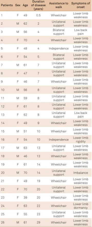
Among 26 HAM/TSP patients, 16 (61.5%) were female, me
-dian age of 56 (range: 39-70 yrs. old). he time of the symptoms ranged from 6 months to 29 years (median = 8 years). he most
commonsymptoms at the onset of the disease included lower
limb muscle weakness in 21 (84%) patients and sphincter dis
-turbance in 20 (77%) patients. Two patients reported lower back pain (Table 1). he majority (92.3%) needed assistance for loco
-motion. Tables 2 and 3 show muscle strength (MRC) and spas
-ticity (Ashworth modiied scale) quantitative measurements. he majority of the studied muscle groups displayed active move
-ment with gravity eliminated (MRC = 2), except for knee lexors and left tibialis anterior (dorsalis lexor) (MRC = 1). Few mus
-cles presented normal power (Table 2). he worst median spas
-ticity predominated in bilateral hip adductors and plantar lex
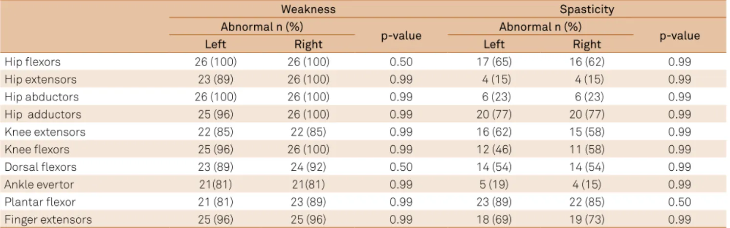
-ors groups (Table 3). here was no statistical diference between right and left muscles strength and spasticity (Tables 2 and 3).
All patients had weakness in at least one muscle group, mostly in the right side (Table 4). The majority of lower limb proximal muscles were weak. Spasticity was more common in hip adductors (77% bilateral
-ly) and plantar flexors (left – 88.5% and right – 84.6%) (Table 4). The majority of patients had normal tonus in hip extensor (85%), hip abductors (77%) and knee flexors (left – 54% and right – 42%) (Table 4).
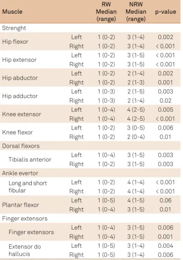
Forty two percent of the cases were restricted to wheel
-chairs. here were signiicant diferences in muscular strength between the patients restricted and non-restricted to wheelchair (p ≤ 0.05), except for the left plantar lexor muscle (Table 5). In the spasticity evaluation, there was no signiicant diference between the two groups (Table 6).
he functional evaluation (Barthel Index) showed medi
-an of 70 (r-ange: 20-100) in the 26 HAM/TSP patients, indicat
-ing minimal dependence dur-ing daily activities. he majority
of patients (69%) demonstrated minimal dependency, 15%
moderate dependence, 8% severe dependence and 8% were independent. he Figure shows the proportion of the cases and the performed functional activities on Barthel Index. he greatest diiculties included mobility, ascending and de
-scending stairs and bladder control.
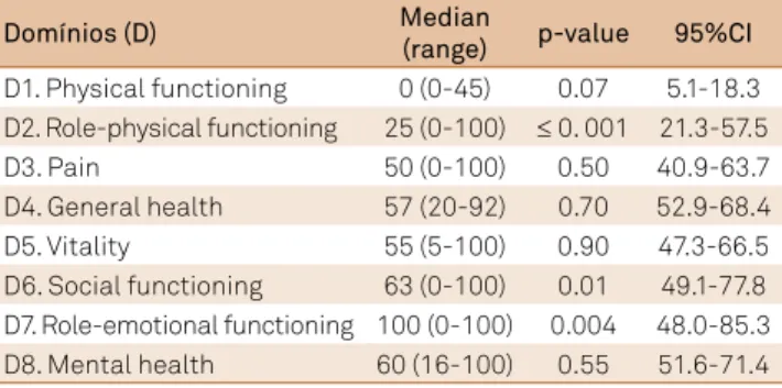
In relation to quality of life, Table 7 shows median and range of the SF-36 survey. From the eight domains, physi
-cal functioning (D1) and role-physi-cal functioning (D2) had the lowest scores.
Correlation between clinical characteristics, functional abilities and quality of life
he functional evaluation (Barthel) demonstrated cor
-relation with reduced muscle strength bilaterally (r = 0.6;
p ≤ 0.001), indicating that there was a relationship between
muscle weakness and dependence during daily activities. No statistical correlation was observed between the functional evaluation (Barthel) and muscle tone.
here was correlation between reduced muscle strength bilaterally and the domains physical functioning (r = 0.422; p = 0.03) and social functioning (r = 0.41; p = 0.04) of life quality. he muscular tonus did not show statistical correlation with life quality (SF-36).
Table 1. Clinical and epidemiological indings in 26 HAM/TSP patients.
Patients Sex Age
Duration of disease
(years)
Assistance to walk
Symptoms of onset
1 F 49 0.5 Wheelchair Lower limb weakness
2 M 62 2 Unilateral support Lower limb weakness
3 M 56 4 Bilateral
support
Low back pain
4 F 70 4 Wheelchair Lower limb weakness
5 F 48 4 Independence Lower limb weakness
6 F 54 5 Bilateral support Lower limb weakness
7 M 61 7 Unilateral support Lower limb weakness
8 F 47 7 Unilateral support Lower limb weakness
9 F 46 7 Wheelchair Lower limb weakness
10 M 56 8 Unilateral
support
Lower limb weakness
11 M 59 8 Unilateral support Lower limb weakness
12 F 61 8 Unilateral support Lower limb weakness
13 F 62 8 Unilateral
support
Low back pain
14 F 49 9 Wheelchair Lower limb weakness
15 M 51 10 Wheelchair Lower limb weakness
16 F 54 10 Independence Lower limb rigidity
17 M 63 13 Unilateral support Lower limb weakness
18 M 46 13 Wheelchair Lower limb weakness
19 F 61 14 Wheelchair Lower limb weakness
20 M 70 14 Unilateral
support Imbalance
21 F 49 19 Wheelchair Lower limb weakness
22 F 70 20 Unilateral support Lower limb weakness
23 F 39 20 Wheelchair Lower limb
weakness
24 F 63 22 Wheelchair Lower limb dormancy
25 F 55 23 Unilateral support Lower limb weakness
26 M 61 29 Wheelchair Lower limb
he measurement of functional evaluation (Barthel) demon
-strated correlation with the domains physical functioning (r = 0.6; p = 0.001) and social functioning (r = 0.524; p = 0.006) of SF-36 sur
-vey. here was no correlation between the Barthel Index, duration of the disease (r = -0.279; p = 0.17) and age (r = 0.016; p = 0.490).
DISCUSSION
his study identiied and quantiied the muscle groups compromised by weakness and spasticity as measured by mod
-iied Ashworth scale in lower limbs and its inluence on activi
-ties of daily living and quality of life of patients with HAM/TSP. In addition, this report attempts to evaluate whether hyperto
-nicity or weakness better predicts the need for a wheelchair.
Weakness and tonus
he results show intense weakness in the lower limbs, as
-sessed by manual muscle testing (MMT) in all groups evaluated.
he highest frequency of muscle strength loss occurred bilat
-erally in the hip lexors, hip abductors, plantar lexors and in
-ger extensors. Diference was found in muscle strength of the wheelchair patients group compared to non-wheelchair which suggests that muscle weakness is the most damaging factor in the active movement of these individuals, thus determining the restriction of patients to wheelchairs. Muscle tonus was not as strongly involved as weakness in the majority of the lower limb muscle groups. herefore, factor was not responsible for the mo
-tion restric-tion observed in most individuals. Only two muscle groups, hip adductors and plantar lexors, showed signiicantly more intense spasticity. However, it has not directly inluenced the restriction of patients to wheelchairs.
Functional abilities
In the assessment of functional abilities based on Barthel Index, minimal functional dependence was found in the majority of the patients with HAM/TSP similar to previ
-ous study14. Although HAM/TSP is a severe and disabling Table 2. Muscle strength (MRC) in 26 HAM/TSP patients.
Muscles Median (range) p-value
Left Right
Hip lexor 2 (0-4) 2 (0-4) 0.99
Hip extensor 2 (0-5) 2 (0-5) 0.99
Hip abductors 2 (0-4) 2 (0-3) 0.99
Hip adductors 2 (0-5) 2 (0-4) 0.58
Knee extensors 3 (0-5) 3 (0-5) 0.57
Knee lexors 1 (0-5) 1 (0-4) 0.58
Dorsal lexors
Tibialis anterior 1 (0-5) 2 (0-5) 0.89
Ankle evertor
Long and short ibular 2 (0-4) 2 (0-4) 0.58
Plantar lexors 3 (0-5) 3 (0-5) 0.17
Finger extensors
Finger extensor 2 (0-5) 2 (0-5) 0.99
Extensor do halluces 3 (0-5) 3 (0-5) 0.99
Table 3. Muscle spasticity (Ashworth modiicated) in 26 HAM/TSP patients.
Muscles Median (range) p-value
Left Right
Hip lexor 1 (0-4) 1 (0-4) 0.19
Hip extensors 0 (0-4) 0 (0-4) 0.99
Hip abductors 0 (0-4) 0 (0-4) 0.99
Hip adductors 2 (0-4) 2 (0-4) 0.99
Knee extensors 1 (0-4) 1 (0-4) 0.26
Knee lexors 0 (0-4) 0 (0-4) 0.99
Dorsal lexors
Tibialis anterior 1 (0-4) 1 (0-4) 0.34
Ankle evertor
Long and short ibular 0 (0-4) 0 (0-4) 0.50
Plantar lexors 2 (0-4) 2 (0-4) 0.77
Finger extensors
Finger extensor 1 (0-4) 1+ (0-4) 0.50
Extensor do hallucis 1 (0-4) 1+ (0-4) 0.20
Table 4. Frequency of muscle weakness and spasticity in 26 HAM/TSP patients.
Weakness Spasticity
Abnormal n (%)
p-value Abnormal n (%) p-value
Left Right Left Right
Hip lexors 26 (100) 26 (100) 0.50 17 (65) 16 (62) 0.99
Hip extensors 23 (89) 26 (100) 0.99 4 (15) 4 (15) 0.99
Hip abductors 26 (100) 26 (100) 0.99 6 (23) 6 (23) 0.99
Hip adductors 25 (96) 26 (100) 0.99 20 (77) 20 (77) 0.99
Knee extensors 22 (85) 22 (85) 0.99 16 (62) 15 (58) 0.99
Knee lexors 25 (96) 26 (100) 0.99 12 (46) 11 (58) 0.99
Dorsal lexors 23 (89) 24 (92) 0.50 14 (54) 14 (54) 0.99
Ankle evertor 21(81) 21(81) 0.99 5 (19) 4 (15) 0.99
Plantar lexor 21 (81) 23 (89) 0.99 23 (89) 22 (85) 0.50
neurological disease, mild dependence in carrying out daily activities can be explained by the lack of concomitant involve
-ment of upper limbs. here was a correlation between mus
-cle strength impairment (MRC) and ability to perform daily activities (Barthel Index). his inding suggests that muscle weakness interferes with the functional independence of HAM/TSP patients. On the other hand, no correlation was observed between the degree of spasticity (Ashworth modi
-ied) and functional ability in daily activities (Barthel). his can be explained by the fact that most of the muscle groups
had normal tonus or slight spasticity. he most important limitations in quality of life were related to physical function and role-physical function. hese two domains showed mod
-erate correlation with the Barthel Index, considering that these aspects were impaired by motor incapacity.
Gait impairment
his study highlights that gait impairment was deter
-mined mainly by weakness of the lower limbs compared to spasticity. his fact is justiied by the diference found in
Table 5. Comparison of strength between HAM/TSP patients restricted to wheelchair and not restricted to wheelchair.
Muscle
RW Median (range)
NRW Median (range)
p-value Strenght
Hip lexor Left 1 (0-2) 3 (1-4) 0.002
Right 1 (0-2) 3 (1-4) < 0.001
Hip extensor Left 1 (0-2) 3 (1-5) < 0.001
Right 1 (0-2) 3 (1-5) < 0.001
Hip abductor Left 1 (0-2) 2 (1-4) 0.002
Right 1 (0-2) 2 (1-3) 0.001
Hip adductor Left 1 (0-3) 2 (1-5) 0.003
Right 1 (0-3) 2 (1-4) 0.02
Knee extensor Left 1 (0-4) 4 (2-5) 0.005
Right 1 (0-4) 4 (2-5) < 0.001
Knee lexor Left 1 (0-2) 3 (0-5) 0.006
Right 1 (0-2) 2 (0-4) 0.01
Dorsal lexors
Tibialis anterior Left 1 (0-4) 3 (1-5) 0.003
Right 1 (0-2) 3 (1-5) 0.003
Ankle evertor Long and short ibular
Left 1 (0-2) 4 (1-4) < 0.001 Right 1 (0-2) 4 (1-4) < 0.001
Plantar lexor Left 1 (0-5) 4 (1-5) 0.06
Right 1 (0-4) 3 (1-5) 0.01
Finger extensors
Finger extensors Left 1 (0-4) 3 (1-5) 0.006
Right 1 (0-4) 3 (1-5) 0.001
Extensor do hallucis
Left 1 (0-5) 3 (1-4) 0.004
Right 1 (0-5) 3 (1-4) 0.006
Restricted to Wheelchair; NRW: Not Restricted to Wheelchair.
Table 6. Comparison of spasticity between HAM/TSP patients restricted to wheelchair and not restricted to wheelchair.
Muscle
RW Median
(range)
NRW Median
(range)
p-value Spasticity
Hip lexor Left 1+ (0-4) 0 (0-3) 0.053
Right 2 (0-4) 0 (0-3) 0.639
Hip extensor Left 0 (0-3) 0 (0-1) 0.148
Right 0 (0-3) 0 (0-1) 0.148
Hip abductor Left 0 (0-4) 0 (0-3) 0.149
Right 0 (0-4) 0 (0-3) 0.149
Hip adductor Left 3 (0-4) 1+ (0-3) 0.023
Right 3 (0-4) 1+ (0-3) 0.023
Knee extensor Left 1+ (0-4) 1 (0-3) 0.187
Right 1+ (0-4) 1 (0-3) 0.122
Knee lexor Left 0 (0-4) 0 (0-2) 0.151
Right 0 (0-4) 0 (0-2) 0.122
Dorsal lexors Tibialis anterior
Left 1 (0-4) 1 (0-3) 0.615
Right 1 (0-4) 1 (0-3) 0.825
Ankle evertor Long and short ibular
Left 0 (0-4) 0 (0-2) 0.181
Right 0 (0-4) 0 (0-1+) 0.064
Plantar lexor Left 2 (0-4) 1+ (0-3) 0.138
Right 3 (0-4) 1+ (0-3) 0.221
Finger extensors Finger extensors
Left 1+ (0-4) 1 (0-2) 0.255
Right 1+ (0-4) 1 (0-2) 0.221
Extensor do hallucis
Left 1+ (0-4) 1 (0-2) 0.075
Right 1+ (0-4) 1 (0-3) 0.278
Restricted to Wheelchair; NRW: Not Restricted to Wheelchair.
Figure. The independent execution of functional tasks on Barthel Index in 26 HAM/TSP cases.
100
70.0
23.1
Feeding Dressing 100
Toileting 77.0
Grooming 11.5
Mobility 51,5
Transfer 38.5
Bathing 11.5
Stais
80.1
reduced muscle strength in the patients restricted to wheel
-chairs compared to ambulatory patients. he intense hyper
-tonicity observed in the hip adductors and plantar lexor muscle groups did not directly inluence the restriction of pa
-tients to wheelchairs, but they may be responsible for com
-promise of the antagonist muscle strength by disuse15. he
spasticity of adductors can be responsible for abductor weak
-ness causing enhanced swing of the pelvis during ambulation phase and consequent gait instability16. Otherwise, a previ
-ous study showed a strong correlation between community ambulation and plantar lexors and knee extensors strength5.
In fact, the reduced strength impairs gait capacity and gradu -ally interferes on the need for locomotion assistance, starting
with unilateral support until the wheelchair. Ten years after disease onset, 30% of patients are bedridden and 45% need some assistance for walking6. Biomechanical changes
relat-ed to weakness and spasticity can contribute to imbalance and falls, leading to gradual loss of the ability to walk. Gait restriction can generate a vicious cycle of inactivity and con
-sequently increase muscle weakness.
Assessment of individual muscles strength and spasticity
Assessment of individual muscles strength and spastic
-ity represents a challenge, since the measurement of both is diicult to discriminate in people with neurological impair
-ments. However, it was observed that the most important weakness (dorsalis lexor and knee lexor muscles) in the studied patients did not correspond to the muscles most af
-fected by spasticity (hip adductor groups and plantar lex
-or), allowing for the assessment of muscle strength and rest
-ing tonus. he use of antispasmodic drugs for all patients may have induced a reduction in spasticity and made it eas
-ier to evaluate muscle strength. he hip adductor groups and plantar lexor showed signiicant degree of spasticity, even taking the drug, indicating that these groups have a
higher involvement.
Walking ability and dependence during activities
he association of muscle changes found in our study may explain the loss of walking ability and dependence conducting daily activities. Another aspect is spasticity of
plantar lexors, which can restrict the dorsilexors, both contributing to changes in the stance phase and swing. In the stance phase, initial foot is impaired causing a short
-ening of step, knee hyperextension and pelvic tilt, spend
-ing more energy at gait. In the sw-ing phase, disability of knee lexion occurs with consequent drag toe, shortening the stride and reducing gait velocity17. Associated with this,
hip adductor spasticity and weakness of the abductor gen
-erate an exagg-erated lateral pelvic tilt and imbalance dur -ing gait16. he correlation found in our study between the
impairment of muscle strength (MRC) of the patients and their ability to perform daily ctivities (Barthel) reinforces the argument that muscle weakness interferes in the func
-tional abilities of patients with HAM/TSP. he lack of cor
-relation between the degree of spasticity and functional dependence in daily activities occurs due to discrete tonus increase in the most of the examined muscles. In a paretic limb, the spasticity may help in certain functions, such as walking, standing and during transfers18.
he correlation between functional abilities (Barthel in
-dex) and the domains of functional capacity and social as
-pects (SF 36) suggest that motor impairment has a social impact on the quality of life of these individuals. On the oth
-er hand, a previous study showed that physical limitations of the disease also determine emotional impact on the qual
-ity of life of patients with HAM/TSP19. Here the most
impor-tant limitations in the quality of life were related to physical functioning and role-physical functioning, also consistent with the literature20.
Limitations of this study
Limitations of this analysis occur due to the relative
-ly small number of subjects. Although there are millions of people infected by HTLV-1 in the world, only 5% of infected individuals develop neurological disease, thus rendering it an underdiagnosed and negligible disease4. In addition, the
physical limitations resulting from gait disturbance and trou
-ble with the access to public transportation diicult the neu
-rological diagnosis, monitoring and follow up of the patients in developing countries. here is no speciic treatment for the virus, the prognosis of HAM/TSP is poor and often brings up a disastrous impact on patients’ lives. he inancial and so -cial costs to the patient, their family and to the health care
system can be intense due to the fact that it is a chronic pro
-gressive disease. hus, the elaboration of public policies on education and prophylaxis should be developed.
CONCLUSION
We show that the degree of weakness and not spastici
-ty predicted wheelchair coninement. he inevitable picture of motor impairment leads to the decreased ability to per
-form daily activities with impact on the patient’s quality of
Table 7. Quality of life (SF-36) in 26 HAM/TSP cases.
Domínios (D) Median
(range) p-value 95%CI D1. Physical functioning 0 (0-45) 0.07 5.1-18.3 D2. Role-physical functioning 25 (0-100) ≤ 0. 001 21.3-57.5
D3. Pain 50 (0-100) 0.50 40.9-63.7
D4. General health 57 (20-92) 0.70 52.9-68.4
D5. Vitality 55 (5-100) 0.90 47.3-66.5
D6. Social functioning 63 (0-100) 0.01 49.1-77.8 D7. Role-emotional functioning 100 (0-100) 0.004 48.0-85.3
life. On the other hand, the identiication of the speciic mus
-cles afected in HAM/TSP enables early physical therapy in
-tervention with a focus on strengthening of the lower limb muscles, especially the lexor knee and dorsalis lexor groups. he functional approach considering rehabilitation may be crucial to reduce the risk of complications, increase the pa
-tient’s independence performing daily activities (mainly gait disability) and improving the social and functional aspects involved in the quality of life.
Acknowledgments
his work was supported by Fundação de Amparo à
Pesquisa do Estado do Rio de Janeiro(FAPERJ) and a Msc fel
-lowship from Coordenação de Aperfeiçoamento de Pessoal de
Nível Superior (CAPES) to R.C.C. We also thank Rosangela
Martins from Research Division of theHospital Universitário
Clementino Fraga Filho/Universidade Federal Rio de Janeiro
(UFRJ) for bioestatistical analysis.
References
1. Osame M. Review of WHO Kagoshima meeting and diagnostic guidelines for HAM/TSP. In: Blattner W, editor. Human retrovirology: HTLV. New York: Raven; 1990. p. 191-7.
2. Castro-Costa CM, Araújo AQ, Menna-Barreto M, Penalva-de-Oliveira AC. [Guide of clinical management of HTLV patient: neurological aspects]. Arq Neuropsiquiatr. 2005;63(2B):548-51. Portuguese. doi:10.1590/S0004-282X2005000300036
3. Gessain A, Cassar Olivier. Epidemiological aspects and world distribution of HTLV-1 infection. Front Microbiol. 2012;3:388. doi:10.3389/fmicb.2012.00388
4. Casseb J. Is human T cell lymphotropic type 1 (HTLV-1) associated myelopathy/tropical spastic paraparesis (HAM/TSP) syndrome a neglected disease? PLoS Negl Trop Dis. 2009;3(11):e487. doi:10.1371/journal.pntd.0000487
5. Franzoi AC, Araújo AQ. Disability and determinants of gait performance in tropical spastic paraparesis/HTLV-I associated myelopathy (HAM/TSP). Spinal Cord. 2007;45(1):64-8. doi:10.1038/sj.sc.3101919
6. Gessain A, Gout O. Chronic myelopathy associated with human T-lymphotropic virus type I (HTLV-I). Ann Intern Med. 1992;117(11):933-46. doi:10.7326/0003-4819-117-11-933 7. Martins JV, Baptista AF, Araújo AQ. Quality of life in patients
with HTLV-I associated myelopathy/tropical spastic paraparesis. Arq Neuropsiquiatr. 2012;70(4):257-61. doi:10.1590/S0004-282X2012005000006
8. Medical Research Council of the United Kingdom. Aids to examination of the peripheral nervous system. Palo Alto, Califórnia: Pedragon House; 1978. (Memorandum, n. 45).
9. Daniels L, Worthingham C. Muscle testing: techniques of manual examination. Philadelphia: WB Saunders; 1986.
10. Ashworth B. Preliminary trial of carisoprodal in multiple sclerosis. Practioner. 1964;192:540-2.
11. Bohannon RW, Smith MB. Interrater reability of a modiicated Ashworth scale of muscle spasticity. Phys Ther. 1987;67:206-7. 12. Mahoney FI, Barthel DW. Functional evaluation: Barthel index. Md
State Med J. 1965;14:61-5.
13. Ciconelli RM, Ferraz MB, Wilton S, Meinão I, Quaresma MR. Tradução para língua portuguesa e avaliação do questionário genérico de avaliação de qualidade de vida SF-36 (Brasil SF-36). Rev Bras Reumatol. 1999;39(3):143-50.
14. Carod-Arthal FJ, Mesquita HM, Ribeiro LS. Manifestaciones neurológicas y discapacidad em pacientes que padecen mielopatía associada al HTLV-I. Neurologia. 2007;22(2):78-84.
15. Sheean G, McGuire JR. Spastic hypertonia and movement disorders: pathophysiology, clinical presentation, and qualiication. PM R. 2009;1(9):827-33. doi:10.1016/j.pmrj.2009.08.002
16. William WC. De Jong´s The neurologic examination. 7rd ed. Philadelphia: Lippincott Williams & Wilkins; 2013. 17. Jansen K, De Groote F, Aerts W, De Schutter J, Duysens J,
Jonkers I. Altering length and velocity feedback during a neuro-musculoskeletal simulation of normal gait contributes to hemiparetic gait characteristics. J Neuroeng Rehabil. 2014;11(1):78. doi:10.1186/1743-0003-11-78
18. Ward AB. Spasticity treatment with botulinum toxins. J Neural Transm (Vienna). 2008;115(4):607-16. doi:10.1007/s00702-007-0833-2
19. Coutinho IJ, Galvão-Castro B, Lima J, Castello C, Eter D, Grassi MFR. Impact of HTLV-associated myelopathy/T tropical spastic paraparesis (HAM/TSP) on activities of daily living (ADL) in HTLV-1 infected patients. Acta Fisiatr. 2011;18(1):6-10. doi:10.5935/0104-7795.20110001