DOI: 10.1590/0004-282X20160043
REVIEW
The Hitchhiker’s guide to the rhinencephalon
Um guia prático sobre a anatomia do rinencéfalo
Laura Silveira-Moriyama1,2,3,4, Philip Glass3,5, Suraj Rajan6, Rafael Carvalho1, Fabiano Reis3, Carlos A. A.
Penatti1, Valeria Muio1
he interest of the medical community in the rhinenceph
-alon has grown exponentially in the past decades. Much of this popularity stems from the inding of severe hyposmia both in Parkinson’s disease1 and Alzheimer’s disease2, two of
the most prevalent neurodegenerative conditions, as well as in schizophrenia3 and many other neurological and psychi
-atric conditions. Although now largely acknowledged in the specialized medical literature, such deicits are only infre
-quently detected in clinical practice, mainly due to a combi
-nation of lack of awareness and technical diiculties access
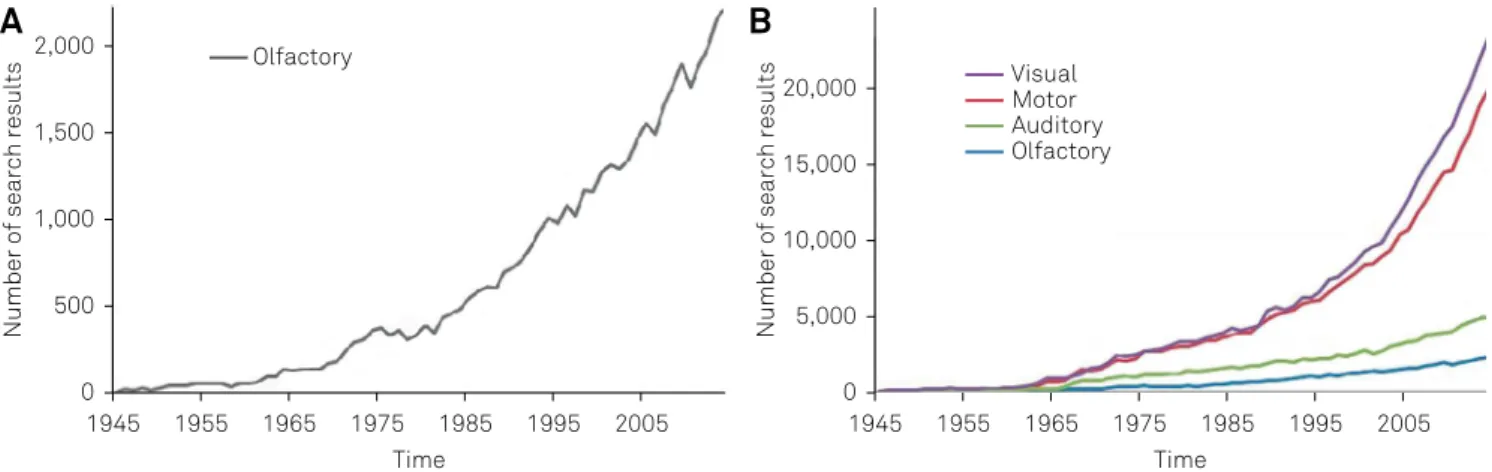
-ing commercially available smell tests. Despite the steadfast increase in the number of publication regarding the sense of olfaction, this ield is still incipient when compared to the study of other senses or the motor system (Figure 1). In ad
-dition, the anatomy and the physiology of the olfactory sys
-tem are still largely mysterious to the average clinician, and
detailed anatomical information regarding the rhinencepha
-lon is largely found in specialized books, usually inaccessible to readers from the developing countries. In this review we aimed to provide the practicing clinicians and the interest
-ed researchers with a brief, objective and practical overview of the anatomy of the rhinencephalon, using an open-access platform to facilitate access to this information, in the hope that it will help fuel research in this area and improve the clin
-ical care of patients sufering from olfactory disorders.
THE FLOW OF THE OLFACTORY INFORMATION
Key authorities4,5,6 split the olfactory areas into periph
-eral and central divisions, analogous to the connections de
-scribed for other sensory systems. his follows the proposed
1Universidade Nove de Julho, Programa de Pós-graduação em Medicina, Sao Paulo SP, Brazil; 2UCL Institute of Neurology, Reta Lila Weston Institute, London, UK;
3Universidade Estadual de Campinas, Faculdade de Ciências Médicas, Campinas SP, Brazil;
4Universidade de São Paulo, Faculdade de Medicina, Departamento de Neurologia, Sao Paulo SP, Brazil; 5Universidade Estadual do Sudoeste da Bahia, Faculdade de Medicina, Vitoria da Conquista BA, Brazil; 6Premier Health Miami Valley Hospital, The Clinical Neuroscience Institute, Dayton OH, USA.
Correspondence: Laura Silveira-Moriyama; Postgraduate Research Program in Medicine, Universidade Nove de Julho (Uninove); Rua Vergueiro, 235/249; 01504-001 São Paulo; SP, Brasil; E-mail: [email protected]
Conflict of interest: There is no conflict of interest to declare. Received 02 January 2016; Accepted 07 January 2016.
ABSTRACT
Pathology of the rhinencephalon has been a subject of interest in the fields of neurodegenerative diseases, trauma, epilepsy and other neurological conditions. Most of what is known about the human rhinencephalon comes from comparative anatomy studies in other mammals and histological studies in primates. Functional imaging studies can provide new and important insight into the function of the rhinencephalon in humans but have limited spatial resolution, limiting its contribution to the study of the anatomy of the human rhinencephalon. In this study we aim to provide a brief and objective review of the anatomy of this important and often overlooked area of the nervous system.
Keywords: olfactory cortex; anatomy; neuroanatomy.
RESUMO
As patologias do rinencéfalo tem sido assunto de interesse para os estudiosos das doenças neurodegenerativas, do traumatismo cranio-encefálico, epilepsia e outras doenças neurológicas. A maior parte do conhecimento sobre a anatomia do rinencéfalo vem de estudos de anatomia comparativa com outros mamíferos e estudos histológicos em primatas. Estudos de imagem funcional, apesar de proporcionarem informações úteis e interessantes a respeito do funcionamento do rinencéfalo em humanos, sofrem de resolução espacial limitada, e portanto contribuem de maneira restrita ao estudo dos limites das áreas anatômicas. Neste artigo buscamos proporcionar ao neurologista e neurocientista interessado uma revisão prática e objetiva da anatomia desta área importante e muitas vezes esquecida do sistema nervoso.
low of the olfactory information (Figure 2). he most pe
-ripheral element in the olfactory system, the olfactory re
-ceptor, was not described in detail until de 1990s. In 1991 Buck and Axel7 published a seminal paper which described
a multigene family encoding olfactory receptors (ORs), which comprised 1–5% of the human genome. his discov
-ery granted them the Nobel Prize in Physiology or Medicine in 20048 and further contributed to the popularity of olfac
-tory science. Following Buck and Axel’s description, it be
-came clear that each neuron in the olfactory mucosa could express one single type of olfactory receptor (OR), although the same receptor can be found in various neurons wide
-ly distributed in the olfactory mucosa. A single odorant
molecule has diferent parts, which are recognized by a set of diferent ORs and activate a set of cells in the muco
-sa9. hese cells are not simply receptors, but real olfactory neurons and their axons form the olfactory nerve bundles, which cross the skull base in the cribriform plate and syn
-apse in the olfactory bulb. Within the olfactory bulb, there are spheres of neuropil formations called glomeruli, which are formed by axons of the various sensory neurons express
-ing the same kind of OR. In the glomerulus, these axons syn
-apse onto the dendrites of the projection (mitral and tufted cells) and modulatory cells (most of which secrete GABA and/or dopamine). Figure 3 illustrates these structures.
From the olfactory bulb the information lows to the pri
-mary olfactory cortex, which includes the anterior olfactory nucleus, piriform cortex, olfactory tubercle, a small part of the amygdala and the anterior part of the entorrhinal cor
-tex. he role of the primary olfactory cortex in humans is not fully understood, but subjects with lesions of this area fail to
Figure 2. Schematic view of the levels of processing in the rhinencephalon. From bottom to top, the levels are showed in sequential order from more peripheral to more central.
Figure 3. Simplified structure of the olfactory mucosa. Olfactory receptor neurons (ORN) have their cilia in the olfactory mucus and contact with odorant substances, which yield action potentials. This information is transmitted through their axons across the cribriform plate and synapse into the olfactory bulb.
Figure 1. Search results on PubMed (www.ncbi.nlm.nih.gov/pubmed) using search terms (“visual”, “motor”, “auditory” and “olfactory”). (A) shows the number of search results for the term “olfactory” alone, using the number of citations up to two thousand, and demonstrating the exponential increase of search results using the term “olfactory”. (B) shows the comparative number of citations for the terms “olfactory”, “motor”, “auditory” and “visual”, clearly demonstrating that despite the increase, the number of search results is still very small when compared to the terms “visual” and “motor”, and almost half of the volume for term “auditory”.
Olfactory
Visual Motor Auditory Olfactory
Number of search results
Time 2,000
1,500
1,000
500
0
1945 1955 1965 1975 1985 1995 2005
Number of search results
Time 20,000
15,000
10,000
5,000
0
1945 1955 1965 1975 1985 1995 2005
identify odours appropriately, even if they can detect that a smell is present10,11,12,13,14,15, suggesting this area is important for encoding odour identity, a notion supported by functional imaging studies16,17,18,19,20.
he primary olfactory cortex sends projections to vari
-ous brain regions, including diencephalic structures (thala
-mus and hypothala-mus), limbic cortex (mainly larger parts of the amygdala and also the hippocampus) and neocortex (particularly the olfactory part of the orbitofrontal cortex). he clinician can easily observe this network as the often impressive hedonic and occasionally autonomic responses to smells, and by the close link between olfaction and mem
-ory which are often referred to particular smells. Olfact-ory stimuli are able to generate changes in emotions, behaviour and autonomic functions that are largely unconscious and often powerful.
UNIQUE CHARACTERISTICS OF THE OLFACTORY SYSTEM
In comparison to other sensory systems, olfaction has unique characteristics, which need to be appreciated for the basic understanding of the olfactory pathways. Unlike other senses, the receptors for olfaction are not mere receptor cells that connect to a bipolar neuron. hey are true neurons pres
-ent in the human surface epithelium. his results on them being more vulnerable to insult than neurons in the central nervous system. To compensate, the olfactory neurons can be generated in life through mitotic divisions of the basal cells present in the olfactory epithelium. heir natural turn
-over if of approximately 30 days21. he presence of pluripo
-tent cells in the olfactory epithelium and elsewhere in the ol
-factory system has been the source of great interest22.
Contrasting with other senses, the irst interneuronal synapse does not happen in the spinal cord or brain stem, but in the mitral layer of the olfactory bulb. Unlike other sensory modalities, olfactory projections travel directly to the cere
-bral hemispheres, without thalamic relay, and while in other senses the diencephalic relay stations project to a single de
-lineated cortical region, the olfactory information is widely projected to a network of distinct regions of the limbic cor
-tex that altogether make up the primary olfactory cor-tex (POC). he topographic organization for the analysis of olfac
-tory stimuli is still obscure. here seem to be “loose olfac-tory maps” linking certain neuronal populations in the olfactory bulb and primary olfactory cortex with broad but grouped olfactory stimuli23 but these “maps” are not as well-deined
and anatomically organized as those for visual, auditory and somatic information.
he low and integration of the olfactory information is rather complex and poorly understood. here is a pro
-jection from the POC to the thalamus, but this is not es
-sential for relay of sensory information into the neocortex,
since the POC also projects directly to the orbitofrontal cortex and other cortical regions24. he number of recipro
-cal and collateral projections of the olfactory system is un
-usually high. he olfactory bulb (OB) and areas within the POC have numerous internal connections and in addition they further project to the striatum, thalamus, hypothala
-mus and orbito-frontal cortex4,6,24,25, which project back to
the POC. Furthermore, the POC and higher areas send in
-formation back to the OB as well26. Among these many neu
-ron pathways and back projections, POC-derived synaptic output to hypothalamus and medial-orbito-frontal cortices have been known to enhance odor-driven social stimuli in appetitive and aversive behaviors27.
THE AREAS OF THE RINENCEPHALON
Olfactory mucosa
he olfactory mucosa is located in the medial and lateral walls of the nasal cavity. It is thicker than the respiratory mu
-cosa and occupies an area of approximately 1 cm2 on each side of the nose28. he thickness, extent and integrity of the
olfactory mucosa decreases signiicantly with age, and neu
-roepithelial degeneration seems to be an inevitable feature of human aging with signiicant reduction in olfaction29,30,31. Exposure to viral and bacterial infections, head injury, neu
-rodegenerative disorders and chemical exposures also can damage the nasal mucosa32.
he main components of the olfactory mucosa are olfac
-tory receptor neurons (ORN), columnar cells, basal cells mi
-crovillar cells and tubo-alveolar cells. ORN are neurons with bipolar appearance that have dendrites immersed in the ol
-factory mucus where they come into contact with odorant molecules. he thin axons of the few millions28 of ORN form
the bundles of the olfactory nerve which travel through the cribriform plate and synapse in the olfactory bulb. Columnar cells provide support for the receptor neurons33.
he function of microvillar cells is still largely unknown but they may be involved in the cell death and regeneration of ORN34. Tubo-alveolar cells of Bowman’s glands secrete a
serous luid which may regulate olfactory transduction33.
Last but not least, basal cells are stem cells situated deep in the olfactory epithelium. hey undergo mitotic division and diferentiate to replace lost receptor neurons, support
-ing cells35 and olfactory ensheating cells. hese olfactory re
-placing mechanisms are crucial for the regeneration of the olfactory nerve after trauma36.
Olfactory bulb
herefore caution must be taken when extrapolating this data to humans. In rodents, six well-deined layers are de
-scribed in the OB. In humans they are not as clearly deined, although the main cells types of each can be identiied37.
he olfactory nerve layer is the most supericial layer of the OB. It is composed of unmyelinated axons of olfactory re
-ceptor neurons (ORN) and express olfactory marker protein (OMP); therefore it can be easily immunolabelled and iden
-tiied38. he glomerular layer has a distinctive appearance
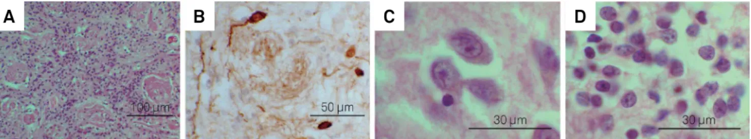
and is clearly visible in humans. It consists of axons of ORN that give rise to spherical neuropil formations (the glomeruli) that vary in vertebrates from 30 to 200 microns in diameter39 (Figure 4A). In the glomeruli the axons of the ORN synapse onto the arborized dendrites of mitral cells, and also with the modulatory periglomerular cells. Most of the periglomerular cells secrete GABA and/or dopamine. Figure 4B shows dopa
-minergic periglomerular cells stained by immunohistochem
-istry for tyrosine hydroxylase.
he external plexiform layer is formed by dendrites of the principal neurons and granule cells and it has few cell bodies in rodents. In humans, it continues with the mitral layer and thus shows histologically with greater cell somata. he mi
-tral cell layer is composed of large pyramidal glutaminergic cells which are the main projection outlets from the olfac
-tory bulb40. In humans this layer is thin and poorly demar -cated. he total number of mitral cells decreases signiicant
-ly with age41,42. he internal plexiform layer is another layer
with few cells bodies: it mainly combines the dendritic pro
-cess of granule cells and the axons of the mitral and tufted cells. Similar to the external plexiform layer, in humans it also merges with the mitral cell layer37.
he granule cell layer has the most numerous cell struc
-tures in the rodent olfactory bulb, with a few million neu
-rons. In humans, mitral and granule cells are more nu
-merous and they make up half of the volume of the OB41.
he granule cells have no distinguished axons and its long GABAergic dendrites project internally to the bulb, mainly in the plexiform layer.
Within the olfactory bulbs, quite visible islands of large pyramidal cells can be seen. hese are parts of the anterior olfactory nucleus and are therefore functionally related to the primary olfactory cortex. hese large neurons contrast with the smaller neurons from the granule cells layer and can be easily identiied (Figures 4C and 4D).
Primary olfactory cortex
he primary olfactory cortex (POC) is deined as the area that receives direct projections from the olfactory bulb. It is made up of ive main regions, which can be further subdi
-vided. Each region is described below and, because these re
-gions can be found under diferent acronyms in the literature, their various nomenclatures are listed in Table, except for the anterior cortical nucleus of the amygdala and the periamyg
-daloid cortex, which are discussed in the text. hese areas
Figure 4. Illustrative photomicrographs showing: histology of glomerular layer (A) and periglomerular dopaminergic cells (B), and appearance of large neurons in islands of the anterior olfactory nucleus inside the olfactory bulb (C), compared to the smaller cells in the granule cell layer (D). Scale bar: 100 µm in A, 50 µm in B, and 30 µm in C and D. (A), (C) and (D) used haematoxylin and eosin stain, and (B) used immunohistochemistry for tyrosine hydroxylase counter stained by haematoxylin.
A
B
C
D
Table. Different acronyms for the subdivisions of primary olfactory cortex found in the literature. The table displays the most commonly used name on the first column, and other common acronyms in the second column, followed by the number of citations found using the acronym as a search term on the Google Scholar (www.googlescholar.com) database.
Most commonly used acronym Other acronyms found in the literature Cites
Piriform cortex (25000 cites)
Pyriform cortex 6,09
Prepyriform cortex 2,31
Piriform area 544,00
Pyriform area 391,00
Prepiriform area 134,00
Lateral olfactory gyrus 97,00
Prepyriform area 95,00
Anterior olfactory nucleus (6,990 cites) Retrobulbar region 613,00
Anterior olfactory cortex 142,00
are not well delimited anatomically or histologically, there
-fore their recognition in histological preparations or in brain imaging largely depends on the appropriate knowledge of their relationship with main anatomical landmarks in the re
-gion, and the comparison with seminal literature on the sub
-ject. Figure 5 shows a coronal section of a half-brain with the location of one of the structures and various landmarks in the region. Figure 6 presents illustrative MRI coronal images with the main landmarks and the location of some areas of the POC.
Anterior olfactory nucleus
Despite being a prominent and extensive olfacto
-ry structure this area has been poorly studied in humans
and “existing research is dispersed and obscured by many diferent nomenclatures and approaches”43. Its solid ana
-tomical location in rodents is quite diferent from that in primates, where it is dispersed as discontinuous islands of large neurons (Figure 4D) within the OB and OT and a more delineated portion in the forebrain, all showing great inter-individual variability41.
here is also controversy as to whether it is better termed a nucleus or cortex44,45. Haberly argues that the physiological
organization of the olfactory pathway, if considered in paral
-lel with the other sensory pathways, would place the olfactory bulb as the primary olfactory cortex (as it is the irst and most simple structure for the coding of smell patterns) and the an
-terior olfactory cortex and other areas of the POC would then
AC: Anterior commissure; Acc : accumbens nucleus; Aco : anterior cortical nucleus of the amygdala; CC: corpus callosum; Cd: Caudate head; GP: Globus pallidus; IC: internal capsule; PAC: periamygdaloid cortex; PiF; PiT: frontal and temporal parts of piriform cortex; Pu: Putamen; Tu: olfactory tubercle.
Figure 6. Coronal images extracted from T1-weighted magnetic resonance imaging showing location of some areas of the primary olfactory cortex. Inserts show the coronal slices with main landmarks labelled in white on the left half, and the rhinencephalon areas labelled in black and marked with arrows on the right. For a reference for coronal slices see the Paxinos & Mai atlas5,
coronal slices numbered from the anterior commissure (AC). Insert (A) approximately 1cm anterior from AC, insert (B) slightly anterior to AC, and insert (C) slightly posterior from AC.
A
B
C
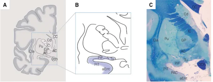
AC: Anterior commissure; aEnt: anterior entorhinal cortex; CC: corpus callosum; Cd: Caudate head; Cla: claustrum; GP: Globus pallidus; IC: internal capsule; Och: optic chiasm; PAC: periamygdaloid cortex; PiF; PiT: frontal and temporal parts of piriform cortex; Pu: Putamen; Tu: Olf tubercle.
Figure 5. Illustrative images showing location of some areas of the primary olfactory cortex. Insert (A) shows a schematic drawing with the main landmarks which are easily visible both in histology slides and neuroimaging, while insert (B) shows the corresponding location of the olfactory tubercle, piriform cortex and anterior portion of the entorhinal cortex (blue highlight). Insert (C) shows a coronal section of a half brain stained for luxol fast blue and counterstained with cresyl violet, showing a slightly more posterior location in which the periamygdaloid cortex can be visualized as well.
be the secondary olfactory cortex. On the other hand standard nomenclature dictates that cortical regions must be clearly di
-visible into a minimum of at least three tangential layers, and the anterior olfactory nucleus has only two.
Olfactory tubercle
he olfactory tubercle is just anterior to the olfactory trigone, and is bordered by the medial and lateral olfactory tracts. It is also referred to as the anterior perforated sub
-stance because of the perforating arteries that transpose it on their way to the subcortical regions. In contrast to rodents and carnivorous mammals where it is trilaminated and quite well developed, in humans it is made up of a loosely lami
-nated allocortical region. In humans its function is not clear, and some evidence suggests it might be linked to the bas
-al ganglia, as it is rich in acetylcholinesterase and its proile of iron, glutamic acid dehydrogenase, succinate dehydroge
-nase, enkephalin, substance P and epidermal growth factor content are typical of pallidal tissue4.
Piriform cortex
he term “piriform cortex” originates from the nomencla
-ture “prepiriform” based on the fact that this distinct region of allocortex is rostral to the “pyriform lobe” present in most carnivores4. In rodents the piriform cortex is divided into an
-terior and pos-terior portions, which have no clear morpho
-logical boundaries. In humans, there is a curvature of the hemisphere with the development of the fetal temporal lobe. In comparison to rodent cerebral hemispheres, the piriform cortex displays a C-type curve. he more anterior portion of the piriform correspond to the human frontal piriform (PiF), and the more posterior portions corresponding to the tem
-poral piriform (PiT)4,46 (Figure 5).
Anterior cortical nucleus of the amygdala and the periamygdaloid cortex
he anterior cortical nucleus of the amygdala (ACo) is a very small region of the amygdala, lateral to the PiT. Studies in macaques show that this region, as well as the adjoining periamygdaloid cortex, receive direct projections from the ol
-factory bulb47,48. In humans the periamygdaloid cortex is al
-most indistinguishable from the anterior cortical nucleus of the amygdala and the whole area is sometimes referred to as the cortico-amygdaloid transition area, or amygdalo-piri
-form transition area amygdala4. Mai5 clearly delimits the ACo
as well as a PCo, but other anatomists consider this contro
-versial. In the lower mammals the posterior cortical nucleus of the amygdala receives projections from the accessory ol
-factory nucleus24, which is a relay for pheromone perception
from the vomeronasal organ. he vomeronasal organ is ves
-tigial in humans and its connections and functions have not been well established, making the anatomy and function of the PCo a controversial subject in human anatomy. Other ac
-ronyms used for this region in humans of other species include
“cortical amygdaloid nucleus” and “semilunar gyrus”, although these names may not apply entirely to human anatomy.
Entorhinal cortex
he more anterior portions of the entorhinal cortex re
-ceive direct input from the olfactory bulb5,47,48 and entorhi
-nal activation has been demonstrated in functio-nal magnetic resonance image studies25. he limits of the olfactory areas
of the entorhinal cortex in humans are not well deined, but studies indicate it is likely to represent less than 15% of the entorhinal cortex49. Figure 5B shows a coronal section of the brain including the anterior portion of the entorhinal cortex.
Olfactory projections beyond the POC
In 1943, Allen showed that potentials could be evoked in the orbital frontal cortex by electrical stimulation of the piri
-form cortex, and that after ablation of this connection the response was abolished50. his led to the notion of the orbi -tofrontal cortex as the isocortical area related to olfaction. hese projections from primary olfactory areas have now been extensively studied in rodents, carnivores and mon
-keys51,52,53,54 mostly by electrophysiological methods, delim
-iting the olfactory area in the latero-posterior orbital fron
-tal cortex of the monkey (the posterior part of Area 12 of Walker52. Injections of anterograde axonal tracers in the piri
-form cortex of monkeys were able to label axons in several regions of the agranular insula/posterior orbital cortex that project back to the primary olfactory cortex48. Functional im
-aging studies have also demonstrated odorant-induced orbi
-tofrontal cortex activation, mainly in the orbi-tofrontal gyri55,
but the precise location of the input is still a challenge due to limitation of spatial resolution of functional imaging56.
In addition to the neocortex, the POC also projects to var
-ious areas in the limbic system. In rats there are projections from the POC to various areas of the amygdala, including cen
-tral nucleus (intermediate, lateral and capsular), basal nucleus (parvicellular division of the basal nucleus) and nucleus of the lateral olfactory tract. he POC also projects to CA1, subicu
-lum and dentate gyrus24. he amygdala is particularly involved
in the afective aspects of olfaction, being activated by olfac
-tory stimuli with emotional valence55. he hypothalamus also
receives an extensive input from the POC in less developed mammals. In humans, its role in olfaction is obscure, but re
-cent research using functional neuroimaging suggests a pos
-sible involvement in afective processing of olfactory informa
-tion57 and sexual behavior58. he POC projects to the ventral
striatum59 and medial-dorsal thalamic nucleus60,61,62. he thala -mus then projects on to the orbitofrontal cortex53.
FINAL REMARKS
Knowledge of the structures involved in human olfac
is described comes from comparative anatomical studies in rodents, where olfaction plays a much more important role in survival and the sheer size of the rhinencephalon is much larger than in humans. herefore, caution is needed when translating indings from rodents and other mammals to
microsmatic animals like man, which have a less developed sense of smell. Functional imaging studies, as well as clini
-cal olfactory studies in subjects with delimited lesions of the rhinencephalon are likely to help delineate the olfactory ar
-eas in humans.
References
1. Doty RL. Olfactory dysfunction in Parkinson disease. Nat Rev Neurol. 2012;8(6):329-39. doi:10.1038/nrneurol.2012.80
2. Velayudhan L, Gasper A, Pritchard M, Baillon S, Messer C, Proitsi P. Pattern of Smell Identification Impairment in Alzheimer’s Disease. J Alzheimers Dis. 2015;46(2):381-7. doi:10.3233/JAD-142838 3. Rupp CI. Olfactory function and schizophrenia: an update. Curr Opin
Psychiatry. 2010;23(2):97-102. doi:10.1097/YCO.0b013e328336643f 4. Gloor P. The temporal lobe and the limbic system. New York: Oxford
University Press; 1997. The olfactory system.
5. Paxinos G, Mai J. Atlas of the human brain. 2nd ed. New York: Elsevier Academic Press; 2004.
6. Price JL. Olfaction. In: Paxinos, G. editor. The human nervous system. San Diego: Elsevier Academic Press; 2004. p. 1197-211.
7. Buck L, Axel R. A novel multigene family may encode odorant receptors: a molecular basis for odor recognition. Cell. 1991;65(1):175-87. doi:10.1016/0092-8674(91)90418-X
8. Buck LB. Unraveling the sense of smell (Nobel lecture). Angew Chem Int Ed Engl. 2005;44(38):6128-40. doi:10.1002/anie.200501120 9. Malnic B, Hirono J, Sato T, Buck LB. Combinatorial receptor codes for
odors. Cell. 1999;96(5):713-23. doi:10.1016/S0092-8674(00)80581-4 10. Eskenazi B, Cain WS, Novelly RA, Friend KB. Olfactory functioning in temporal lobectomy patients. Neuropsychologia. 1983;21(4):365-74. doi:10.1016/0028-3932(83)90023-4
11. Eichenbaum H, Morton TH, Potter H, Corkin S. Selective olfactory deficits in case H.M. Brain. 1983;106 (2):459-72. doi:10.1093/brain/106.2.459
12. Eskenazi B, Cain WS, Novelly RA, Mattson R. Odor perception in temporal lobe epilepsy patients with and without temporal lobectomy. Neuropsychologia. 1986;24(4):553-62. doi:10.1016/0028-3932(86)90099-0
13. Jones-Gotman M, Zatorre RJ. Olfactory identification deficits in patients with focal cerebral excision. Neuropsychologia. 1988;26(3):387-400. doi:10.1016/0028-3932(88)90093-0 14. Zatorre RJ, Jones-Gotman M. Human olfactory discrimination after
unilateral frontal or temporal lobectomy. Brain. 1991;114 (1A):71-84. 15. Gonçalves Pereira PM, Insausti R, Artacho-Pérula E, Salmenperä
T, Kälviäinen R, Pitkänen A. MR volumetric analysis of the piriform cortex and cortical amygdala in drug-refractory temporal lobe epilepsy. AJNR Am J Neuroradiol. 2005;26(2):319-32. 16. Gottfried JA, Deichmann R, Winston JS, Dolan RJ. Functional
heterogeneity in human olfactory cortex: an event-related functional magnetic resonance imaging study. J Neurosci. 2002;22(24):10819-28. 17. Zelano C, Bensafi M, Porter J, Mainland J, Johnson B, Bremner E
et al. Attentional modulation in human primary olfactory cortex. Nat Neurosci. 2005;8(1):114-20. doi:10.1038/nn1368
18. Gottfried JA. A truffle in the mouth is worth two in the bush: odor localization in the human brain. Neuron. 2005;47(4):473-6. doi:10.1016/j.neuron.2005.08.002
19. Gottfried JA, Winston JS, Dolan RJ. Dissociable codes of odor quality and odorant structure in human piriform cortex. Neuron. 2006;49(3):467-79. doi:10.1016/j.neuron.2006.01.007 20. Li W, Luxenberg E, Parrish T, Gottfried JA. Learning to smell
the roses: experience-dependent neural plasticity in human
piriform and orbitofrontal cortices. Neuron. 2006;52(6):1097-108. doi:10.1016/j.neuron.2006.10.026
21. Mackay-Sim A. Neurogenesis in the adult olfactory neuroepithelium. In: Doty RL. Handbook of olfaction and gustation. Philadelphia: Marcel Dekker; 2003. p. 93-113.
22. Brann JH, Firestein SJ. A lifetime of neurogenesis in the olfactory system. Front Neurosci. 2014;8:182. doi:10.3389/fnins.2014.00182 23. Uchida N, Takahashi YK, Tanifuji M, Mori K. Odor maps in the
mammalian olfactory bulb: domain organization and odorant structural features. Nat Neurosci. 2000;3(10):1035-43. doi:10.1038/79857
24. Price JL, Russchen FT, Amaral DG. The amygdaloid complex. In: Björklund A, Hökfelt T, Swanson LW. Handbook of chemical neuroanatomy. Amsterdam: Elsevier Science; 1987. p. 279-388. 25. Sobel N, Johnson BN, Mainland JD, Yousem DM. Functional
neuroimaging of human olfaction. In: Doty RL. Handbook of olfaction and gustation. Philadelphia: Marcel Dekker; 2003.
26. Cleland L. Central olfactory structures. In: Doty RL. Handbook of olfaction and gustation. Philadelphia: Marcel Dekker; 2003. p. 165-80. 27. Choe HK, Reed MD, Benavidez N, Montgomery D, Soares N, Yim
YS et al. Oxytocin Mediates Entrainment of Sensory Stimuli to Social Cues of Opposing Valence. Neuron. 2015;87(1):152-63. doi:10.1016/j.neuron.2015.06.022
28. Moran DT, Rowley JC 3rd, Jafek BW, Lovell MA. The fine structure of the olfactory mucosa in man. J Neurocytol. 1982;11(5):721-46. doi:10.1007/BF01153516
29. Nakashima T, Kimmelman CP, Snow JB. Structure of human fetal and adult olfactory neuroepithelium. Arch Otolaryngol. 1984;110(10):641-6. doi:10.1001/archotol.1984.00800360013003 30. Suzuki Y, Takeda M, Obara N, Suzuki N, Takeichi N. Olfactory
epithelium consisting of supporting cells and horizontal basal cells in the posterior nasal cavity of mice. Cell Tissue Res. 2000;299(3):313-25. doi:10.1007/s004410050030
31. Doty RL, Kamath V. The influences of age on olfaction: a review. Front Psychol. 2014;5:20. doi:10.3389/fpsyg.2014.00020
32. Menco M. Morphology of the mammalian olfactory epithelium: form, fine structure, function and pathology. In: Doty RL. Handbook of olfaction and gustation. Philadelphia: Marcel Dekker; 2003. p. 17-49. 33. Jafek BW. Ultrastructure of human nasal mucosa. Laryngoscope.
1983;93(12):1576-99. doi:10.1288/00005537-198312000-00011 34. Montani G, Tonelli S, Elsaesser R, Paysan J, Tirindelli R. Neuropeptide
Y in the olfactory microvillar cells. Eur J Neurosci. 2006;24(1):20-4. doi:10.1111/j.1460-9568.2006.04878.x
35. Huard JM, Youngentob SL, Goldstein BJ, Luskin MB, Schwob JE. Adult olfactory epithelium contains multipotent progenitors that give rise to neurons and non-neural cells. J Comp Neurol. 1998;400(4):469-86. doi:10.1002/(SICI)1096-9861(19981102)400:4<469::AID-CNE3>3.0.CO;2-8 36. Beites CL, Kawauchi S, Crocker CE, Calof AL. Identification and
molecular regulation of neural stem cells in the olfactory epithelium. Exp Cell Res. 2005;306(2):309-16. doi:10.1016/j.yexcr.2005.03.027 37. Smith RL, Baker H, Kolstad K, Spencer DD, Greer CA. Localization
of tyrosine hydroxylase and olfactory marker protein
38. Smith RL, Baker H, Greer CA. Immunohistochemical analyses of the human olfactory bulb. J Comp Neurol. 1993;333(4):519-30. doi:10.1002/cne.903330405
39. Kratskin B. Anatomy and neurochemistry of the olfactory bulb. In: Doty RL. Handbook of olfaction and gustation. Philadelphia: Marcel Dekker; 2003. p. 139-64.
40. Chen WR, Xiong W, Shepherd GM. Analysis of relations between NMDA receptors and GABA release at olfactory bulb reciprocal synapses. Neuron. 2000;25(3):625-33. doi:10.1016/S0896-6273(00)81065-X
41. Bhatnagar KP, Kennedy RC, Baron G, Greenberg RA. Number of mitral cells and the bulb volume in the aging human olfactory bulb: a quantitative morphological study. Anat Rec. 1987;218(1):73-87. doi:10.1002/ar.1092180112
42. Meisami E, Mikhail L, Baim D, Bhatnagar KP. Human olfactory bulb: aging of glomeruli and mitral cells and a search for the accessory olfactory bulb. Ann N Y Acad Sci. 1998;855:708-15. doi:10.1111/j.1749-6632.1998.tb10649.x
43. Brunjes PC, Illig KR, Meyer EA. A field guide to the anterior olfactory nucleus (cortex). Brain Res Brain Res Rev. 2005;50(2):305-35. doi:10.1016/j.brainresrev.2005.08.005
44. Ekstrand JJ, Domroese ME, Johnson DM, Feig SL, Knodel SM, Behan M et al. A new subdivision of anterior piriform cortex and associated deep nucleus with novel features of interest for olfaction and epilepsy. J Comp Neurol. 2001;434(3):289-307. doi:10.1002/cne.1178 45. Haberly LB. Parallel-distributed processing in olfactory cortex:
new insights from morphological and physiological analysis of neuronal circuitry. Chem Senses. 2001;26(5):551-76. doi:10.1093/chemse/26.5.551
46. Leon M, Johnson B. Functional units in the olfactory system. Proc Natl Acad Sci USA. 2006;103(41):14985-6. doi:10.1073/pnas.0607416103
47. Turner BH, Gupta KC, Mishkin M. The locus and cytoarchitecture of the projection areas of the olfactory bulb in Macaca mulatta. J Comp Neurol. 1978;177(3):381-96. doi:10.1002/cne.901770303
48. Carmichael ST, Clugnet MC, Price JL. Central olfactory connections in the macaque monkey. J Comp Neurol. 1994;346(3):403-34. doi:10.1002/cne.903460306
49. Insausti R, Marcos P, Arroyo-Jiménez MM, Blaizot X,
Martínez-Marcos A. Comparative aspects of the olfactory portion of the entorhinal cortex and its projection to the hippocampus in rodents, nonhuman primates, and the human brain. Brain Res Bull. 2002;57(3-4):557-60. doi:10.1016/S0361-9230(01)00684-0 50. Allen WF. Distribution of cortical potentials resulting from
insufflation of vapors into the nostrils and from stimulation of the
olfactory bulbs and the pyriform lobe. Am J Physiol - Leg Content. 1943;139(4):553-5.
51. Tanabe T, Iino M, Takagi SF. Discrimination of odors in olfactory bulb, pyriform-amygdaloid areas, and orbitofrontal cortex of the monkey. J Neurophysiol. 1975;38(5):1284-96.
52. Tanabe T, Yarita H, Iino M, Ooshima Y, Takagi SF. An olfactory projection area in orbitofrontal cortex of the monkey. J Neurophysiol. 1975;38(5):1269-83.
53. Yarita H, Iino M, Tanabe T, Kogure S, Takagi SF. A transthalamic olfactory pathway to orbitofrontal cortex in the monkey. J Neurophysiol. 1980;43(1):69-85.
54. Naito J, Kawamura K, Takagi SF. An HRP study of neural pathways to neocortical olfactory areas in monkeys. Neurosci Res. 1984;1(1):19-33. doi:10.1016/0168-0102(84)90027-0
55. Zald DH, Pardo JV. Emotion, olfaction, and the human amygdala: amygdala activation during aversive olfactory stimulation. Proc Natl Acad Sci USA. 1997;94(8):4119-24. doi:10.1073/pnas.94.8.4119 56. Gottfried JA, Zald DH. On the scent of human olfactory
orbitofrontal cortex: meta-analysis and comparison to non-human primates. Brain Res Brain Res Rev. 2005;50(2):287-304.
doi:10.1016/j.brainresrev.2005.08.004
57. Bensafi M, Rouby C, Farget V, Bertrand B, Vigouroux M, Holley A. Autonomic nervous system responses to odours: the role of pleasantness and arousal. Chem Senses. 2002;27(8):703-9. doi:10.1093/chemse/27.8.703
58. Savic I, Berglund H, Gulyas B, Roland P. Smelling of odorous sex hormone-like compounds causes sex-differentiated hypothalamic activations in humans. Neuron. 2001;31(4):661-8. doi:10.1016/S0896-6273(01)00390-7
59. Fuller TA, Russchen FT, Price JL. Sources of presumptive glutamergic/aspartergic afferents to the rat ventral striatopallidal region. J Comp Neurol. 1987;258(3):317-38. doi:10.1002/cne.902580302
60. Russchen FT, Amaral DG, Price JL. The afferent input to the magnocellular division of the mediodorsal thalamic nucleus in the monkey, Macaca fascicularis. J Comp Neurol. 1987;256(2):175-210. doi:10.1002/cne.902560202
61. Kuroda M, Price JL. Synaptic organization of projections from basal forebrain structures to the mediodorsal thalamic nucleus of the rat. J Comp Neurol. 1991;303(4):513-33. doi:10.1002/cne.903030402 62. Kuroda M, Murakami K, Kishi K, Price JL. Distribution of the piriform