O
RIGINALA
RTICLE Revista Brasileira de FisioterapiaAbstract
Objectives: The aim of this study was to evaluate pain symptoms, teeth clenching, quality of sleep, sensitivity to pain in the main masticatory and stabilizer muscles, and quality of life among women with temporomandibular disorder (TMD). Methods: Forty-five women were evaluated and divided into two groups. Group I included 27 women (mean age 30.1±5.8 years) with a diagnosis of TMD and Group II (control) included 18 healthy women (mean age 23.4±2.3 years). The intensity of pain symptoms (headache, neck pain), teeth clenching and trouble sleeping was evaluated using a visual analog scale (VAS). The pain thresholds of the masseter, anterior temporalis, upper trapezius and sternocleidomastoid muscles were evaluated using a dolorimeter. Quality of life was evaluated using SF-36. Statistical analysis was performed and the significance level was α≤0.05. Results: The results showed that the women with TMD presented more intense headache (p<0.001), neck pain (p<0.001), teeth clenching (p<0.001) and trouble sleeping (p<0.001). They also presented lower pain threshold in the masseter (p<0.001), anterior temporalis (p<0.001), upper trapezius (p<0.001) and sternocleidomastoid (p<0.001) muscles and lower quality of life in all evaluated domains (p<0.05) when compared with the control group. Conclusions: Women with TMD had greater intensity of pain symptoms, teeth clenching, trouble sleeping, sensitivity to pain in the masticatory and neck muscles and lower quality of life, compared with women without TMD.
Key words: temporomandibular joint disorder; pain; masticatory muscles; quality of life.
Resumo
Objetivos: Este estudo teve como objetivo avaliar sintomas de dor, apertamento dos dentes, qualidade do sono e sensibilidade dolorosa nos principais músculos mastigatórios e estabilizadores cervicais e qualidade de vida de mulheres com Disfunção Temporomandibular (DTM). Métodos: Foram avaliadas 45 mulheres, divididas em dois grupos. O grupo I, composto por 27 mulheres (30,1±5,8anos) com diagnóstico de DTM e o grupo II, controle, composto por 18 mulheres saudáveis (23,4±2,3 anos). A intensidade dos sintomas de dor, cefaleia, cervicalgia, de apertamento dos dentes e dificuldade de dormir foram avaliados por escala visual analógica (EVA), o limiar de dor dos músculos masseter, temporal anterior, trapézio superior e esternocleidomastoideo, com dolorímetro e a qualidade de vida, pelo SF-36. Foi realizada análise estatística e o nível de significância foi α=0,05. Resultados: Os resultados mostram que mulheres com DTM têm sintomas mais intensos de cefaleia (p<0,001), cervicalgia (p<0,001), intensidade de apertamento dos dentes (p<0,001) e dificuldade de dormir (p<0,001). Também apresentam limiar de dor mais baixo nos músculos masseter (p<0,001), temporal anterior (p<0,001), trapézio superior (p<0,001), esternocleidomastoideo (p<0,001) e pior qualidade de vida em todos os domínios avaliados (p<0,05), quando comparados com o grupo controle. Conclusões: Mulheres com DTM têm maior intensidade dos sintomas de dor, apertamento dos dentes, dificuldade de dormir, maior sensibilidade dolorosa em músculos mastigatórios e cervicais e pior qualidade de vida quando comparadas com mulheres sem DTM.
Palavras-chave: disfunção da articulação temporomandibular; dor; músculos mastigatórios; qualidade de vida.
Received: 01/04/2008 – Revised: 10/10/2008 – Accepted: 21/01/2009
1 Department of Physical Therapy, Faculdades Adamantinenses Integradas (FAI), Adamantina (SP), Brazil 2 Department of Physical Therapy, Centro Universitário Hermínio Ometto (UNIARARAS), Araras (SP), Brazil
3 Department of Physical Therapy, Fonoaudiologia e Terapia Ocupacional, Universidade de São Paulo (USP-SP), São Paulo (SP), Brazil 4 Department of Surgery, Prosthetics and Maxillofacial Traumatology, USP
Correspondence to: Bruno Gonçalves Dias Moreno, Rua Pérola, 89, Eldorado, CEP 17800-000, Adamantina (SP), Brazil, e-mail: [email protected]
Clinical and quality-of-life assessment among
women with temporomandibular disorder
Avaliação clínica e da qualidade de vida de indivíduos
com disfunção temporomandibular
Moreno BGD1, Maluf SA2, Marques AP3, Crivello-Júnior O4
Introduction
Temporomandibular Disorder (TMD) can be deined as a set of clinical manifestations of poor mandibular function, which may or may not be associated with pain. hese mani-festations are caused by agents that attack the morphological or functional integrity of the temporomandibular system1. he
American Academy of Temporomandibular Disorders charac-terizes its etiology as multifactorial, but the exact role of these agents in the pathophysiology of TMDs varies greatly, given the large number of asymptomatic individuals who clinically have one or more potentially triggering or perpetuating factors2,3.
he signiicant number of patients with TMD and the di-versity of symptoms require adequate knowledge of the disease and careful study for classiication. It is diicult to classify the symptoms not only for statistical and didactic purposes, but also for monitoring in outpatient clinics, where the number of patients is high. herefore, classiication indices were created, including the Helkimo Index4.
Tension-type headaches and migraines are the most common causes of complaint of pain, which afects the adult population5.
he correlation between headache and TMD has been shown in various epidemiological and clinical studies6,7, but its relationship
with bruxism is inconclusive8. Parafunctional habits such as
brux-ism and teeth clenching are considered important factors in the etiology of TMD, but should be studied separately for a better un-derstanding of their role in the manifestation of symptoms9. A
com-mon consequence of these conditions is the increase in tension in the masticatory muscles, associated with the increase in muscle tonus. Neck disorders are also present in large numbers of patients with TMD, but these conditions also afect the general population10.
herefore, controlled studies are important for a better understand-ing of the role of neck disorders in patients with TMD.
Besides pain, patients with headaches or TMD often have dif-iculty sleeping. Studies suggest that the mentioned conditions can be also consequences of sleep disorders11. Due to the physical
and mental impairment caused by TMD, evaluation of the im-pact on quality of life of these people deserves special attention. TMD patients have clinical characteristics in common with other chronic disease patients, such as high-intensity pain, behavioral and psychological disorders12. he aim of the present study was to
evaluate symptoms of pain, teeth clenching, quality of sleep and pain sensitivity in the main masticatory and stabilizer muscles and the quality of life of women sufering from TMD.
Methods
Forty-ive women took part in this cross-sectional study. hey were divided into two groups: group I and group II. Group I was
composed of 27 women aged between 19 and 40 years (30.1±5.8) with a diagnosis of TMD, referred by the Department of Surgery, Prosthetics and Maxillofacial Traumatology, Faculty of Dentistry, Universidade de São Paulo. he inclusion criteria for this group were: Helkimo Index III and parafunctional habit of teeth clench-ing. Patients who had more than two dental laws, direct or surgical trauma in the orofacial region, systemic or degenerative disease and ongoing dental, psychological or physical therapy treatment were excluded. Group II was composed of 18 healthy, female volunteers aged between 19 and 28 years (23.4±2.3) with no complaints of musculoskeletal pain. Participants were se-lected among the staf and students of the university where all of the evaluation procedure took place. Women who had any other musculoskeletal disease, history of TMD symptoms, or who were undergoing any kind of treatment were excluded. One of the pa-tients in group I was excluded due to a fracture in the irst two cervical vertebrae. All participants signed a consent form, and the research project was approved by CAPPesq of Universidade de São Paulo, protocol number 103/04.
Variables
Pain, headache, neck pain, teeth clenching and trouble sleeping were evaluated by visual analog scale (VAS) which consists of a 10cm horizontal line in which the left end repre-sents no pain and the right end, the worst pain imaginable. he participants were instructed to place a vertical line at the point on the line to indicate the pain intensity. he VAS is a simple and reliable instrument to evaluate pain in both clinical and research situations13.
Pain threshold
his relects the lowest intensity of stimulation in which the individual perceives pain. Fischer dolorimeter14 was used and, in
this procedure, perpendicular pressure to the skin’s surface was applied at a velocity of 1cm/s over the muscle motor points. he participants were in the supine position with the head slightly turned away from the evaluated side until they reported that the feeling of pressure became pain, while a manometer recorded the pressure level. Lower values indicate a lower pain threshold. he motor point was used as reference to ensure reproducibility and also with the advantage of always evaluating the same place. In group I, dolorimetry was performed on the side of the reported symptoms, and in Group II, the left side was standardized.
Quality of life
Portuguese language by Ciconelli15. his questionnaire
con-sists of 36 items, stratiied into eight domains: physical func-tion (10 items), role physical (4 items), pain (2 items), general health status (5 items), vitality (4 items), social function (2 items), role emotional (3 items), mental health (5 items) and a question concerning a comparative evaluation between the current health and the previous year’s health. he values range from 0 to 100, and the higher the score, the better the quality of life.
Statistical analysis
Descriptive statistical analysis was initially performed to calculate the mean and standard deviation for each meas-ured variable for both groups. Subsequently, the non-para-metric Mann-Whitney test was performed; it is indicated for comparison of two sample groups, when the samples show different patterns of variation16. The sample size
cal-culation was done using 80% of statistical power to detect a difference of 20% between groups, considering a confidence interval of 95%.
Table 1. Visual Analog Scale (VAS) for each analyzed symptom
presented as mean (SD).
Variables (cm) Group I N=26
Group II N=18
Mann-Whitney test TMD Pain
Mean (SD) 7.7 (1.5) 0.1 (0.3)
p<0.001*
Headache
Mean (SD) 6.8 (2.5) 3.7 (2.5)
p<0.001*
Neck Pain
Mean (SD) 6.4 (2.7) 2.7 (2.3)
p<0.001*
Teeth clenching
Mean (SD) 6.5 (2.8) 0.5 (0.6)
p<0.001*
Trouble sleeping
Mean (SD) 5.2 (1.0) 0.4 (0.1)
p<0.001*
* statistically significant values.
Table 2. Dolorimetry of the anterior temporal, masseter, superior
trapezoid, sternocleidomastoid muscles presented as mean (SD).
Dolorimetry (Kg/cm2) Group I
N=26
Group II N=18
Mann-Whitney test Temporalis
Mean (SD) 2.4 (0.7) 3.8 (0.9)
p<0.001*
Masseter
Mean (SD) 2.0 (0.6) 3.3 (0.4)
p<0.001*
Trapezius
Mean (SD) 2.1 (0.6) 3.0 (0.3)
p<0.001*
Sternocleidomastoid
Mean (SD) 1.6 (0.4) 2.6 (0.3)
p<0.001*
*statistically significant values.
Results
he intensity of symptoms in both groups can be seen in Table 1. hese are more pronounced in group I with a statis-tically signiicant diference (p<0.001), but group II also had complaints of neck pain and headaches. Table 2 presents the dolorimetry values of the anterior temporalis, masseter, up-per trapezius and sternocleidomastoid muscles in groups I and II. he pain threshold was considered positive if values were below 2.6 kg/cm2 (Marques et al.15). he
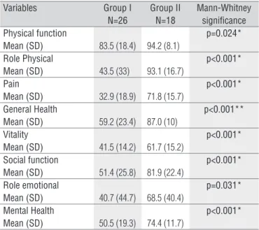
sternocleido-mastoid muscle showed the lowest values in both groups but all muscles presented a statistically signiicant diference (p<0.001). he quality of life of both groups is described in Table 3. Group I shows lower values indicating lower quality of life, with a statistically signiicant diference in the eight domains evaluated (p<0.05), drawing attention to the pain and role physical domains in which the diference was more pronounced.
Discussion
he aim of this study was to evaluate pain symptoms, teeth clenching, quality of sleep, sensitivity to pain and quality of life of women sufering from TMD, and the results indicate more intense symptoms, more intense pain and worse quality of life in women with TMD. he results indicate that pain levels and pain sensitivity in patients with TMD were signiicantly higher. Studies have shown that this population has less neck mobility, painful points elicited by palpation in the shoulder and neck muscles, lower pain tolerance17 and more reports of stress and
depression than people without TMD18.
Studies on headaches suggest a strong connection between signs of TMD and tension-type headache19. he same authors
argue that the headache associated with TMD may represent changes in pain sensitivity. Studies also indicate a signiicant association of neck disorders and temporomandibular joint disorders and suggest that individuals with TMD have less mo-bility and more intense pain elicited by palpation of the neck muscles than people without DTM10.
Dolorimetry is often used in the evaluation of individu-als with chronic pain, including patients with ibromyalgia20.
In the present study, the dolorimetry was used to evaluate the pain threshold of the masticatory and neck muscles. Of the muscles evaluated, the sternocleidomastoid showed the greatest pain sensitivity, i.e. the lowest pain threshold in both groups. Furthermore, both the TMD patients and the partici-pants from the control group reported neck pain and head-aches. Because the sternocleidomastoid muscle is one of the muscles responsible for neck mobility, it can be assumed that
there is a relationship between these symptoms. Jensen21 states
that cervicogenic headache has a much higher prevalence than reported by some epidemiological studies; the same author at-tributes this to the medical community’s lack of skill to evalu-ate the musculoskeletal system.
Parafunctional habits are pointed out as important etio-logical factors in TMD. hey also contribute to the appearance of neck injuries2,23. Moderate teeth clenching force is strongly
related to signs and symptoms of the temporomandibular joint24. his further suggests that it can compromise the quality
of sleep of TMD patients.
The present study indicates that headache and neck pain were also present in the control group but with sig-nificantly lower intensity than in the TMD group, therefore supporting the clinical evidence of other studies that sug-gest a lower pain threshold in this population13,25. Studies
suggest that a hyperexcitability in the central nociceptive system may contribute to the development or maintenance of chronic pain in TMD26. It is also possible to infer that the
increased sensitivity of the masticatory and neck muscles can be directly related to neck symptoms, headache and to the intensity of teeth clenching.
Although there is evidence that TMDs or any other pain-ful facial condition have some impact on quality of life, few studies document the use of speciic tools or even multidi-mensional tools to measure this impact27. Studies described
a signiicant reduction in the quality of life of patients with facial pain28,29. Bernhardt et al.30 reported a lower quality of
life in women when compared to men with TMD. his difer-ence was associated with the increase in pain sensation dur-ing palpation of the masticatory muscles and with a greater impact related to the limitations imposed more by physical than emotional aspects. he present study found no signii-cant diference between the physical and emotional aspects, but it did ind similar results to those previously described, indicating a lower quality of life in patients with TMD in all evaluated domains. Although the questionnaire used here is not speciic to TMD, it presents the advantage of being easy
to administer and understand and not as extensive as other questionnaires created for the same purpose15.
Another important aspect to be considered is the relation-ship between TMD and emotional disorders. Pallegama et al.25
found a high rate of anxiety in patients with TMD and neck pain when compared to a group without TMD. he authors concluded that anxiety could directly inluence the onset of neck pain.
Conclusion
he results of the present study indicate that women with TMD, classiied as Helkimo III, have stronger symptoms of pain, headache, neck pain, teeth clenching and trouble sleep-ing when compared to women without TMD. hey also had more painful sensitivity in the masseter, anterior temporalis, upper trapezius and sternocleidomastoid muscles and lower quality of life than women without TMD.
Table 3. Quality of life evaluated by SF-36. Mean (SD).
Variables Group I N=26
Group II N=18
Mann-Whitney significance Physical function
Mean (SD) 83.5 (18.4) 94.2 (8.1)
p=0.024*
Role Physical
Mean (SD) 43.5 (33) 93.1 (16.7)
p<0.001*
Pain
Mean (SD) 32.9 (18.9) 71.8 (15.7)
p<0.001*
General Health
Mean (SD) 59.2 (23.4) 87.0 (10)
p<0.001**
Vitality
Mean (SD) 41.5 (14.2) 61.7 (15.2)
p<0.001*
Social function
Mean (SD) 51.4 (25.8) 81.9 (22.4)
p<0.001*
Role emotional
Mean (SD) 40.7 (44.7) 68.5 (40.4)
p=0.031*
Mental Health
Mean (SD) 50.5 (19.3) 74.4 (11.7)
p<0.001*
* statistically significant values.
1. Munhoz WC, Marques AP, de Siqueira JT. Evaluation of body posture in individuals with internal temporomandibular joint derangement. Cranio. 2005;23(4):269-77.
2. Reher P, Harris M. Dor facial idiopática, parte 1: definição, classificação e etiologia. Rev Hosp Clín Fac Med São Paulo. 1998;53(4):189-94.
3. Fricton J. Myogenous temporomandibular disorders: diagnostic and management considerations. Dent Clin North Am. 2007;51(1):61-83.
4. Helkimo M. Studies on function and dysfunction of the masticatory system. I. An epidemiological investigation of symptoms of dysfunction in Lapps in the north of Finland. Proc Finn Dent Soc. 1974;70(2):37-49.
5. Rasmussen BK, Jensen R, Schroll M, Olesen J. Epidemiology of headache in a general population – a prevalence study. J Clin Epidemiol. 1991;44(11):1147-57.
6. Pettengill C. A comparison of headache symptoms between two groups: a TMD group and a general dental practice group. Cranio. 1999;17(1): 64-9.
7. Molina OF, dos Santos JJr, Nelson SJ, Grossman E. Prevalence of modalities of headaches and bruxism among patients with craniomandibular disorder. Cranio. 1997;15(4):314-25.
8. Rauhala K, Oikarinen KS, Raustia AM. Role of temporomandibular disorders (TMD) in fascial pain: occlusion muscle and TMJ pain. Cranio. 1999;17(4):254-61.
9. Seligman DA, Pullinger AG, Solberg WK. The prevalence of dental attrition and its association with factors of age, gender, occlusion, and TMJ symptomatology. J Dent Res. 1988 67(10):1323-33.
10. Stiesch-Scholz M, Fink M, Tschernitschek H. Comorbidity of internal derangement of the temporomandibular joint and silent dysfunction of the cervical spine. J Oral Rehabil. 2003;30(4):386-91.
11. Baba K, Haketa T, Sasaki Y, Ohyama T, Clark GT. Association between masseter muscle activity levels recorded during sleep and signs and symptoms of temporomandibular disorders in healthy young adults. J Orofac Pain. 2005;19(3):226-31.
12. Turner JA, Dworkin SF, Mancl L, Huggins KH, Truelove EL. The roles of beliefs, catastrophizing, and coping in the functioning of patients with temporomandibular disorders. Pain. 2001;92(1-2):41-51.
13. Dixon JS, Bird HA. Reproducibility along a 10-cm vertical visual analogue scale. Ann Rheum Dis. 1981;40(1):87-9.
14. Fischer AA. Pressure algometry over normal muscles. Standard values, validity and reproducibility of pressure threshold. Pain. 1987;30(1): 115-26.
15. Ciconelli RM, Feraz MB, Santos W, Meinão I, Quaresma MR. Tradução para a língua portuguesa e validação do questionário genérico de avaliação de qualidade de vida SF-36. Rev Bras Reumatol. 1999;39(3):143-9.
16. Zar JH. Biostatistical analysis. 4ª ed. New Jersey: Prentice Hall; 1999.
17. Dworkin SF, LeResche L. Research diagnostic criteria for temporomandibular disorders: review, criteria, examinations and specifications, critique. J Craniomandib Disord. 1992;6(4):301-55.
18. Visscher CM, Lobbezoo F, de Boer W, van der Meulen M, Naeije M. Psychological distress in chronic craniomandibular and cervical spinal pain patients. Eur J Oral Sci. 2001;109(3):165-71.
19. Liljeström MR, Le Bell Y, Anttila P, Aromaa M, Jämsä T, Metsähonkala L, et al. Headache children with temporomandibular disorders have several types of pain and other symptoms. Cephalalgia. 2005;25(11):1054-60.
20. Marques AP, Ferreira EA, Matsutani LA, Pereira CA, Assumpção A. Quantifying pain threshold and quality of life of fibromyalgia patients. Clin Rheumatol. 2005;24(3):266-71.
21. Jensen S. Neck related causes of headache. Aust Fam Physician. 2005;34(8):635-9.
22. Litonjua LA, Bush PJ, Andreana S, Tobias TS, Cohen RE. Effects of occlusal load on cervical lesions. J Oral Rehabil. 2004;31(3):225-32.
23. Lee WC, Eakle WS. Possible role of tensile stress in the etiology of cervical lesions of teeth. J Prosthet Dent. 1984;52(3):374-80.
24. Carlsson GE, Egermark I, Magnusson T. Predictors of signs and symptoms of temporomandibular disorders: a 20-year follow-up study from childhood to adulthood. Acta Odontol Scand. 2002;60(3):180-5.
25. Pallegama RW, Ranasinghe AW, Weerasinghe VS, Sitheeque MA. Influence of masticatory muscle pain on electromyographic activities of cervical muscles in patients with myogenous temporomandibular disorders. J Oral Rehabilit. 2004;31(5):423-9.
26. Sarlani E, Greenspan JD. Why look in the brain for answers to temporomandibular disorder pain? Cells Tissues Organs. 2005;180(1): 69-75.
27. Oliveira AS, Bermudez CC, Souza RA, Souza CMF, Dias EM, Castro CES, et al. Impacto da dor na vida de portadores de disfunção temporomandibular. J Appl Oral Sci. 2003;11(2):138-43.
28. Murray H, Locker D, Mock D, Tenenbaum HC. Pain and the quality of life in patients referred to a craniofacial pain unit. J Orofac Pain. 1996;10(4):316-23.
29. Von Korff M, Dworkin SF, Le Resche L, Kruger A. An epidemiologic comparison of pain complaints. Pain. 1988;32(2):173-83.
30. Bernhardt O, Gesch D, Schwahn C, Mack F, Meyer G, John U, et al. Risk factors headache, including TMD signs and symptoms, and their impact on quality of life. Results of the Study of Health in Pomernia (SHIP). Quintenssence Int. 2005;36(1):55-64.