1
Arquivos Brasileiros de Cardiologia - Volume 84, Nº 6, Junho 2005
Original Article
Functional Assessment of the Left Atrial Appendage
at Transesophageal Echocardiography Before and
After Percutaneous Valvotomy in the Mitral Stenosis
Solange Bernardes Tatani, Orlando Campos Filho, Claudio Henrique Fischer,
Valdir Ambrósio Moisés, José Augusto Marcondes de Souza, Cláudia Maria Rodrigues Alves,
Angelo Amato Vincenzo de Paola, Antonio Carlos C. Carvalho
Universidade Federal de São Paulo - UNIFESP - São Paulo, SP - Brazil
Mailing address: Solange Bernardes Tatani - Rua Gil Fernandes, 183 04148-020 – São Paulo, SP – Brazil
E-mail: [email protected], [email protected] Sent for publishing on 06/03/2004
Accepted on 11/08/2004
The preferential location of the thrombi in the left atrial ap-pendage has been known for many decades1,2, as well as the
association between mitral stenosis and left atrial thrombosis, especially under atrial fibrillation3. However, only recently, with
the arrival of the transesophageal echocardiography, it was possible a better understanding on the physiological and abnormal aspects of the function of the left atrial appendage and its relationship with thromboembolic phenomena.
Pollick & Taylor4 and Pozzoli et al.5, by means of the
transeso-phageal echocardiography, that, in patients with a normal heart and sinus rhythm, the left atrial appendage shows a characteristic pattern of function: its apex is highly contractile, frequently obli-terating, whereas its base practically does not constrict. Those authors described the regular pattern of biphasic flow of that structure with a anterograde component directed towards the main cavity of the left atrium, just after the P wave of the electro-cardiogram (appendicular systole), immediately followed by a re-trograde flow directed to the inside of the appendage (stage of appendicular relaxation). They also demonstrated that, under pa-thological conditions, especially under atrial fibrillation, the for-mation of thrombi in the left atrial appendage is associated to the dilatation and loss of its contractile ability, with slowness of the blood flow in its inside. From those studies the concept of dys-function left atrial appendage was created, by establishing a relation with embolic potential.
Despite the mitral stenosis is knowingly associated to a grea-ter possibility of thromboembolic phenomena resulting from the blood stasis on the atriun, few studies took only the Doppler echocardiographic analysis of left atrial appendage into conside-ration. Hwang et al6 and Tukek et al7, based on the
transesopha-geal echocardiography, observed a greater compromising of the left atrial appendage function in the mitral rheumatic disease with atrial fibrillation, in relation to the patients with non-rheumatic atrial fibrillation. However, few studies have asses-sed the functional behavior of the left atrial appendage after percutaneous mitral valvotomy.
The present study had as objective to assess the possible varia-tions of the function of the left atrial appendage, which were produced by the valvular dilatation in patients with symptomatic mitral ste-nosis, under a sinus rhythm, systematically submitted to the transe-sophageal echocardiography, before and after the well-succeeded percutaneous mitral valvotomy.
Objective
To assess the effects of the relief of the mitral stenosis by percutaneous ballon valvotomy in the function of the left atrial appendage.
Methods
Twelve patients with symptomatic mitral stenosis, in sinus rhythm, were studied. They were submitted to the transesopha-geal echocardiogram before and after effective percutaneous ballon valvotomy. Concerning the left atrial appendage, the peak flow velocities and the respective integral of the anterograde and re-trograde flow, in addition to the ejection fraction calculated through the planimetry of the area of that structure, were analyzed at the pulsatile Doppler.
Results
There was a significant increase of the anterograde flow veloci-ties of the left atrial appendage after percutaneous ballon valvo-tomy (pre: mean of 0.30 m/s; post: mean of 0.47 m/s; p<0.05) and their respective integrals. The same happened with the retrograde flow velocities (pre: mean of 0.35 m/s, post: mean of 0.53 m/s; p<0.05). There was a tendency of increase of the ejection fraction of the left atrial appendage after the procedure (pre: mean of 20%, post: mean of 31%; p=0.08).
Conclusion
The effective opening of the stenosed mitral orifice resulting from the percutaneous ballon valvotomy determined an improvement of the flow pattern of the left atrial appendage, which can poten-tially contribute for the reduction of the embolic risk.
Key words
2
Arquivos Brasileiros de Cardiologia - Volume 84, Nº 6, Junho 2005
Functional Assessment of the Left Atrial Appendage at Transesophageal Echocardiography Before and After Percutaneous Valvotomy in the Mitral Stenosis
Methods
Twelve consecutive patients (10 women, mean 24 years old, varying between 15 to 38 years old) with symptomatic mitral stenosis, in sinus rhythm, submitted to a well-succeeded percu-taneous mitral valvotomy, assessed through hemodynamic criteria. The patients were submitted to transesophageal echocardiogra-phy in the selection stage for the valvular percutaneous dilatation, up to 15 days before the procedure. After the valvotomy, a new transe-sophageal echocardiography was performed before discharge.
The study was approved by the Ethics Committee of our ins-titution and all patients informed their consent.
The patients included in the study were carriers of symptomatic mitral stenosis, under sinus rhythm, with indication of valvular opening, submitted to the percutaneous ballon valvotomy, whose success was determined by hemodynamic parameters: a significant increase of the mitral valvular area (at least twice the pre-procedure area), a significant decrease (p<0.05) of the transvalvular mid-diastolic gradient and mitral insufficiency resulting from the pro-cedure not greater than 2 in 4.
The acquisition of the echocardiographic images was done with an ATL equipped with a 5 MHz biplanar transesophageal transdu-cer. The patients were kept in lateral decubitus and received topic anesthesia of the oropharynx through nebulization of lidocaine at 10%. The introduction of the esophageal probe and the acquisition of the images were carried out through the conventional technique8.
The curves of flow velocity of the left atrial appendage were obtained by positioning the volume sample of the pulsatile Doppler inside its cavity, approximately at 1cm from its base. In all cases low wall filters were used and we opted for the longitudinal plan for allowing a better aligning of the ultrasonic beam with the blood flow of the appendage, which warranted a lower dispersion of the ultrasonographic signal.
The analysis of the left atrial appendage function included the study of the appendicular flow pattern and the ejection fraction of that structure4 concerning the blood flow, the peak flow velocity
and the integrals of the anterograde (wave A) and retrograde (wave R) of the velocity curve of the blood flow of the LAA (fig. 1) were analyzed, and concerning the ejection fraction, which was calculated from the planimetry of the maximum and minimum areas of that structure, obtained in appendicular diastole and systole, to the transver-sal cross-section (fig. 2), by means of the following relation:
diastolic area – systolic area x 100 (%) diastolic area
The maximum area (diastolic) was measured at the beginning of the P wave of the electrocardiogram, and the minimum area (systolic) was measured at the end of the dissection of the left atrial appendage (which means, at the beginning of the QRS complex of the electrocardiogram), opting for the transversal cross-section because it allows for a view of its whole extent.
The measurements of the variables analyzed resulted from the arithmetic means of the values obtained by the analysis of the three consecutive heart beats.
All echocardiographic exams were performed by a single exa-miner. The exams were recorded in videotape and submitted to the analysis of a single observer, which did not participate in the realization of the transesophageal exams and also in the valvular dilatation procedure.
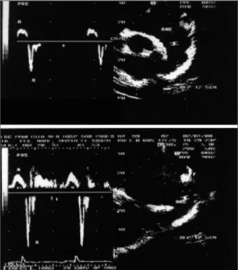
Fig. 1 – Curves of flow velocity of the left atrial appendage obtained through the study with pulsatile Doppler, at the cross section of two chambers, with the volume sample positioned inside the cavity. Anterograde (A) and retrograde (R) waves that compose the appendicular flow are observed. In the upper image, there is an example of curve before valvotomy and in the lower one, a curve after the mitral dilatation.
Fig. 2 – Calculation of the ejection fraction of the left atrial appendage. The planimetry in the diastole (D) and in the systole (S) was made, guided by the electrocardiographic derivation.
For the analysis of the results a non-parametric test was applied, taking into consideration the nature and distribution of the variable involved in. The test of Wilcoxon was applied for two non-indepen-dent samples when comparing the variables analyzed with the transesophageal echocardiography, for each patient, before and after the mitral valvotomy. The mean, minimum and maximum values found were described for each variable. The level for rejection of the hypothesis of nullity was fixed at 0.05 (p<0.05).
Results
3
Arquivos Brasileiros de Cardiologia - Volume 84, Nº 6, Junho 2005
Functional Assessment of the Left Atrial Appendage at Transesophageal Echocardiography Before and After Percutaneous Valvotomy in the Mitral Stenosis
15 days (average of 5 days). The interval between the valvotomy and the realization of a new transesophageal study varied from 1 to 6 days (average of 3 days). There was no significant difference between the pre- (values between 70 and 115 bpm; mean value of 88 bpm) and post-valvular dilatation (values between 70 and 125 bpm; mean value of 93 bpm) heart rate.
After percutaneous ballon valvotomy, there was a significant increase of the valvular area (pre: average of 0.88 cm2 and post:
mean of 2.8 cm2; p=0.0004), with a significant decrease of the
mean diastolic gradients (p=0.0003). There was a discreet valvu-lar insufficiency undertaking the mitral valve in 8 patients, tricuspid in 10 and aortic in 3, isolated or associated, and those that did not significantly change after the valvular dilatation procedure.
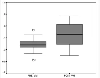
Before the mitral valvotomy, the anterograde component of flow of the left atrial appendage (wave A) showed peak flow veloci-ties that varied from 0.13 to 0.53 m/s (mean of 0.30 m/s), with the respective integral varying from 0.01 to 0.04 m (mean of 0.03 m). After the procedure, the peak flow velocity varied from 0.10 to 0.77 m/s (mean of 0.47 m/s) (fig. 3) and the integral from 0.01 to 0.08 m (mean of 0.04 m), which corresponded to a significant increase of the anterograde velocities (p=0.01) and their integrals (p=0.04), induced by the percutaneous ballon valvotomy, cor-responding to an mean increase of, respectively, 57% and 33% of those variables in relation to the pre-procedure values.
In relation to the retrograde component of the flow of the left atrial appendage, the peak flow velocity before the valvotomy varied from 0.23 to 0.51 m/s (mean of 0.35 m/s) and the integral from 0.02 to 0.04 m (mean of 0.03 m). After the valvular dilata-tion, the peak flow velocity varied from 0.20 to 0.88 m/s (mean of 0.53 m/s) (fig. 4) and the integral from 0.01 to 0.08 m (mean of 0.04 m). There was a significant difference between the values of the p and post-percutaneous ballon valvotomy maximum re-trograde velocity (p=0.04), with an mean increase of 51%. There was not any statistic difference in relation to the values of the integral of the retrograde flow in the two periods considered (p=0.59).
The aspect of the spectral curve of the flow of the left atrial appendage, before and after the mitral valvotomy, is displayed in the figure 1.
In the pre-valvotomy stage, the ejection fraction varied from 9 to 34% (mean of 20%) and after the procedure, from 14 to 60% (mean of 31%). Such differences only show a tendency for the increase of such variable, without any significance from the statistic point of view, when compared to the data gauged before and after the mitral valvotomy (p=0.08).
Discussion
This study objectively shows the functional changes of the left atrial appendage induced by the relief of the left atrial hyper-tension after percutaneous ballon valvotomy of patients with mitral stenosis. It also highlights the importance of the transesophageal technique as an especially useful instrument in the observation of those aspects, which are impossible of being approached through other diagnostic methods.
In patients with normal left atrium and sinus rhythm, Pollick & Taylor4 observed at the transesophageal echocardiography that
the ejection fraction of the left atrial appendage is of approximately 55%, with average values of maximum anterograde velocity of 0.48 m/s. In the patients with mitral stenosis in this research, before the percutaneous ballon valvotomy, the transesophageal study of the left atrial appendage showed an average ejection fraction of 20%, with average anterograde maximum velocities of 0.30 m/s. Those data suggest the existence of dysfunction of the left atrial appendage in the mitral stenosis and reflect a compro-mising of the function ability of that structure. Similar data were described by Hwang et al6, characterizing a compromising of the
intrinsic contractile function of the left atrial appendage in the mitral stenosis. Those authors observed more dilated left atrial appendages, with lower ejection fractions and lower velocities in patients with mitral rheumatic disease in sinus rhythm, when compared with patients with non-rheumatic atrial fibrillation. Such dysfunction of the left atrial appendage in the mitral stenosis can be explained by the degeneration of the myocardial fibers with a consequent diffused interstitial fibrosis of this structure, which result from repeated crises of the rheumatic activity. Madden9 has
already demonstrated that the tissue of the left atrial appendage in the mitral stenosis presents degeneration of the myocardial fibers and diffused interstitial fibrosis.
Fig. 3 - Box-plot of the values of the maximum velocity (m/s) of wave A of the appendicular flow, before (PRE VM) and after well-succeeded valvotomy (POST VM).
PRE_VM POST_VM
Fig. 4 - Box-plot of the values of the maximum velocity (m/s) of wave R of the appendicular flow, before (PRE VM) and after well-succeeded valvotomy (POST VM).
4
Arquivos Brasileiros de Cardiologia - Volume 84, Nº 6, Junho 2005
Functional Assessment of the Left Atrial Appendage at Transesophageal Echocardiography Before and After Percutaneous Valvotomy in the Mitral Stenosis
After the percutaneous ballon valvotomy, a significant impro-vement of the left appendicular flow of the patient in the present series was observed, which superimposed the values found in normal individuals4, and only a tendency for a better contractile
performance of that structure, regarding the increase, however insignificant, of its ejection fraction after the procedure. Similar results were obtained by Porte et al.10.
In the patients with mitral stenosis, the increase of the left atrial pressure causes increase of the resistance against the function of the appendicular chamber, with a consequent reduction of its flow, which contributes for the deterioration of the contractile function of the left atrial appendage. With the effective opening of the mitral valvular area resulting from the valvotomy, the decrease of the left atrial pressure determines a reduction in the atrial post-load, which makes easy the appendicular function in the atrial systole, thus justifying our findings.
The fact that there has been an improvement of the flow, without a significant increase of the ejection fraction of the left atrial appendage, suggests that the flow of that structure is more dependent of the left intra-atrial pressure than the recovery of the appendicular contractile function. Those observations are corro-borated by Hoit et al.11, who in experimental studies also did not
find any relation between the velocity and the contractile function of the left atrial appendage.
It is known that there are embryologic, structural and functional differences between the left atrial appendage and the body of the left atrium. The appendicular chamber is the remaining of the
embryonic left atrium that develops during the 3rd week of gestation,
whereas the main cavity of the left atrium develops later, from the pulmonary veins. Gall et al.12 demonstrated similarities in the
structure of the left atrial appendage with the skeletal muscle, which can explain the contractile nature of that chamber and maybe different answers to the inflammatory rheumatic process. Functionally it is also known that the left atrial appendage differs from the body of the atrium for being presented as a larger com-placence chamber13, which may lead to different adaptative
proces-ses in relation to the left atrium.
So, it can be acknowledged that such anatomic-functional peculiarities of the left atrial appendage can determine differences in the functional response of that chamber, in a different way from the body of the left atrium in the rheumatic process, facing the therapeutic procedure used.
Maybe the full restoration of the contractile ability of the left atrial appendage, in terms of ejection fraction, happens in a later stage to the relatively early period when the post-valvotomy tran-sesophageal exam was performed, by means of a slower process of remodeling.
Summarizing, the comparative Doppler echocardiographic data of the present study document the early recovering, although partial, of the dysfunction of the left atrial appendage. There will be necessary studies with a greater sample and a follow-up for a longer time after the valvular dilatation procedure to assess the relation of these functional aspects of the left atrial appendage with a possible reduction of the embolic events.
1. Jordan RA, Scheifley CH, Edwards JE. Mural thrombosis and arterial embolism in mitral stenosis: a clinicopathologic study of fifty-one cases. Circulation. 1951; 3: 363-7.
2. Shresta NK, Moreno FL, Narciso FV, Torres L, Calleja HB. Two-dimensional echo-cardiographic diagnosis of left atrial thrombus in rheumatic heart disease: a clini-copathologic study. Circulation. 1983; 67: 341-146.
3. Braunwald E. Valvular heart disease. In: Heart Disease. A Textbook of Cardiovas-cular Medicine. 2.ed. Philadelphia, Saundres, 1984. P.1043-110.
4. Pollick C, Taylor D. Assessment of left atrial appendage function by transesopha-geal echocardiography. Implications for the development of thrombus. Circulation. 19991; 84: 223-31.
5. Pozzoli M, Febo O, Torbicki A, et al. Left atrial appendage dysfunction: a cause of thrombosis? Evidence by transesophageal echocardiography-Doppler studies. J Am Soc Echocardiogr.1991; 4: 435-41.
6. Hwang JJ, Li YH, Lin JM, et al. Left atrial appendage function determined by tran-sesophageal echocardiography in patients with rheumatic mitral valve disease. Cardiology. 1994; 85:121-8.
References
7. Tukek T, Atilgan D, Akkaya V, et al. Assessment of left atrial appendage function and its relationship to pulmonary venous flow pattern by transesophageal echo-cardiography. Int J Cardiol. 2001; 78: 121-6.
8. Seward J, Khanderia BK, Edwards W, Oh JK, Freeman WK, Tajik AJ. Biplanar transesophageal echocardiography: anatomic correlations, image orientation, and clinical applications. Mayo Clin Proc. 1990; 65: 1193-213.
9. Madden JL. Resection of left auricular appendix: a prophylaxis for recurrent arte-rial embolism. JAMA. 1949; 140: 769-72.
10. Porte JM, Cormier B, Iung B, et al. Early assessment by transesophageal echocar-diography of left atrial appendage function after percutaneous mitral commissu-rotomy. Am J Cardiol.1996; 77: 72-6.
11. Hoit BD, Shao Y, Gabel M. Influence of acutely altered loading conditions on left atrial appendage flow velocities. J Am Coll Cardiol. 1994; 24: 1117-23. 12. Gall JA, Alcorn D, Fernley R, Coghlan JP, Ryan GB. Qualitative and quantitative
analysis of granules in atrial appendage cardiocytes in different physiological states. Cell Tissue Res. 1990; 259: 529-34.