Printed version ISSN 0001-3765 / Online version ISSN 1678-2690 http://dx.doi.org/10.1590/0001-3765201720160519
www.scielo.br/aabc | www.fb.com/aabcjournal
Physical and chemical study of lattice kaolinites and their interaction with orthophosphate
MARCONDES SILVA E SILVA, ANDERSON S. LAGES and GENILSON P. SANTANA
Universidade Federal do Amazonas, Instituto de Ciências Exatas, Departamento de Química, Campus Artur Virgilio Filho, Setor Norte, Av. Gal. Rodrigo Octávio, 6200, Coroado I, 69077-000 Manaus, AM, Brazil
Manuscript received on September 12, 2016; accepted for publication on March 27, 2017
ABSTRACT
This study covers the evaluation of the structure and the capacity in adsorbing orthophosphate ions of kaolinites collected from sampling sites in Manaus (Brazil). The kaolinites were obtained using physical fractioning (sieving/siphoning) techniques and chemical treatment with HCl, H2SO4, H2O2 and KCl. The investigation of the kaolinite lattices involved the Hinckley and Plançon indexes determined from X-ray diffraction data, Fourier transformed infrared spectroscopy, scanning electron microscopy and Mössbauer spectroscopy. The absorption capacity of orthophosphate ions was calculated by Freundlich and Langergren isotherms. A transitional state was observed in the crystallographic structure from kaolinites because of the isomorphic substitution of Al3+ by Fe3+. This isomorphic substitution occurs accompanied by the optical pleochroism behavior, but it also reduces the mean particle sizes and increases the number of structural defects and magnetic properties of these kaolinites. Mössbauer spectroscopy showed that the substituting Fe3+ preferentially occupies octahedral sites. In the kaolinite lattices there are different octahedral sites of Al bounded by cis-OH-Fe3+ and trans-OH-Fe3+ octahedral sites. The kaolinite from the Kao1 sample has a higher number of cis-OH-Fe3+
from octahedral sites and is able to adsorb higher contents of orthophosphate ions than those from samples Kao2 and Kao3.
Key words: Freundlich, isomorphic substitution, Langergren, Manaus, Mössbauer.
Correspondence to: Genilson Pereira Santana E-mail: [email protected]
INTRODUCTION
Kaolinite, a clay mineral formed by octahedral and tetrahedral sites (1:1), is the mineral that is most found in soil and sediment, mainly in humid tropical regions (Corrêa et al. 2008). This clay mineral is a typical 1:1-type phyllosilicate with the chemical composition Al2Si2O5(OH)4. Its surface
comprises aluminol groups (≡Al-OH) situated at
the edges and the hydroxyl-terminated planes of
the clay lamellae (Wei et al. 2014). On its basal surface there are permanent negative charges that are responsible for sorption of cations,(Tombácz et al. 2004) and silanol (≡SiOH) linked to the
aluminol groups through hydrogen atom (Rehim et al. 2010).
Anion exchange capacity (AEC) occuring on the edges of the aluminosilicate sand layer is pH-dependent. Kaolinite AEC increases with decreasing pH but never > 2.00 cmol kg-1 (Dixon 1989). On its
reactivity of the kaolinite (Cui and Weng 2013).
The pH value has great influence on the capacity
adsorption of ions by kaolinite: i) pH <4.0 adsorbs anions, ii) pH ~4.0 is PCZ of the kaolinite it does not adsorb ions and iii) pH> 4.0 adsorbs cations
(Khawmee et al. 2013). Specifically, the adsorption
of phosphate occurs in pH 3.0-5.0 (Kamiyango et al. 2009) Malawi, has revealed high phosphate levels ranging from 0.63 to 5.50 mg/L. These phosphate levels would stimulate excessive growth of plants and toxic cyanobacteria in stagnant receiving water bodies hence posing a threat to aquatic life and water quality. Phosphate removal by kaolinite obtained from Linthipe, Malawi, was investigated as a function of pH, contact time, clay dosage, competing ions and initial phosphate concentration by means of jar tests. Phosphate uptake was pH dependent with adsorption mechanisms on kaolinite and iron oxide surfaces dominant generally below pH 7 and precipitation by calcium ions dominant above pH 7. Maximum phosphate removal occurred at high pH values of 11.22 (97.1% Obviously, the presence of substituents alters the particle size and crystallinity and consequently the ionic adsorption capacity of the kaolinite (Balan et al. 2000).
Since phosphorus is one of the essential elements required for plant growth kaolinite and
its modified forms provide an important control
over the availability of this nutrient to the soil solution. Phosphate adsorption onto minerals is an important process that significantly affects the mobility and bioavailability of phosphorus in natural environments. Furthermore, the kinetics and thermodynamics studies from the adsorption process provide important outcomes on available phosphorus. Previous researchers have reported numerous kinetic models used to describe sorption
processes. These include first-order, and order reversible ones, and first-order and second-order, irreversible ones, pseudo-first-order and
pseudo-second-order ones based on the solution concentration (Adebowale et al. 2008). The major
contribution of this study is to provide data on crystallinity and interaction with orthophosphate by kaolinites extracted from Oxisoil and bottom sediment from the Central Amazonia.
EXPERIMENTAL PART
SAMPLES COLLECTION
Samples of Oxisoil located in S 03°02’55.3” and W 60° 04’ 21,4” (Kao1) and bottom sediments in S 03° 08’ 45.9” and W 60° 01› 05,4» (Kao2) and S 03° 05’ 53.9” and W 59° 58› 15» (Kao3). Soil samples were collected from 40 to 80 cm depths
(C horizon), classified according to their physical
and chemical characteristics in yellow latosols, and sediment samples were collected from the bottom of streams.
PREPARATION OF FINER KAOLINITE
To begin, all samples are dried at room temperature for a week and sieved down to a size < 0.053 mm
to obtain finer particles from the mixed deionized
through a 12 h period of stirring with KCl 1.0 mol L-1, dried at room temperature, crushed in an agate mortar and stored.
MINERALOGICAL COMPOSITION
To develop the study on the mineralogical composition of Kao1, Kao 2 and Kao3 the physical techniques XRD, FTIR and SEM were used. The XRD measurements carried out on a Shimadzu
diffractometer LabX 6000 coupled to a source of
Cu (kα) with l = 1.5418 Å, angle ranging from 5o to 60° (2/θ min-1), time constant at 5 s. The Hinckley and Plançon index was determined in the program Origin 8.0. The chemical analyses were carried
out by X-ray fluorescence (spectrometer Philips
PW2400). Operation of the X-ray spectrometer had been automated and goniometer angles, and kV and mA for samples.
Infrared measurements were carried out on KBr pellet dried at 100 oC at a ratio of 1:300 using
a Fourier Transform Infrared (FTIR) spectrometer (model 640 FTIR, Varian). Conditions for obtaining spectra include a resolution of 4 cm-1, scan number of 20 and a range of 4,000 to 400 cm-1. The SEM electron microscope FEI QUANTA 250 in samples with deposition of a conductive tape, coated with a layer of gold and images recorded under accelerating voltage of 20 kV, a current of 6.0 x10
-11
and 5x10⁻⁷ torr pressure. In addition, mapping of the elements was done by the Energy Dispersive System (EDS) EDAX model.
Iron was studied as kaolinite substituent with
57Fe Mössbauer spectrometer at 298 and 80 K used
in a conventional transmission assembly and a source of 57Co/Rh. The correction of isomer shifts
used αFe. The data were numerically fitted with Lorentzian functions by the method of least squares,
using the computer program NORMOS® 90,
written by R. A. Brand, Laboratorium für Physik Argewandte, Universität Duisburg, D-47048, Duisburg-Germany.
ADSORPTION STUDY USING ISOTHERMS AND KINECTS
The adsorption isotherms for Kao1, and Kao2
Kao3 were carried out in flasks by tanking standard
KH2PO4 solution with concentration at 3 mg L-1 and pH ~ 4.0, adjusted with HNO3 at 0.1 mol L-1. According to Miranda-Trevino and Coles (2003) mainly as a result of the degree of weathering in the different compounds. Nevertheless, the kaolinite structure possesses great advantages in many processes due to its high chemical stability
and low expansion coefficient. As a consequence
of adsorption, the kaolinite structure and the soil solution pH will change. To analyze the adsorption behaviour of kaolinite, Pb, Zn and Cd were studied
at three different concentrations (1, 2 and 3 mmol/l in pH 4.0 value the kaolinite superficial charge is
altered to positive that is ideal for anions adsorption. Each isotherm comprised kaolinite mass from 0.050 to 0.500 g, ranging 0.05 g under agitation on shaker for 2 h and centrifugation at 3,000 rpm for
5 min. The supernatants were filtered on Millipore
cellulose ester membrane with 45 mm in pore diameter and 47 mm (Santos et al. 2007). All the adsorption isotherms developed in triplicate.
The kinetic curves for Kao1, and Kao2 Kao3 were carried out by the flask tanking standard KH2PO4 solution with concentration at 3 mg L-1, pH ~ 4 adjusted with HNO3 to 0.1 mol L-1. Each kinetic curve comprised kaolinite mass of 0.30 g under different times of agitation. The flasks were stirred in a shaker at intervals of 10 min and ranged from 0 to 120 min. After each interval, one
flask was centrifuged at 3,000 rpm for 5 min. The supernatants were filtered on Millipore cellulose
ester membrane with 45 mm in pore diameter and 47 mm (Santos et al. 2007). All the kinetic curves developed in triplicate.
The KH2PO4 contents of the supernatants
spectrophotometry and the ammonium molybdate method at 885 nm.
The equations of Freundlich and Lagergren pseudo-second order adjusted to the results obtained by adsorption isotherms and kinetic curves (Dotto et al. 2011, Souza and Santana 2014):
1. Freundlich equation
1/
=
ne f e
q
k C
Where is amount of phosphorus adsorbed,
Ce is equilibrium concentration of phosphorus in solution, kf and n are empirical constants.
2. Lagergren equation
(
)
10 e− = 10 e− l / 2, 303
log q q log q k t
Where qe and q are phosphorus concentrations adsorbed in equilibrium in time t respectively, kl is the adsorption velocity constant.
RESULTS AND DISCUSSION
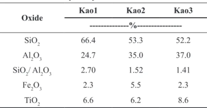
Table I shows that the SiO2 and Al2O3 contents determined in the Kao1, Kao2, and Kao3 are very
different from theoretical values: 46.5 % - SiO2
and 39.5 % - Al2O3 (Aranha et al. 2010). This
difference is explained by the high level weathering
Amazonian region condition. Additionally, Fe2O3,
TiO2, and in minor proportion, MgO, and CaO
were identified. The literature shows that mineral
impurities such as quartz, anatase, rutile, pyrite, limonite, feldspar, mica, montmorillonite, and various iron and titanium oxides in kaolinites are usually found. Si and Al, in the form of hydroxides, can apparently occur as coatings on the kaolinite layers. In fact, the contents of SiO2, Al2O3 and Fe2O3 correspond to kaolinite, quartz and gibbsite (XRD not shown). The presence of Fe and SiO2/Al2O3 ratio indicate that Kao1, Kao2, and Kao3 can have isomorphic substitution of Al3+ by Fe3+ (Melo et al. 2002). The literature explained the TiO2 presence as an aggregate commonly found in kaolinite. The TiO2 content of kaolinite is apparently dependent
on the mineral source. For example, feldspar, such as kaolinite from Manaus, has a low TiO2 content compared to a biotite schist or biotite granite source which shows a relatively high TiO2 (Weaver and Pollard 1973).
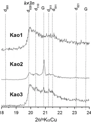
Figure 1 clearly shows that d011 d011 d011
values are distinguishable for the three kaolinites.
The kaolinite XRD presents (hkl) reflections
commonly affected by structural defects. Among such reflections, we can distinguish those wherein
k is multiple of 3, i.e., k = 3n (n = 1, 2, 3, ...) and those wherein k ¹ 3n. The existence within pilings of defects caused by displacements and/or rotations at the plane layers explains the multiple observed k. Therefore, the Hinckley and Liètard indices indicate that three kaolinites have important structural
differences (Table II). Typically the modifications
occur in hkl where k ¹ 3n (n = 1, 2, 3, ...) suggesting loss of the crystallographic order and/or defects in the kaolinite structure caused by displacement type ± b/3 or ± 2/3 rotations. As the proportion of defects is reduced, it suggests the existence of k≠3n with a profile similar to the hkl reflections (Aglietti
and Porto Lopes 1986).
According to the Hinckley index, the structural disorder of kaolinite is more noticeable for Kao1 and Kao2 than for Kao3. The structural disorder suggests the existence of weathering products from feldspars, which are precursors of kaolinites well and/or poorly ordered mainly along the b axis. The Liètard index shows a high number of
TABLE I
Chemical composition of Kao1, Kao2 and Kao3 obtained
by X-ray fluorescence.
Oxide Kao1 Kao2 Kao3
---%---SiO2 66.4 53.3 52.2
Al2O3 24.7 35.0 37.0
SiO2/ Al2O3 2.70 1.52 1.41
Fe2O3 2.3 5.5 2.3
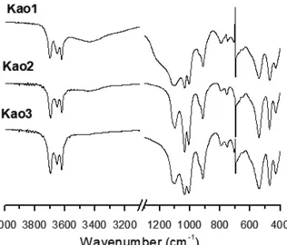
The FTIR spectra shows absorption bands of stretching vibrational OH groups (internal and external) present in the octahedral sites (Figure 2). At 3619-3621 cm-1 a band stretching vibrational absorption parallel to the ab plane is observed, i.e., in the common plane of apical oxygen of the tetrahedral sheet. The vibrational absorption bands in Kao1, Kao2, and Kao3 indicates a similarity to the internal OH groups of the three kaolinites. The presence of an absorption band at 3696 cm-1 characterizes perpendicular vibrational mode typical of the octahedral layer. It suggests a preservation of the OH groups from the kaolinite surface in all samples. It shows Kao1, Kao2, and Kao3 having their hydrogens linked with oxygen atoms bound to octahedral layer surface without considerable change. The vibrational mode of O-H bound to the atom of aluminum octahedral layer
confi rms what we had imagined.
However, the relationship of intensity of the stretch bands 3619-3621 and 3696 cm-1 indicates
diff erent levels of pleochroism for Kao1, Kao2,
and Kao3. Kao1 presents an inversion of the intensity of the bands, i.e., absence of pleochroism
caused due to rotation ±2π/3, as shown in the XRD
(Aglietti and Porto Lopez 1986). The ratio of intensity between the bands at 795 and 755 cm-1 also demonstrates loss of pleochroism. According to Deng et al. (2002), it occurs because of the particle size reduction. The absence of the 3652 cm-1 band in Kao3 shows vibrations directly related to weak hydrogen bonds typical of kaolinite of low crystallinity and the three vibrational modes remaining indicate OH groups coupled with moments of transition in the d001 plane (Farmer 2000, Frost et al. 2001). The shape of the band that corresponds to the angular-displacement vibration in 938 cm-1 indicates a kaolinite with a low number
of hydrogen bonds in octahedral surface (Figure 2). The absorption band in 914 cm-1 relative to the
angular-displacement vibration corresponds to the aluminol (Al-O-H) group. Furthermore, the O-H
TABLE II
Crystallinity index of Kao1, and Kao2 Kao3.
Sample Hinckley Defect* Liètard
(R2) Defect*
Kao1 0.549 Mean 0.026 High
Kao2 0.837 Mean 0.871 Mean
Kao3 0.465 High 0.453 High
*Categorized according to Aparicio et al. 2006.
Figure 1 - X-ray diff raction patterns of kaolinites.
defects for Kao1 and Kao3 and average for Kao2 obeying the following order: Kao1 > Kao3 > Kao2. Alterations in the kaolinite structure occur due to the isomorphic substitution by Fe3+, spaces caused
bond is unchanged for octahedral and tetrahedral sheets layers indicated by the stretching band at 3619-3621 cm-1.
In the 1010 cm-1 region, the Kao1, Kao2, and
Kao3 are varied by absorption band of Si-O stretch
suggesting diff erent particle sizes as well as low
crystallinity (Shoval et al. 1999). It is interesting to note that the concentrations of SiO2 and Al2O3 are
diff erent for Kao2 and Kao3, which suggests two
types of kaolinite. The relative intensities of 468 (band angular vibration of Si-O) and 539 cm-1 (band angular vibration of Si-O-Si) showed that Kao1 has small amounts of Al2O3 suggesting the existence of isomorphic substitution in the octahedral site. Therefore, the changes in the vibrational modes
detected by the loss of pleochroism, diff erences
in the angular vibration suggest that kao1 has changed its crystalline structure due to isomorphic substitution.
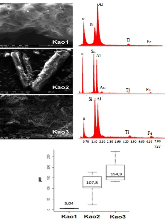
The SEM of Kao1, Kao2 and Kao3 are typical of kaolinite (Figure 3). Kao1 presents groups and stacks in the form of pilasters and crystals in the form of lamellae. This structure called booklet with pictures and cylindrical vacancies, produces porosities in the kaolinite lattices while Kao2 presents rod-shaped forms of “stalactite”,
presenting better crystallinity. Kao3 shows strong placoid sheets, vertical growth toward the crystallographic c axis, showing mineral habit is not
well defi ned. The mapping elements by EDS Kao1,
Kao2 and Kao3 revealed that the predominant elements are Si, Al, O, Fe and Ti. The SEM shows that the morphology and particle sizes from Kao1, Kao2 Kao3 have several levels.
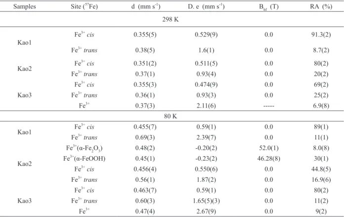
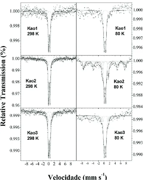
Mössbauer parameters and spectra at 298 K
clearly show that Kao1, and Kao2 Kao3 possesses
paramagnetic Fe3+, suggesting isomorphic
substitution of Fe3+ by Al3+ in the octahedral sites
(Figure 4 and Table III). Furthermore, the widest line (G = 0.62 and 0.71 mm s-1) indicate the presence
of a triclinic kaolinite with layers infl uenced by diff erences in layer stacking of polytypes as well
as by degree of layer stacking order. (Komusinski et al. 1981).
Although the Mössbauer spectra showed that
the majority of kaolinites contain divalent iron in octahedral coordination, there were no values of d = 1.130 ± 0.050 mm s-1 for the Fe2+ substituted octahedral site. In turn, the orientation of the hydroxyl groups of octahedral sheets is determined by the stretching layer as well as by the symmetry
of the crystal fi eld in which the Fe3+
is located.
Mössbauer parameters found for Fe3+ are
characteristic of isomorphic substitution in position
cis and trans-OH (Taneja and Raj 1993). Therefore,
it is not only the structural diff erences of kaolinites caused by defects and diff erent octahedral shapes
(Heller-Kallai and Rozenson 1981). Actually, the paramagnetic behavior observed reveals that kaolinite contains structural defects in its crystal lattice due to isomorphic substitution by Fe3+, causing disorder and changing the spin-spin interaction making the paramagnetic (Gaite et al.
1997). Furthermore, the Mössbauer spectra are distinguishable showing polytypic modifi cations of kaolinite with diff erent line-widths. The presence
of Fe3+ forming OH-cis and OH-trans points out trapped hole centers in kaolinite structure,
trapping locus for electron holes (holes), i.e., the substitution of Al3+ by Fe3+ forms a positive moiety of the trapped separated charge pair produced by
electronic excitation (Coyne and Summers 1991). According to Sato et al. (2004), the holes in kaolinites occur in B- or C-sites which explains the
formation of the chiral structure.
The relative areas of the fitted Mössbauer
spectra indicate that isomorphic substitution
is greater in the octahedral site in cis position, following this order of reason AR-cis/AR-trans:
Kao1 (10.5) > Kao2 (4.0) > Kao3 (2.8). Mössbauer
spectra d and D (e) of paramagnetic Fe3+ from
silicate and/or iron oxides, not identified by previous physical techniques. The 80 K spectra
demonstrate that only Kao2 shows low among
iron oxides as well as a low supermagnetism effect (Rozenson et al. 1982). The Mössbauer parameters
(d, D/e e Bhf) are typical of hematite (α-Fe2O3) and
goethite (α-FeOOH).
The study of the crystal structure observed for Kao1, Kao2 and Kao3 indicate a relation among the degree of substitution of Fe3+ at octahedral sites,
characterized by the presence of Fe3+ at positions
cis/trans-OH causing defects in the crystal structure of kaolinite, changing the crystalline arrangement and size of the average particle. Physical measurements reveal a larger amount of octahedrons consisting of Fe3+ at the position cis to Kao1, differing considerably in relation to Kao2
and Kao3.
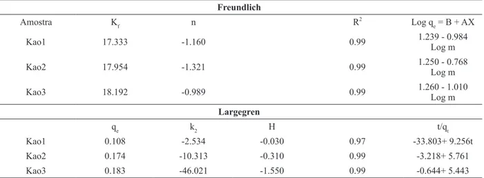
The adsorption process of phosphorus is characterized by having the parameters of adjustments to the Freundlich isotherms values R2 ~ 0.99 indicating that adsorption occurs in the
presence of heterogeneous surfaces on octahedral sites (Table IV). The negative n values suggest low
TABLE III
Mössbauer parameters at 298 and 80K.
Samples Site (57
Fe) d (mm s-1
) D. e (mm s-1
) Bhf (T) RA (%)
298 K
Kao1
Fe3+
cis 0.355(5) 0.529(9) 0.0 91.3(2)
Fe3+
trans 0.38(5) 1.6(1) 0.0 8.7(2)
Kao2 Fe
3+
cis 0.351(2) 0.511(5) 0.0 80(2)
Fe3+
trans 0.37(1) 0.93(4) 0.0 20(2)
Kao3
Fe3+
cis 0.355(3) 0.474(9) 0.0 69(2)
Fe3+
trans 0.36(1) 0.93(3) 0.0 25(2)
Fe3+ 0.37(3) 2.11(6) --- 6.9(8)
80 K
Kao1 Fe
3+
cis 0.455(7) 0.59(1) 0.0 89(1)
Fe3+trans 0.69(3) 2.39(7) 0.0 11(1)
Kao2
Fe3+(α-Fe
2O3) 0.48(2) -0.20(2) 52.0(1) 8.0(8)
Fe3+(α-FeOOH)
0.45(1) -0.23(2) 46.28(8) 30(1)
Fe3+
cis 0.456(4) 0.550(6) 0.0 44.8(5)
Fe3+
trans 0.56(1) 1.87(2) 0.0 16.9(6)
Kao3
Fe3+
cis 0.463(7) 0.59(1) 0.0 80(2)
Fe3+
trans 0.60(3) 1.65(5)(3) 0.0 11(2)
Fe3+
0.47(4) 2.67(9) 0.0 9(2)
Figure 4 - Mössbauer spectra at 298K and 80K of the samples.
adsorption capacity of orthophosphate adsorption for all samples. It is explained by physical
measurements mainly by substitution of Fe3+,
responsible for the diff erentiation of cis, and trans
octahedral sites. The adsorption capacity obeys the following order Kao1> Kao2 > Kao3. Largegren parameters (equation pseudo-second-order) clearly show that Kao1 retains faster orthophosphate on its surface. The increase in the substitution of Fe3+
-cis in the octahedral sites explains the retention of orthophosphate by Kao1 than Kao2 and Kao3.
These fi ndings reveal that the iron in the kaolinite
structure contributed with their chemical nature
and high specifi c area as an important role in the
transport and the removal of orthophosphate.
CONCLUSIONS
The mineral composition comprised kaolinite, quartz, gibbsite and goethite. As major element the
EDS spectra and X-ray fl uorescence showed the
prevalence of Al and Si.
The presence of goethite indicates that Kao1
summarizes Kao1 and Kao2 as poorly crystallized/ ordering, while kao2 as crystallized/ordering. As a result of average particle reduction, FTIR showed a higher inversion of the vibration bands relative to the hydroxyl groups in the range of 3619-3696 cm-1
and 795 and 755 cm-1 suggesting a natural loss of
pleochroism of the Kao1 samples.
SEM Images showed that Kao2 is well-crystallized while Kao1 and Kao3, poorly crystallized. These results explain the values of the Hinckley, and Liétard indices obtained. Generally, samples substituted by Fe3+
form octahedral sites in the position cis-OH and trans-OH. The adsorption
isotherms and kinetics, fitted by Freundlich and
Largegren equations confirm the existence of heteronuclear sites on kaolinite surfaces
The Fe3+-isomorphic substitution alters the magnetic properties of the kaolinite to paramagnetic
according to the Mössbauer spectroscopy. The
number of Fe3+-OH-cis determines the capacity to adsorb orthophosphate.
REFERENCES
ADEBOWALE KO, UNUABONAH EI AND OLU-OWOLABI BI. 2008. Kinetic and thermodynamic aspects of the adsorption of Pb2+
and Cd2+
ions on tripolyphosphate-modified kaolinite clay. Chem Eng J 136: 99-107.
AGLIETTI EF AND PORTO LOPEZ EP. 1986. Mechanochemical effects in kaolinite grinding. ii. Structural aspects. Int J Miner Process 16: 135-146. APARICIO P, GALÁN E AND FERRELL RE. 2006. A new
kaolinite order index based on XRD profile fitting. Clay Miner 41(4): 811-817.
ARANHA DA PAZ SP, ANGÉLICA RS AND DE FREITAS NEVES R. 2010. Síntese hidrotermal de sodalita básica a partir de um rejeito de caulim termicamente ativado. Quim Nova 33(3): 579-583.
BALAN E, ALLARD T, BOIZOT B, GUILLAUME M AND MULLER JP. 2000. Quantitative measurement of paramagnetic Fe3+
in kaolinite. Clay Clay Miner 48(4): 439-445.
CORRÊA MM, KER JC, BARRÓN V, TORRENT J, CURI N AND TORRES TCP. 2008. Caracterização física, química, mineralógica e micromorfológica de horizontes coesos e fragipãs de solos vermelhos e amarelos do ambiente tabauleiros costeiros. R Bras Ci Solo 32: 297-313. COUCEIRO PRC AND SANTANA GP. 1999. Caulinita em
solo da Amazônia: Caracterização e Permutabilidade. Acta Amaz 29: 267-275.
COYNE LM AND SUMMERS DP. 1991. Surface activation of air oxidation of hydrazine on kaolinite. 2. Consideration of oxidizing/reducing entities in relationship to other compositional, structural, and energetic factors. Langmuir 7(8): 1675-1688.
CUI Y AND WENG L. 2013. Arsenate and phosphate adsorption in relation to oxides composition in soils: LCD modeling. Environ Sci Technol 47(13): 7269-7276. DENG Y, WHITE GN AND DIXON JB. 2002. Effect of
Structural Stress on the Intercalation Rate of Kaolinite. J Colloid Interf Sci 250(2): 379-393.
TABLE IV
Parameters of Freundlich adsorption kinetics and Largegren (equation pseudo-second-order). Freundlich
Amostra Kf n R2
Log qe = B + AX
Kao1 17.333 -1.160 0.99 1.239 - 0.984
Log m
Kao2 17.954 -1.321 0.99 1.250 - 0.768
Log m
Kao3 18.192 -0.989 0.99 1.260 - 1.010
Log m
Largegren
qe k2 H t/qt
Kao1 0.108 -2.534 -0.030 0.97 -33.803+ 9.256t
Kao2 0.174 -10.313 -0.310 0.99 -3.218+ 5.761
DIXON JB. Kaolin and serpentine group minerals. 1989. In: Dixon JB and Weed SB (Eds), Minerals in Soil Environments, 2nd
ed., Madison: Soil Science Society of America, p. 467-525.
DOTTO GL, VIEIRA MLG, GONÇALVES JO AND PINTO LAA. 2011. Remoção dos corantes azul brilhante, amarelo crepúsculo e amarelo tartrazina de soluções aquosas utilizando carvão ativado, terra ativada, terra diatomácea, quitina e quitosana: estudos de equilíbrio e termodinâmica. Quim Nova 34(7): 1193-1199.
FARMER VC. 2000. Transverse and longitudinal crystal modes associated with OH stretching vibrations in single crystals of kaolinite and dickite. Spectrochimica Acta A 56: 927-930.
FROST RL, KRISTÓF J, HORVÁTH E AND KLOPROGGE JT. 2001. The Modification of Hydroxyl Surfaces of Formamide-Intercalated Kaolinites Synthesized by Controlled Rate Thermal Analysis. J Colloid Interf Sci 239(1): 126-133.
GAITE JM, ERMAKOFF P, ALLARD T AND MULLER JP. 1997. Paramagnetic Fe3+: a sensitive probe for disorder in kaolinite. Clay Clay Miner 45(4): 496-505.
HELLER-KALLAI L AND ROZENSON I. 1981. The use of
Mössbauer spectroscopy of iron in clay mineralogy. Phys
Chem Miner 7: 223-238.
KAMIYANGO MW, MASAMBA WRL, SAJIDU SMI AND FABIANO E. 2009. Phosphate removal from aqueous solutions using kaolinite obtained from Linthipe, Malawi. Phys Chem Earth 34(13-16): 850-856.
K H A W M E E K , S U D D H I P R A K A R N A , KHEORUENROMNE I AND SHING B. 2013. Surface charge properties of kaolinite from Thai soils. Geoderma 192: 120-131.
KOMUSINSKI J, STOCH L AND DUBIEL SM. 1981. Application of electron paramagnetic resonance and
Mössbauer spectroscopy In The investigation of
kaolinite-group minerals. Clay Clay Miner 29(1): 23-30.
MELO VF, SCHAEFER CEGR, SINGH B, NOVAIS RF AND FONTES MPF. 2002. Propriedades químicas e cristalográficas da caulinita e dos óxidos de ferro em
sedimentos do grupo Barreiras no município de Aracruz, Estado do Espírito Santo. R Bras Ci Solo 26: 53-64. MIRANDA-TREVINO JC AND COLES CA. 2003. Kaolinite
properties, structure and influence of metal retention on pH. Appl Clay Sci 23(1-4): 133-139.
REHIM MHA, YOUSSEF AM AND ESSAWY HA. 2010. Hybridization of kaolinite by consecutive intercalation: Preparation and characterization of hyperbranched poly(amidoamine)–kaolinite nanocomposites. Mat Chem Phys 119(3): 546-552.
ROZENSON I, SPIRO B AND ZAK I. 1982. Transformation of iron-bearing kaolinite to iron-free kaolinite, goethite, and hematite. Clay Clay Miner 30(3): 207-214.
SANTOS MDLS, MUNIZ K, FEITOSA FAN AND BARROS NETO B. 2007. Estudo das diferentes formas de fósforo nas águas da plataforma continental do Amazonas. Quim Nova 30(3): 569-573.
SATO H, ONO K, JOHNSTON CT AND YAMAGISHI A. 2004. First-principle study of polytype structures of 1:1 dioctahedral phyllosilicates. Am Mineral 11-12: 1581-1585.
SHOVAL S, YARIV S, MICHAELIAN KH AND GÉRARD PANCZER L. 1999. A Fifth OH-Stretching Band in IR Spectra of Kaolinites. J Colloid Interf Sci 212(2): 523-529. SOUZA WB AND SANTANA GP. 2014. Mineralogy,
zinc kinetic adsorption and sequential extraction of contaminated soil in Manaus, Amazon. Ciênc Rural 44(5): 788-793.
TANEJA SP AND RAJ D. 1993. Mössbauer and X-ray studies
of soils. Nucl Instrum Methods Phys Res B76: 233-235. TOMBÁCZ E, NYILAS T, LIBOR Z AND CSANAKI C.
2004. Surface charge heterogeneity and aggregation of clay lamellae in aqueous suspensions. Prog Coll Pol Sci 125: 206-215.
WEAVER CE AND POLLARD LD. 1973. The chemistry of clay minerals. Amsterdam: Elsevier Science, p. 212. WEI S, TAN WF, LIU F, ZHAO W AND WENG LP. 2014.