www.scielo.br/aabc
Removal of the cortical projections alters expression of NOS
in the different cell types of the superficial layers
of the superior colliculus in rats
FRANK TENÓRIO1, ARTHUR GIRALDI-GUIMARÃES2 and ROSALIA MENDEZ-OTERO2
1Departamento de Farmacologia e Psicobiologia, Instituto de Biologia Roberto Alcântara Gomes, UERJ 20551-030 Rio de Janeiro, Brazil
2Instituto de Biofísica Carlos Chagas Filho, UFRJ, 21941-590 Rio de Janeiro, Brazil
Manuscript received on September 13, 2002; accepted for publication on September 20, 2002; contributed byRosalia Mendez-Otero*
ABSTRACT
Nitric oxide has several biological roles and nitric oxide synthase (NOS) is expressed in the nervous system, and co-localizes with NADPH-diaphorase. The superficial layers of the superior colliculus (SC), which receive retinal and cortical inputs, present NADPH-d staining in a sub-population of neurons that include all cell types. We have previously shown, by NADPH-diaphorase, that eye enucleation alters the intracellular distribution of NOS. Here, we studied the effect of cortical ablation on NOS expression by neurons in collicular superficial layers. Our results show that cortical ablation alters the proportion of different NOS-positive cell types, but not the intracellular distribution of the enzyme.
Key words:superior colliculus, nitric oxide, visual cortex, NADPH-diaphorase.
INTRODUCTION
Nitric oxide (NO) is a gas that plays several roles in the central nervous system. Nitric oxide synthase (NOS) is highly expressed in the nervous system and NADPH-diaphorase staining is highly corre-lated with NOS activity (Bredt et al. 1991, Daw-son et al. 1991, Hope et al. 1991, Matsumoto et al. 1993). Despite of being a constitutive enzyme, neu-ronal NOS activity and its sub cellular distribution appears to be actively regulated at multiple levels (Vincent 1995).
Results form different laboratories suggest that retinal projections and/or its neural activity might have a role in the expression of this enzyme in the
*Member of Academia Brasileira de Ciências Correspondence to: Frank Tenório
E-mail: [email protected]
visual system. Light deprivation enhances NOS activity in the lateral geniculate nucleus (LGN) of mice and rabbits (Ientile et al. 1996) and enhances NADPH-d staining in the LGN of cats (Gunluk et al. 1994). Monocular enucleation also alters NOS distribution in the monkey visual cortex (Aoki et al. 1993).
collicu-collicular projections was disturbed by removal of the visual cortex.
MATERIALS AND METHODS
Lister rats from our breeding colony were used. All chemicals were purchased from Sigma Chemical Co. (St. Louis, MO). Newborn animals (P0) were anaesthetized by hypothermia to be submitted to vi-sual cortex removal surgery (n=5). An incision was made at the top of the head. The skull was opened and the region corresponding to the left occipital cortex was aspirated with a pipette tip attached to a pump. The left SC was the experimental one and the right SC of the same animal was used as the control. The animals were sutured and returned to their mother after recovering from the surgery. In addition, control animals (n=9) were used for com-parison. At ages between P30 and P60, animals were anaesthetized with sodium pentobarbital (50mg/kg) and intracardially perfused with 0.9% saline solution followed by 4% paraformaldehyde (PF) in 100mM phosphate buffer (pH 7.4). After dissection, the brains were immersed in 100mM phosphate buffer containing 20% sucrose, overnight at 4oC and then sectioned at 60µm on a cryostat at –20oC in the
coronal or parasagital planes. Sections containing the SC were collected in 50 mM Tris buffer (pH 7.4), washed twice in this buffer and submitted to NADPH-d histochemistry technique. After 1h at 37oC under constant shaking, reaction was inter-rupted, sections were washed twice in Tris buffer and mounted in gelatinized slides. The slides with sections were dehydrated and coversliped. Photomi-crographs were made using a Zeiss Axioplan micro-scope. Cell counts were performed under a 40X objective. Four fields in sections of 4 animals of each group were used. Piriform and wide field ver-tical cells were included in the same group (ganglion cells type I and II). The differences between the con-trol group and ablated animals were analyzed using
RESULTS AND DISCUSSION
Previously we have shown that eye enucleation do not interfere with the temporal sequence of NOS ex-pression in the rat SC superficial layers, although it alters the intracellular distribution of this enzyme by these cells (Tenório et al. 1998). After removal of the retinal projections the SC superficial layers still receive projections from the ipsilateral visual cor-tex. The removal of these collicular afferents could origin different changes, from those seem in enucle-ated animals, in the pattern of NOS expression by neurons of the SC superficial layers.
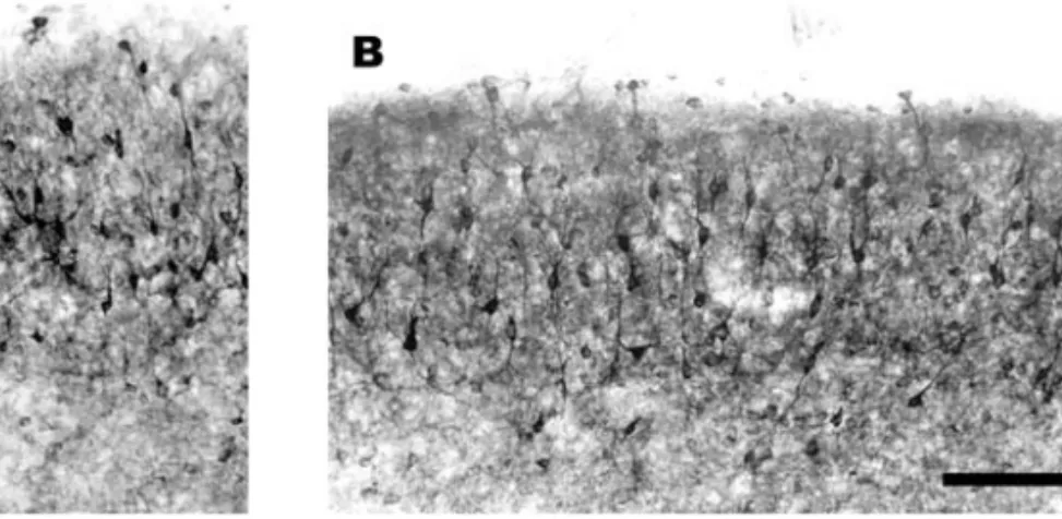
Fig. 1 – Photomicrograph of the superior colliculus of an animal P31 whose cortex was removed at P0. (A) SC contralateral to the removed cortex. (B) SC ipsilateral to the removed cortex. No significant difference was observed in the NADPH-d pattern. NOS was present in the cell bodies and in dendrites of the cells. The neuropil is also stained in both SCs. Scale bar: 100µm.
TABLE I
Cellular types in control and experimental animals.
Cell type Control (%) n Ablated (%) n
Marginal cell 22,89 195 22,00 190
Narrow field vertical cell* 42,96 366 23,25 200 Type I and II ganglion cell* 10,21 87 23,45 202
Type III ganglion cell 3,64 31 3,45 30
Stellate cell* 3,05 26 9,05 78
Surface horizontal cell* 1,64 14 2,70 24
Deep horizontal cell* 2,58 22 5,00 44
Non-classified cells 13,03 111 11,10 96
Total 100,00 852 100,00 860
P < 0,001 (χ2to k independent samples) differences between the control group and ablated animals. *correspond to statistical differences.
The difference in the results between the abla-tion of retinal afferents and cortical inputs shows that these projections might have different influences on the expression of NOS by SC cells and it is possible to suggest that this difference might be correlated with different functional roles of these afferents. In fact, it has been shown that the ablation of visual cortex facilitated induction of LTP in the SC super-ficial layers, indicating that the corticocollicular pro-jection could have a modulatory function, blocking the enhancing of the synaptic transmission
deter-in this co-expression. However, results from other laboratories allow us to make some inferences. In cats, the GABAergic neurons have the cortical pro-jections as a very important synaptic source (Mize 1988). These cells were described as horizontal I, granule I and granule II and, by the morphological similarities, they probably correspond to the super-ficial horizontal and the ganglion cells, respectively, described in rats and found in our work. Our results showed that cortical ablation induces an increase in the proportion of the NADPH-d stained cells of these groups, in relation to control animals. Then it is possible to suggest that the absence of cortical projections, which is an important source of synap-tic input to these cells, directly induces an increase in the NOS expression by them. However, there are a few points against this conclusion. First, the affer-ent inputs to the deep horizontal cells, the proportion of which also increases, are unknown. Second, al-though there are a large number of reports involving the study of the sensory processing in the SC, the complete synaptic circuitry underlying SC function remains not completely understood. So, it is also possible that the effects we described after removal of the cortical projection are due to indirect effects of the absence of these projections onto SC cells.
RESUMO
O óxido nítrico apresenta diversos papéis biológicos e a óxido nítrico sintase (ONS) é expressa no sistema nervoso, co-localizando-se com a NADPH-diaforase. As camadas superficiais do colículo superior (CS), as quais recebem aferências retinianas e corticais, possuem marcação para NADPH-diaforase numa sub-população de neurônios que inclui todos os tipos celulares. Previamente demostramos, por NADPH-diaforase, que a enucleação ocular altera a distribuição intracelular da ONS. Neste trabalho estu-damos o efeito da ablação cortical na expressão da ONS por neurônios das camadas superficiais coliculares. Nos-sos resultados demonstraram que a ablação cortical altera
visual, NADPH-diaforase.
REFERENCES
Aoki C, Fenstemaker S, Lubin M and Go C-G. 1993. Nitric oxide synthase in the visual cortex of monocular monkeys as revealed by light and elec-tron microscopic immunocytochemistry. Brain Res 620: 97-113.
Bolz J, Hübener M, Kehrer I and Novak N.1991. Structural organization and development of identified projection neurons in primary visual cortex. In Bag-noli P and Hodos W(Eds), The Changing Visual System, New York: Plenum, pp. 233-246.
Bredt DS, Glatt CE, Hwang PM, Fotuhi M, Dawson
TM and Snyder SH.1991. Nitric oxide synthase protein and mRNA are discretely localized in neu-ronal populations of the mammalian CNS together with NADPH diaphorase. Neuron 7: 615-624.
Dawson VL, Dawson TM, London ED, Bredt DS and
Snyder SH.1991. Nitric oxide mediates glutamate neurotoxicity in primary cortical cultures. Proc Natl Acad Sci USA 88: 6368-6371.
Gunluk AE, Bickford ME and Sherman SM.1994. Rearing with monocular lid suture induces abnormal NADPH-diaphorase staining in the lateral geniculate nucleus of cats. J Comp Neurol 350: 215-228.
Hirai H and Okada Y.1993. Ipsilateral corticotectal pathway inhibits the formation of long-term poten-tiation (LTP) in the rat superior colliculus through GABAergic mechanism. Brain Res 629: 23-30.
Hope BT, Michael GJ, Knigge KM and Vincent SR. 1991. Neuronal NADPH diaphorase is a nitric oxide synthase. Proc Natl Acad Sci USA 88: 2811-2814.
Ientile R, Picciurro V, Pedale S, Nucci C, Malecka
B, Nistico G and Macaione S.1996. Nitric oxide enhances amino acid release from immature chick embryo retina. Neurosci Lett 219: 79-82.
Langer TP and Lund RD.1974. The upper layers of the superior colliculus: A Golgi study. J Comp Neurol 158: 405-435.
Forstermann Y.1993. A correlation between sol-uble brain nitric oxide synthase and NADPH-diaphorase activity is only seen after exposure of the tissue to fixative. Neurosci Lett 155: 61-64.
Mize RR.1988. Immunocytochemical localization of gamma-aminobutyric acid (GABA) in the cat supe-rior colliculus. J Comp Neurol 276: 169-187.
Mize RR, Spencer RF and Sterling P.1982. Two types of GABA-accumulation neurons in the superfi-cial gray layer of the cat superior colliculus. J Comp Neurol 206: 180-192.
Okada Y. 1974. Distribution ofγ-aminobutyric acid (GABA) in the layers of superior colliculus of the rabbit. Brain Res 75: 362-365.
Soares-Mota M, Henze I and Mendez-Otero R. 2001. Nitric oxide synthase-positive neurons in the rat superior colliculus: colocalization of NOS with NMDAR1 glutamate receptor, GABA and parvalbu-min. J Neurosci Res 64: 501-507.
Tenório F, Giraldi-Guimarães A and
Mendez-Otero R.1996. Morphology of NADPH-diaphor-ase-positive cells in the retinoceptive layers of the developing rat superior colliculus. Int J Dev Neurosci 14: 1-10.
Tenório F, Giraldi-Guimarães A, Santos HR, Cintra
WM and Mendez-Otero R.1998. Eye enucleation alters intracellular distribution of NO synthase in the superior colliculus. Neuro Report 9: 145-148.