Acta Cir. Bras. 2018;33(9):744-752 DOI: http://dx.doi.org/10.1590/s0102-865020180090000002
Gustavo Ithamar Souto MaiorI, Guilherme Veras MascenaII, Valéria Wanderley Pinto Brandão
MarquisIII, Carlos Alberto Figueiredo FilhoIII, Alexandre Rolim da PazIV, Líbia Cristina Rocha Vilela
MouraV, Carlos Teixeira BrandtVI
Role of moxifloxacin-dexamethasone in cardiac
histomorphometric findings among Wistar rats from
infected mothers
1Abstract
Purpose: To investigate cardiac changes in young rats, whose mothers underwent autogenic
fecal peritonitis, during organogenesis phase and to evaluate the role of intravenous administration of moxifloxacin and dexamethasone in preventing infection-related cardiac changes.
Methods: A prospective histomorphometric study was performed on 29 hearts of Wistar
four-month old rats. Animals were divided into three groups: Negative Control Group (NCG) included 9 subjects from healthy mothers; Positive Control Group (PCG) included 10 subjects from mothers with fecal peritonitis (intra-abdominal injection of 10% autogenic fecal suspension in the gestational period) and did not receive any treatment; and Intervention Group (IG), with 10 animals whose infected mothers received moxifloxacin and dexamethasone treatment 24 hours after induction of fecal peritonitis.
Results: Nuclear count was higher in the IG group as compared to PCG (p = 0.0016) and in
NCG as compared to PCG (p = 0.0380). There was no significant difference in nuclear counts between NCG and IG.
Conclusion: Induced autogenic fecal peritonitis in pregnant Wistar rats determined myocardial
changes in young rats that could be avoided by the early administration of intravenous moxifloxacin and dexamethasone.
Key words: Peritonitis. Myocardium. Pregnancy. Dexamethasone. Rats.
IFellow PhD degree, Postgraduate Program in Tropical Medicine, Health Sciences Center, Universidade Federal de
Pernambuco (UFPE), Recife-PE, Brazil. Acquisition and interpretation of data, manuscript writing.
IIFellow PhD degree, Postgraduate Program in Surgery, Health Sciences Center, UFPE, Recife-PE, Brazil. Statistical analysis,
critical revision.
IIIFellow PhD degree, Postgraduate Program in Tropical Medicine, Health Sciences Center, UFPE, Recife-PE, Brazil.
Acquisition of data, critical revision.
IVFellow PhD degree, Postgraduate Program in Pathology, Health Sciences Center, Universidade Estadual de Campinas
(UNICAMP), Campinas-SP, Brazil. Acquisition and interpretation of data.
VPhD, Head, Postgraduate Program in Tropical Medicine, Health Sciences Center, UFPE, Recife-PE, Brazil. Interpretation
of data, final approval.
VIPhD, Head, Postgraduate Program in Surgery, Health Sciences Center, UFPE, Recife-PE, Brazil. Interpretation of data,
Medical School from Campina Grande (FCM-CG), Paraíba.
Thirty-two Wistar rats (Rattus
Novergicus Albinus Rodentia Mammalia) were included in the study. Three female adult rats (mothers) with approximately three months of age (300g), without any disease or injury, and
29 young rats (offspring) with 4 months of age
were evaluated and monitored.
The animals were housed and cared in accordance with the “Guide for the Care and Use of Laboratory Animals” and the study was
conducted respecting the ethical principles in animal experimentation. The animal ethics committee (CEUA/CESED) approved the study protocol under number 5804122015. Animals
were provided by the breeding colony of FCM -CG.
Young rats were divided into three
groups: Negative Control Group (NCG; n = 9), offspring of rats without induced peritonitis (healthy controls); Positive Control Group (PCG; n = 10), offspring of rats who underwent to induction of autogenic fecal peritonitis but did not receive any treatment; and the Intervention Group (IG; n = 10), offspring of rats with induced autogenic fecal peritonitis who were treated with the intravenous administration of moxifloxacin (40mg/Kg) and dexamethasone (0.2mg/kg) 24 hours after maternal infectious
insult.
Animals that at the time of induction of peritonitis showed signs of any organic fluid in
the peritoneal cavity aspirate, evidence of an
inflammatory process in the stool, including pus or blood, or which developed malnutrition,
were excluded.
The pregnant rats were kept in polypropylene cages measuring 43 x 43 x 20 cm at a controlled temperature of 23 ± 1°C and 12-hour light/dark cycles. Access to food and drink was ad libitum, with food consisting of a nutritionally balanced rodent diet.
Body weight, respiratory rate and temperature were monitored in a daily basis
■
Introduction
Congenital heart disease is the most common detected anomaly at birth and is associated with high morbidity and mortality in childhood and adolescence1. The elucidation of mechanisms underlying cardiac development
is crucial not only for the identification of abnormalities during organogenesis but also to adopt strategies of early intervention2.
The association of ocular and encephalic
congenital anomalies in concepts with maternal bacterial endotoxemia has already been demonstrated in pre-clinical study3.
Induction of peritonitis in rats on the ninth day of pregnancy, using a 10% filtered autogenic fecal suspension (4ml/kg), was associated with
the onset of encephalomalacia and cataract. On
the other hand, intraperitoneal (IP) injection of moxifloxacin and dexamethasone 48-72h after the induction of maternal infection was effective in preventing these complications4.
This research, however, neither evaluated the histological repercussions of the maternal
infection on the offspring nor the intravenous (IV) route for antimicrobial therapy, which is the most used in clinical practice in cases of peritonitis in pregnant women.
The purpose of this study was to
investigate cardiac histomorphometry in
four-month-old Wistar female rats, whose mothers
underwent peritonitis with a 10% autogenic
fecal suspension in the early phase of pregnancy (ninth day), during which maximum cardiac
organogenesis takes place, and to evaluate the histological cardiac findings of intravenous administration of a combination of moxifloxacin and dexamethasone 24 hours after induction
of maternal abdominal infection.
■
Methods
This was a prospective, longitudinal,
by the same veterinarian. They were also evaluated for the amount and color of the hair
in order to detect early signs of infection. The birth of the puppies took place approximately 21 days after pregnancy. All the others survived and showed good development. After the three-week period of breastfeeding, each of
the three mothers was separated from their
offspring. The male puppies were separated
from the females as well.
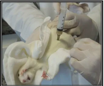
In two pregnant Wistar rats, fecal peritonitis was induced with 4ml/kg of 10%
autogenous fecal suspension in the abdominal
cavity in the left lower quadrant on the 9th day of pregnancy (Figure 1). Fecal suspension was
obtained from two grams of feces collected from each rat. This sample was dissolved in 20
ml of 0.9% saline solution, homogenized and gauze filtered for removing large particles.
Figure 1 – Fecal peritonitis induction. Injection of 4
ml/kg of 10% autogenous fecal suspension filtered into the left iliac quadrant of the Wistar rat on the
9th day of pregnancy.
At 4 months of age, young animals (n=29), 15 males and 14 females were euthanized with intravenous ketamine hydrochloride, 50 mg/kg, and xylazine, 10 mg/ kg. The thoracic cavity was accessed through a midline incision with a 15cold scalpel blade. The
remaining layers of the thoracic wall and the
sternum were sectioned with surgical scissors.
The heart and great vessels were carefully
removed in block (Figure 2), embedded in 10% formaldehyde and sent for embedment in paraffin and sectioned for the histological slides of approximately 5 mm thick, from the tip of the heart to the base.
Figure 2 – Heart removal technique. Median
thoracotomy by 15 scalpel blade. Isolation and block removal of the heart and great vessels with posterior placement in 10% formaldehyde.
After washing in running water
for one hour, to remove possible formalin
precipitations, the material was submitted to a chemical process that allowed adequate and standardized stiffness of the samples for the histological sections.
The chemical process followed an
alcoholic sequence for dehydration, in order to render the solutions miscible and avoid tissue retraction. After dehydration, the tissue specimens were immersed in xylene solution, which removes the alcohol. Alcohol is immiscible in molten paraffin, besides being submitted to diaphanization of the tissue. All tissue material was immersed in successive baths of fused paraffin at 60-63°C to remove all xylene and to harden the tissue for the histological sections. After chemical tissue
processing, all material was sent for inclusion
preheated molds.
The immersed material was placed
in the mold with the molten paraffin, cooled
and then removed from the mold, forming
the so-called histological paraffin blocks. Those blocks were cooled at low temperature, promoting further solidification of the paraffin and better grinding of the block during the microtomy. The blocks were stored in a freezer with temperature -3ºC and kept for at least 30
minutes before slicing. The histological slides
were identified and serial histological sections were made from the paraffin block, with the
purpose of assembling a slide for analysis. The
thickness of the obtained cuts was 5 μm. The cuts already laid on the blade were kept on the stand for about 20 to 30 minutes. Excess paraffin was removed from the “freshly caught” slices by heat in a sterilization oven and dried at about 60-65°C for about 30 minutes.
Subsequently, the preparation
(submission of chemical substances) of the
histological section was made for staining in Hematoxylin and Eosin. The entire process was performed in automatic equipment for staining histological slices, Leica ST5010 model. After
staining was completed, the slides analyses
were performed and the heart sections were observed under a microscope with x400 magnification. Representative samples of each section of the left ventricle - free wall - of each mouse, from the respective groups, were
randomly selected for analysis.
Histomorphometric analyzes were performed with the aid of a Leica DM 2000 microscope (x40 magnification lens) attached to a digital camera (Leica DFC 425, Leica Microsystems, Wetzlar, Germany) and coupled to a computer equipped with an image analyzer program. In this research,
the histomorphometric measurements
were performed using the Image J 1.51, Java 1.8.0_101 (64-bit) digital image analysis software. This program is widely used in scientific research involving quantification
and analysis of images5-10, with programming
platform based on the Java language. In a systematic way, all the photomicrographs were
processed according to the same algorithm.
Initially, the photomicrography was converted
to 8-bits. Then in the “processes” tab, image was rendered binary. The data of each image
(number, size and nuclear area) were obtained
through the “Nucleus Counter” plugin. The “Threshold” command was used to separate
the background of the image from the particles of interest. A cut-off point was determined for all particles with an area of less than 100 pixels or greater than 5000 pixels. It was not imposed any restrictions on shape. The cut-off point eliminated artifacts and any coalesced
cells.
The results were analyzed using GraphPad Prism (Prism 7 for Mac OS X, Version 7.0c, March 1, 2017 - GraphPad Software, Inc., USA). The quantitative parameters
were expressed by their means and standard
deviation and represented in box-plot graphs. Each parameter was tested for Gaussian distribution using the Kolmorogov-Smirnov test. For the comparative analysis of the multiple means among the groups that passed the normality test, one-way ANOVA and the Tukey multiple comparisons post-test were
used. Comparisons of non-parametric means
were performed using the Kruskal-Wallis test. Dunn’s post-test was used to identify which groups differed from each other. Values of p
<0.05 were used to reject the null hypothesis.
■
Results
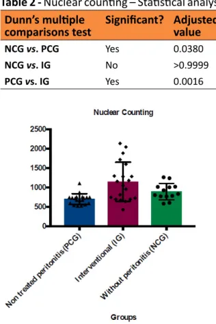
The nuclear count differed among
the groups (Kruskal-Wallis - p = 0.0015) and was significantly higher in the NCG when compared to the PCG (p = 0.0380). In the IG, a
higher nuclear count was also observed when
Table 1 - Nuclear counting - Descriptive
statistics.
Without
Peritonitis
(NCG)
Non Treated
Peritonitis
(PCG)
Intervention
Group (IG)
Minimum 584 526 429
25%
Percentile 769.5 594 702.3
Median 833 727 1103
75%
Percentile 1018 746 1525
Maximum 1264 1110 2136
Midium 894.2 700.1 1150
Std.
Deviation 211 137.7 503.9
Std. Error of Mean
58.5 31.6 112.7
Kruskal-Wallis test (p=0.0015)
Table 2 - Nuclear counting – Statistical analyses.
Dunn’s multiple
comparisons test Significant?
Adjusted p value
NCG vs. PCG Yes 0.0380
NCG vs. IG No >0.9999
PCG vs. IG Yes 0.0016
Figure 3 - Nuclear counting per field evaluated in
the investigated groups.
The percentage of the photomicrograph
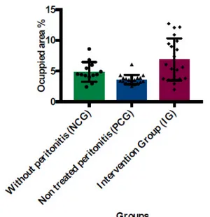
area occupied by the nuclei differed among the groups (Kruskal-Wallis - p = 0.0007) and was significantly higher in the NCG when compared to the PCG (p = 0.0448) and in the IG group when compared to the PCG (p = 0.0006). There was no significant difference between NCG and IG groups (Tables 3 and 4, Figure 4).
Table 3 - Area of photomicrography (%)
occupied by nuclei - Descriptive statistics.
Without
Peritonitis
(NCG)
Non Treated
Peritonitis
(PCG)
Intervention
Group (IG)
Minimum 2.4 2.6 2.0
25%
Percentile 4.2 3.1 3.8
Median 4.5 3.6 5.9
75%
Percentile 5.8 3.9 9.8
Maximum 8.7 6.1 12.8
Midium 4.9 3.7 6.9
Std.
Deviation 1.6 0.8 3.4
Std. Error
of Mean 4.5 0.2 0.8
Kruskal-Wallis test (p=0.0007)
Table 4 - Area of photomicrography (%) occu
-pied by nuclei – Statistical analyses.
Dunn’s
multiple
comparisons test
Significant? Adjusted p value
NCG vs. PCG Yes 0.0448
NCG vs. IG No >0.9999
Figure 4 - Area of photomicrography (%) occupied by nuclei.
The nuclear size differed among the groups (ANOVA - p = 0.0207). This variable was significantly higher in IG when compared to PCG (p = 0.0170). There was no significant difference between NCG and IG groups, nor between NCG and PCG groups (Tables 5 and 6,
Figure 5).
Table 5 - Nuclear size (pixels) – Descriptive
statistics.
Without
Peritonitis
(NCG)
Non Treated
Peritonitis
(PCG)
Intervention
Group (IG)
Minimum 380584 472049 485281
25%
Percentile 497572 509866 541215
Median 577524 540007 626429
75%
Percentile 623857 595697 678605
Maximum 719346 616406 769391
Midium 570701 546566 617717
Std.
Deviation 100882 45246 85052
Std. Error
of Mean 27980 10380 19018
ANOVA (p=0.0207)
Table 6 - Nuclear size - Statistical analyses.
Tukey’s multiple
comparisons test
Significant Adjusted P value
NCG vs. PCG No 0.6665
NCG vs. IG No 0.2167 PCG vs. IG Yes 0.0170
Figure 5 - Nuclear size (pixel).
■
Discussion
Cardiac histological changes in Wistar rats have been demonstrated in infarct models11. However, evidence linking experimentally induced maternal peritoneal
infection to fetal cardiovascular repercussions
is scarce.
In the present study, ImageJ software
was used to perform the histomorphometric
analysis and to investigate fetal cardiac alterations caused by maternal bacterial
endotoxemia in pregnant Wistar rats.
The nuclear count and the percentage of
occupied nucleus area was significantly higher
in the NCG compared to PCG and PCG than in
and greater apoptotic activity, as determined by maternal peritonitis. Therefore, the absence of a significant difference in nuclear counts between the IG and NCG groups indicated the efficacy of the treatment (intravenous moxifloxacin and dexamethasone), dose (40 mg / kg and 0.2 mg / kg respectively) used in the GI, to control the maternal infectious
process.
No significant difference was found in nuclear NCG size when compared to PCG or IG. However, the nuclear size in PCG was lower than in IG. It is possible that the greater dispersion of the NCG (amplitude of 117 - 190) compared to the PCG (amplitude of 144 - 357), compared to a small sample, justifies the absence of significant statistical difference between the groups evidenced by the Tukey multiple comparison test (p = 0.6665).
Fluoroquinolone antibacterial compounds are treatment options of increasing importance for bacterial infections. In recent
years, immunomodulatory treatments of
infectious diseases have attracted the attention of researchers, increasing their expectations for additional benefits apart from that of the antibiotics. Accumulated knowledge attributes the beneficial effect of dexamethasone to its potential to reduce inflammatory mediators. In vitro studies support the finding that
dexamethasone promotes phagocytosis by human monocytes, not only of S. aureus, but of other macromolecules as well, and thereby
may contribute to tissue repair after immune-mediated tissue damage or infection12.
In experimental studies with murine,
the age of the animals is as important as the
weight to evaluate a therapeutic intervention.
The three mother pregnant rats in the present study had similar ages, approximately
12-month old.
In human beings as maternal ages increases, the rates of cesarean section are
high, and fetal death and neonatal morbidity
and mortality also grow. The incidence and mortality rate due to maternal sepsis have
increased in the last decade. In the US, the maternal mortality rate increased from 12 per 100,000 live births in 1990 to 14 per 100,000 live births in 201513,14.
Immunosenescence, a decline in the
immune system with increasing age is one of
the factors that justify the increase in maternal
mortality in recent years. The relevance of these
data led the United Nations (UN) to determine that reducing maternal mortality by 75 percent in 15 years would be one of the main goals for
the millennium. This goal was not achieved by
most industrialized countries15. Although the
results of this research had been obtained in
young rats from adult mothers with peritonitis, it is possible that the cardiac alterations found
in rats from elderly mothers needs further
investigation.
In the face of maternal sepsis,
newborns have high rates of adverse events.
Infection of the amniotic fluid can occur at any stage of gestation, but it is more frequent near
childbirth or in the puerperium. Aerobic and
anaerobic germs, gram positive and negative, are the most commonly found flora - mixed flora15.
In late neonatal sepsis, changes in
heart rate, reduced heart rate variability and
decelerations are observed, often up to 12 to 24 hours before the clinical diagnosis of sepsis.
The analysis of heart rate variability is usually
attributed to a dysfunction of the sinus node cells caused by the release of cytokines and alterations in cellular transduction processes,
or changes in the central and autonomic
nervous system, with consequent impairment of the feedback control circuit for heart rate variation16. However, the reduction in PCG
count and nuclear area in comparison to NCG raises the hypothesis that, as evidenced in
peritonitis in the organogenesis, there may be histomorphometric changes in the cardiac
conduction system. In this situation, the sinus node would play an active role in the genesis of
arrhythmias. Changes in heart rate variability would be caused directly by involvement of the
sinus node by maternal infection.
The association of peritoneal infection
in pregnant rats with the appearance of
encephalomalacia and ocular alterations (cataract) in their offspring has already been
demonstrated3. The similarity of the degree
of differentiation of the neuronal cells of the brain with the cells of the cardiac conduction
system reinforces the suspicion that the cardiac
histomorphometric findings of the free wall of the left ventricle, evidenced in this research, can be extended to the cardiac conduction tissue.
The results support a better
understanding of the fetal cardiac repercussion
caused by the bacterial translocation of the
maternal abdominal cavity into the fetal blood
circulation. In a scenario of experimental infection, involving the maternal-fetal
binomial, this research originally showed that early treatment of maternal peritoneal
infection with intravenous corticoid-associated antibiotic may avoid fetal heart changes.
■
Conclusions
In pregnant adult rats that underwent
to peritonitis with 4 ml/kg of 10% autogenous fecal filtrate solution, a statistically significant reduction in nuclear counts and percentage of the area occupied by tissue nuclei was
observed from the histomorphometric analysis
with the ImageJ software in the young rats.
These changes were prevented with early and
intravenous administration of moxifloxacin 40 mg/kg and dexamethasone 0.2 mg/kg. No significant difference was observed in nuclear size when comparing PCG groups with NCG,
however, it was observed that the nuclear size in the IG was greater than in the PCG.
■
References
1. Khalil A, Suff N, Thilaganathan B, Hurrell A, Cooper D, Carvalho JS. Brain abnormalities
and neurodevelopmental delay in congenital
heart disease: systematic review and
meta-analysis. Ultrasound Obstet Gynecol.
2014;43(1):14–24. doi: 10.1002/uog.12526. 2. Chen H, Vanburen V. A provisional
gene regulatory atlas for mouse heart
development. PLoS One. 2014;9(1):e83364. doi: 10.1371/journal.pone.0083364.
3. Brandt CT, Melo MCSC, Gadelha DNB, Gadelha NNCB, Oliveira TKB, Falcão MPMM.
Brain damage and congenital cataract due
to autogenously fecal peritonitis in pregnant Wistar rats. Acta Cir Bras. 2014;29(10):681– 7. PMID: 25318001.
4. Nery D, Gadelha B, Brandt CT. Severe autogenously fecal peritonitis in Wistar rats with permanent bilateral carotid. Response to intraperitoneal moxifloxacin combined
with dexamethasone. Acta Cir Bras.
2014;29(2):76–81. PMID: 24604309.
5. Daunoravicius D, Besusparis J, Zurauskas E, Laurinaviciene A, Bironaite D, Pankuweit
S, Plancoulaine B, Herlin P, Bogomolovas
J, Grabauskiene V, Laurinavicius A. Quantification of myocardial fibrosis by digital image analysis and interactive stereology. Diagn Pathol. 2014;9:114. PMID: 24912374.
6. Lousinha A, Antunes E, Borrecho G, Oliveira MJ, Brito J, dos Santos JM. Histomorphometric evaluation of the small
coronary arteries in rats exposed to industrial
noise. Int J Mol Sci. 2015;16(5):10095–104. PMID: 26227715.
7. Rossini KF, Andrea De Oliveira C, Rebelato J, Augusto M, Esquisatto M, Catisti R. Gestacional protein restriction increases cardiac connexin 43 mRNA levels in male adult rat offspring. Arq Bras Cardiol. 2017;109(1):63–70. PMID: 28678925. 8. Grishagin I V. Automatic cell counting with
ImageJ. Anal Biochem. 2015;473:63–5. PMID: 25542972.
Methods. 2012;7:671–5. PMID: 22930834. 10. Girish V, Vijavalakshmi A. Affordable image
analysis using NIH Image/Image J. Indian J Cancer. 2004; 41(1):47. PMID: 15105580. 11. Zaki SM, Abdalla IL, Sadik AO El, Mohamed EA,
Kaooh S. Protective role of N-acetylcysteine on isoprenaline-Induced myocardial injury:
histological, immunohistochemical and morphometric study. Cardiovasc Toxicol.
2018;18(1):9-23. PMID: 28439707.
12. Skiadas I, Pefanis A, Papalois A, Kyroudi A, Triantafyllidi H, Tsaganos T, Giamarellou H. Dexamethasone as adjuvant therapy to
moxifloxacin attenuates valve destruction in experimental aortic valve endocarditis
due to Staphylococcus aureus. Antimicrob
Agents Chemother. 2007;51(8):2848-54. PMID: 17562794.
13. Chescheir NC. Enough already! Obstet Gynecol. 2015;125(1):2–4. PMID: 25560096. 14. Creanga AA, Berg CJ, Syverson C, Seed K,
Bruce FC, Callaghan WM. Pregnancy-related
mortality in the United States, 2006-2010. Obstet Gynecol. 2015;125(1):5–12. PMID: 25560097.
15. Lisonkova S, Potts J, Muraca GM, Razaz N, Sabr Y, Chan S, Kramer MS. Maternal age and severe maternal morbidity : a population-based retrospective cohort study. PLos Med. 2017;14(5):e1002307. PMID: 28558024. 16. Griffin MP, O’Shea TM, Bissonette EA, Harrell
FE, Lake DE, Moorman JR. Abnormal heart rate characteristics preceding neonatal sepsis and sepsis-like illness. Pediatr Res. 2003;53(6):920–6. PMID: 12646726.
Correspondence:
Carlos Teixeira Brandt
Avenida Senador Argemiro de Figueiredo, 1901 58.411-020 Campina Grande – PB Brasil Tel.: (55 83)98105-5920
carlosbrandt@bol.com.br
Received: May 10, 2018 Review: July 12, 2018 Accepted: Aug 10, 2018
Conflict of interest: none
Financial source: Faculty of Medical Sciences of Campina Grande
1Research performed at Experimental Research Unit, UNIFACISA, Medical Sciences, Faculty of Campina Grande (FCM), Paraiba, Brazil. Part of Master degree thesis, Postgraduate Program