Fundação Cardiovascular São Francisco de Assis - Hospital São Francisco de Assis Mailing address: Otoni Moreira Gomes – Rua Jacuí, 1191 – 31110-050 – Belo Horizonte, MG, Brazil – E-mail:
English version by Stela Maris C. e Gandour
Objective - To study the effect of propafenone on the contractile function of latissimus dorsi muscle isolated from rats in an organ chamber.
Methods - We studied 20 latissimus dorsi muscles of Wistar rats and divided them into 2 groups: group I (n=10), or control group - we studied the feasibility of muscle contractility; group II (n=10), in which the contra-lateral muscles were grouped - we analyzed the effect of propafenone on muscle contractility. After building a mus-cle ring, 8 periods of sequential 2-minute baths were per-formed, with intervals of preprogrammed electrical stimu-lation using a pacemaker of 50 stimuli/min. In group II, propafenone, at the concentration of 9.8 µg/mL, was ad-ded to the bath in period 2 and withdrawn in period 4.
Results - In group I, no significant depression in mus-cle contraction occurred up to period 5 (p>0.05). In group II, a significant depression occurred in all periods, except between the last 2 periods (p<0.05). Comparing groups I and II only in period 1, which was a standard period for both groups, we found no significant difference (p>0.05).
Conclusion – Propafenone had a depressing effect on the contractile function of latissimus dorsi muscle isola-ted from rats and studied in an organ chamber.
Key words: skeletal muscle/drug effect,
propafenone/to-xicity, cardiomyoplasty
Arq Bras Cardiol, volume 78 (nº 3), 304-8, 2002
Ricardo Simões, Eduardo Luis Guimarães Machado, Odilon Gariglio de Alvarenga Freitas, Maria da Consolação Vieira Moreira, Otoni Moreira Gomes
Belo Horizonte, MG - Brazil
Effect of Propafenone on the Contractile Activity of
Latissimus Dorsi Muscle Isolated in an Organ Chamber.
Experimental Study in Rats
One aspect of myocardial failure syndrome is the pro-gressive loss of systolic function, and, therefore, dilation of the cardiac cavities; the results obtained with the pharma-cological treatment are not totally satisfactory 1-10. In an at-tempt to increase systolic contraction, pedicled flaps of ske-letal muscle involving the heart were projected.
In 1985, Carpentier and Chachques 11 reported the first cardiomyoplasty with successful clinical evolution. Latissi-mus dorsi (LD) muscle was chosen because it is wide enou-gh, is conveniently located, and is easily mobilized towards the inside of the chest. Another advantage is its long vas-cular-nervous pedicle, which facilitates electrical conditio-ning with a pacemaker, and the presence of the thoracodor-sal nerve specifically developed for cardiomyoplasty 12,13. This procedure is mainly indicated for patients with New York Heart Association functional class III congestive heart failure, because it provides a myocardial compression effect and also prevents progressive cardiac dilation 11,14,15. Even though a low degree of atrophy and a drop in tension was found in LD muscle, resistance to fatigue was significantly increased when it was electrically conditioned. Adaptation of the skeletal muscle is confirmed by a complete histologi-cal transformation, in which the rapid-contraction fibers ac-quire all the physiological, biochemical, and morphological attributes of the slow-contraction fibers 13,16-18.
was to analyze the effects of propafenone [2’-2 hydroxy – 3 – (propylamine) – propoxy – 3 phenylpropylfenone chloride].
Methods
We used 10 male Rattus norvegicus of the Wistar variety, with ages ranging from 18 to 24 weeks, and weight from 320 to 360 g (mean = 340±14.53 g). They were divided into 2 groups as follows: group I - 10 LD muscles were isolated with a mean weight of 1.25±0.09 g and were used as controls; group II – 10 LD muscles were isolated with a mean weight of 1.27±0.103 g and were studied after addition of propafenone to the perfusion solution. We used, alternately, one of the muscles as group I and that of the opposite side as group II. The rats were anesthetized with diethyl ether in a fume cupboard, and their mean time of anesthetic induction was 14±5 minutes.
We used Krebs-Henseleit solution bubbled in a carbonic mixture to maintain muscle feasibility (95% O2 and 5% CO2) 30.
For measurements and recordings, we used the GRAS-S® transducer (USA) FT 03 model, and a BESE-Bio III bio-monitor (MG, Brazil).
Electrical stimulation was performed with a BRASCOR 12 pacemaker with the following programming: frequency of 50 stimuli/minute, pulse amplitude of 20 mA, and pulse width of 0.5 milliseconds.
We used the Cleveland Clinic Foundation model (USA) organ chamber with a volume capacity of 25 mL for the nourishing solution and a thermoregulated bath, HC-FM-USP model (SP, Brazil).
We isolated the LD muscles bilaterally and implanted pacemaker electrodes in 2 distinct anatomical planes. In the superior surface of the muscle, 1 electrode was fixed along the extension of that plane with 3 suture stitches (nylon 5-0) and connected to the positive pole of the electrical stimu-lator. In the inferior surface of the muscle, the 2nd electrode was fixed along the entire circular extension of the ring and connected to the negative pole.
The muscular ring was modeled using the interposi-tion of an acrylic tube, which was surrounded by the LD muscle. After the electrodes were positioned, the mold was removed, and the insertion of the muscle in the scapula was sectioned. Then, the muscle ring was fixed in the organ chamber by traction straps in the inferior rod and in the ten-siometer, in its superior part (fig. 1) 30. The mean time elap-sed from muscle dissection and isolation until its posi-tioning in the organ chamber was 5.0 ± 2.0 minutes.
As we could not find any previous report about the te-chnique in the consulted literature, we performed the baths with 8 periods of 2 minutes each for obtaining adequate readings for analysis.
The periods consisted of immersion of the muscular ring in previously warmed Krebs-Henseleit solution bub-bled in a carbonic mixture 31, where the muscular ring was maintained at rest for 2 minutes. Then, the muscular struc-ture was removed from the solution to avoid multifocal de-polarization and electrical stimulation. After stabilizing the tracing, the contractile muscle force was recorded. We used
calibration with equivalence of 1.0cm to 2.0g of tension (22-X amplification) and recorded 3 deflections for measure-ment; this way, we obtained the mean of the addition of the readings and tabulation of data for analysis. Period 1 was common for the 2 groups. For group II, propafenone (245 µg) was added to the perfusion solution in the bath of period 2, and this was done according to the recommended dosage of propafenone for arrhythmia conversion in bolus of 1 to 2mg/kg. Period 3 consisted of maintaining the same solu-tion of period 2, without or with propafenone for groups I or II, respectively. From period 4 on, the bathing solutions we-re successively changed until period 8, aiming to promote washing of the drug, when it was present. This procedure was the same for both groups.
At the end of each experiment, the rats and muscles were weighed separately. At the end of periods 1 and 3, the solution present inside the chamber was collected for gas analysis and pH determination for both groups.
In the statistical analysis, we used the following me-thods: analysis of variance, Duncan multiple range test (for multiple comparisons), and the least significant difference (LSD) test (for comparing mean values).
Results
significant difference until period 4 (p>0.05) (tab. I). These data also showed the nonexistence of the so-called “solu-tion effect.” That is, the solu“solu-tion maintained between pe-riods 2 and 3 did not influence the contractile muscle force (p>0.05). From period 5 to period 8, the drop was significant with p<0.05 (tab. I).
Group II exhibited a significant drop in contractile muscle force from period 2 on (p<0.05), except for the last 2 periods (p>0.05) (tab. I).
Figure 2 depicts the means with their respective stan-dard errors, and the difference between the 2 groups can be seen with a more marked drop in the contractile muscle for-ce in group II (p<0.05).
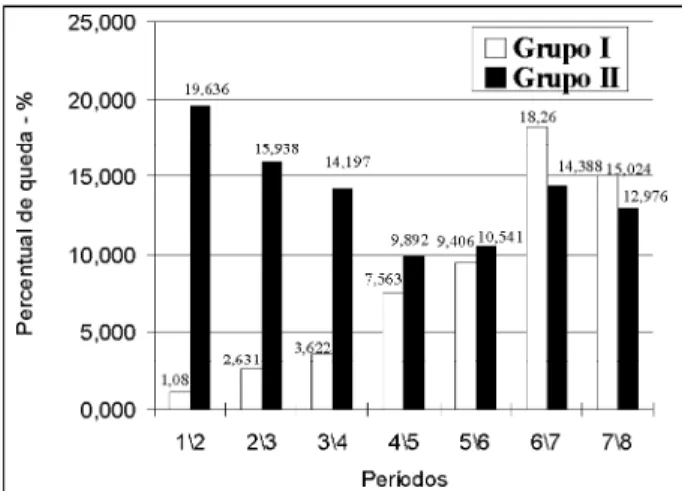
When comparisons of the percentage drop in contrac-tile muscle force were performed between 2 subsequent pe-riods for group I in regard to group II (fig. 3), we observed that this drop occurred until the last period of the experi-ment. The mean percentage of total drop was 45.3% for group I and 65.4% for group II.
Discussion
Left ventricular ejection fraction is one of the major predictors of survival in the analysis of heart disorders, and its drop results from cardiac dilation 19,32,33. Some studies showed the efficacy in generating systolic force by surro-unding the heart with muscular flaps 11,14. Several experi-ments showed the capacity to replace cardiac muscle force, as a pump, synchronous with the QRS complex 11,14,34-41.
Data in the literature have shown that drugs with a pri-mary cardiac effect also act upon skeletal muscle modifying its function 42-44. Dubelaar et al 42 studied the effect of L-car-nitine on LD muscle of dogs and showed an increase in its contractile force. Other drugs, such as the steroids methe-nolon and clenbuterol, may increase skeletal muscle mass and contractile force when associated with the cardiomyo-plasty technique 43,44. Howell et al 45 showed the effects of some drugs, such as isoproterenol, acting on skeletal muscle and producing positive inotropism in the rapid-con-traction fibers and negative inotropism in the slow-con-traction fibers. They also showed that aminophylline and theophylline have an effect on skeletal muscle, which was evidenced through muscle tremor, mainly postural.
Propafenone belongs to the Ic class of antiarrhythmic drugs 24. Its clinical use began in the 1980s due to its charac-teristics as a local anesthetic and stabilizer of myocardial cell membrane. Propafenone has been shown to reduce the am-plitude of the action potential (phase 0), the conduction in the
atrioventricular node, and the ascending velocity of the cur-ve of spontaneous diastolic depolarization (phase 4). Depending on the metabolic pathway of hydroxylation of propafenone, different levels of its metabolites (5 - hydroxy-propafenone and N-dipropylhydroxy-propafenone) may result, promoting a mild beta-blocking action 45-47. Propafenone has a predominantly inhibitory effect on sodium (Na+) channels, and a less effective impact on calcium (Ca++) and potassium (K+) channels 24,25. In reality, its effect on calcium (Ca++) channels was considered nonsignificant. However, the hypothesis that this might be the probable cause of myocardial depression was raised, leading to an increase in final diastolic pressure (DP2) and a reduction in systolic indices 26-28.
Some studies have shown the effect of propafenone on vascular smooth muscle relaxation after endothelial ske-letonizing, confirming its depressing action on the muscle 29. In 1987, Fierro et al 48 reported 1 case of myasthenia-like syndrome induced by propafenone, but they did not define the drug’s action on neuromuscular receptors and myofibrils.
Table I - Comparative analysis of the mean variations of tensions (g) of muscle contractions observed between groups I and II - values in grams
Period
Group 1º 2º 3º 4º 5º 6º 7º 8º
I 2.78 2.75(*) 2.68(*) 2.58(*) 2.39(*) (**) 2.17(*) (**) 1.79(*) (**) 1.52(*) (**) II 2.75 2.21(*)(**) 1.86(*)(**) 1.59(*)(**) 1.43(*)(**) 1.27(*)(**) 1.09(*) 0.95(*)
(*) p<0,05 - significant between the groups; (**) p<0.05 - significant betweens the periods.
T
e
n
s
ã
o
(
g
)
Fig. 2 - Mean values of the tensions of muscle contractions in groups I and II.
References
Some animal species have pharmacokinetic characte-ristics related to the tendency towards a concentration rate (plasma/tissue) below 1 at the skeletal muscle, showing that the m uscle serves as a drug reservoir 49.
A great variety of acute and chronic pathophysiologi-cal and pharmacologipathophysiologi-cal factors may cause changes in skeletal muscle contraction and fatigue and in its adaptive responses 42-45. These facts indirectly support the results obtained in the present study.
Rats were the animals chosen for our study because of the size of and also the constitution of the latissimus dorsi muscle to be analyzed in our study. In small animals, this muscle has a larger number of type I fibers, because it re-quires a greater resistance to fatigue, as the animals perform repetitive movements more frequently.
Side alternation, left and right, for muscles in groups I or II aimed to alter the disposition of muscle fibers for the confection of the muscle ring, so that muscle feasibility would be the same, independent of the side.
A constant temperature of 37ºC, with a variation of 0.5ºC, was maintained, and it was measured in real time with a catheter inserted into the bath solution, reflecting the ba-seline levels of the species studied 39. The pH of the medium was maintained even after addition of propafenone, becau-se metabolic acidosis reduced the drug’s action; however, variations within physiological limits were respected 50,51.
In the study of the action of propafenone, when the analysis of variance indicated a depressing effect, the least significant difference test for comparing mean valueswas used to evaluate this effect between the different periods. The interaction, when significant, indicated the existence of differences in the behavior of the muscle for each period when periods were compared with one another.
In group I, no significant (p>0.05) drop in muscle contraction was observed until period 4 (2.78g, 2.75g, 2.68g, 2.58g, respectively); from period 5 on, the reduction in contractile force became significant (p<0.05) (2.39g, 2.17g, 1.79g, 1.52g, respectively). This may be justified by the progressive loss of muscle feasibility during the experiment. In group II, after addition of propafenone in period 2, a drop in muscle contraction could be observed until period 7 (p<0.05) (2.75g, 2.21g, 1.86g, 1.59g, 1.43g, 1.27g, 1.09g, respectively); no difference between the last 2 periods was observed (p>0.05) (1.09g; 0.95g, respectively). One of the hypotheses would be the possible washing away of propafenone impregnated in LD muscle through the consecutive baths performed. Another hypothesis is the progressive loss of muscle feasibility with a
drop in contractile force, even when immersed in a solution adequate for study.
In regard to the Krebs-Henseleit solution, which is considered adequate for maintaining muscle feasibility, we analyzed the possibility that it could cause alterations in the results, the so-called “solution effect.” To check the effect of the solution on the experiment, the analysis of variance of the results of the means obtained in periods 2, 3, and 4 was performed. In the protocol established, the change of so-lution between periods 2 and 3 in both groups was not per-formed, and the action of propafenone was maintained for approximately 5 minutes. Therefore, we admit that no influence of the solution occurred on the results, because group I maintained the stability when the means were compared until period 4 (p>0.05).
In the multiple comparison of the means of the diffe-rent periods between the groups, we could not reject the hy-pothesis of equality in the first period only, because it was common to both groups. From period 2 on, the difference was maintained significantly until the end of the experiment (p<0.05).
In regard to LD muscle contractile force, the evalua-tion of the percentage of its total drop was 45.3% in group I and 65.4% in group II, evidencing a greater percentage of drop with the use of propafenone.
Considering the limitations of the present study, we may conclude that propafenone determines depression of the LD muscle contractile force in rats, with depression of muscle contraction with no recovery, even after sequential baths with a solution with no drug; the use of propafenone, in an acute administration regimen, may affect muscle performance in the cardiomyoplasty technique. Therefore, the control of ventricular function in patients undergoing cardiomyoplasty and requiring the use of propafenone should be carefully observed during the impregnation phase to decide about risks and benefits.
Based on the results of this study, the following facts should be considered because they may constitute obsta-cles to the immediate clinical application of this technique: LD muscle did not undergo proper preconditioning in the cardiomyoplasty technique; the different types of fibers that constitute LD muscle would be under the influence of the differentiated action of propafenone, as well as that of the dose being used; we still lack adequate definitions of the interference of the species-dependent characteristics for generalizing the conclusions.
1. Kannel WB, Belanger AJ. Epidemiology of heart failure. Am Heart J 1991; 121: 951-7. 2. Benedict CR, Weiner DH, Johnstone DE, et al. Comparative neurohormonal response in patients with preserved and impaired left ventricular ejection fraction: Results of the Studies of Left Ventricular Dysfunction (SOLVD) Registry. The SOLVD Investigators. J Am Coll Cardiol 1993; 22: 146A-53A.
3. Setaro JF, Soufer R, Remetz MS, Perlmutter RA, Zaret BL. Long-term out-come in patients with congestive heart failure and intact systolic left ventricular performance. Am J Cardiol 1992; 69: 1212-6.
variables on the reduction of mortality by treatment with hydralazine and isosorbide dinitrate. Circulation 1987; 75: IV-49-54.
5. The CONSENSUS Trial Study Group. Effects of enalapril on mortality in severe congestive heart failure. N Engl J Med 1987; 316: 1429-35.
6. Swedberg K, Held P, Kjekshus J, Rasmussen K, Rydén L, Wedel H. Effects of the early administration of enalapril on mortality in patients with acute myocardial infarction. Results of the Cooperative New Scandinavian Enalapril Survival Study II (CONSENSUS II). N Engl J Med 1992; 327: 678-84.
7. The SOLVD Investigators. Effect of enalapril on mortality and the development of heart failure in assyntomatic patients with reduced left ventricular ejection fractions. N Engl J Med 1992; 327: 685-9.
8. Cohn JN, Johnson G, Ziesche S, et al. A comparison of enalapril with hydralazi-ne-isosorbide dinitrate in the treatment of chronic congestive heart failure. N Engl J Med 1991; 325: 303-10.
9. Pfeffer MA, Brawnwald E, Moyé LA, et al. Effect of captopril on mortality and morbidity in patients with left ventricular dysfunction after myocardial infarction: results of the Survival and Ventricular Enlargement Trial. N Engl J Med 1992; 327: 669-77.
10. Schwarz F, Mall G, Zebe H, et al. Determinants of survival in patients with congestive cardiomyopathy: quantitative morfologic findings and left ven-tricular hemodynamics. Circulation 1984; 70: 923-8.
11. Carpentier A, Chachques JC. Myocardial substitution with a stimulated skeletal muscle: first successful clinical case (letter). Lancet 1985; 1: 1267. 12. Magovern GJ, Park SB, Christlieb IY, Magovern Jr GJ, Kao RL. Paced
conditio-ned latissimus dorsi for cardiac assist. In: Chiu RC-J, Bourgeois IM, editors. Transformed Muscle for Cardiac Assist and Repair. New York: Futura Publi-shing Co. Inc., 1990: 199.
13. Salmons S, Sréter FA. Significance of impulse activity in transformation of skele-tal muscle type. Nature 1976; 263: 30-4.
14. Chiu RC-J, editor. Biomechanical Cardiac Assist: Cardiomyoplasty and Muscle Powered Devices. New York: Futura Publishing Co. Inc.,1986.
15. Carpentier A, Chachques JC, Grandjean PA. Cardiomyoplasty.1st ed. Mount Kisco (NY): Futura Publishing Co. Inc., 1991.
16. Havenith MG, van-der-Veen FH, Glatz JFC, et al. Monitoring of muscle fiber type of canine Latissimus dorsi muscle during chronic electrical stimulation by en-zyme- and immunohistochemistry. In: Chiu RC-J, Bourgeois IM, editors. Transformed Muscle for Cardiac Assist and Repair. New York: Futura Publi-shing Co. Inc., 1990: 53.
17. Henriksson J, Salmons S, Lowry OH. Chronic stimulation of mamaliam muscle: enzyme and metabolite changes in homogenates and individual fibers. In: Chiu RC-J, Bourgeois IM, editors. Transformed Muscle for Cardiac Assist and Repair. New York: Futura Publishing Co. Inc., 1990: 9.
18. Mannion JD, Shannon J, Chen W, Brown WE, Gale DR. Skeletal muscle-powered assistance for the heart: assessment of a goat model. In: Chiu RC-J, Bourgeois IM, editors. Transformed Muscle for Cardiac Assist Repair. New York: Futura Pu-blishing Company, Inc., 1990: 117.
19. Kannel WB, Plehn JF, Cupples LA. Cardiac failure and sudden death in the Fra-mingham study. Am Heart J 1988; 115: 869-75.
20. CAST Investigators. Prelimirary report: effect of encainide and flecainide on mortality in a randomized trial of arrhythmia suppression after myocardial infarc-tion. N Engl J Med 1989; 321: 406-12.
21. Siebels J, Kuck KH and the CASH Investigators. Implantable cardioverter defi-brillator compared with antiarrhythmic drug treatment in cardiac arrest survi-vors (the Cardiac Arrest Study Hamburg). Am Heart J 1994;1 27: 1139-44. 22. Multicenter Posinfarction Research Group. Risk stratification and survival after
myocardial infarction. N Engl J Med 1983; 309: 331-6.
23. Batsford WP, Mickleborough LL, Elefteriades JA. Cardiac arrhythmias in the he-art failure. Cardiol Clinics. Ed. Philadelphia: WB Saunders 1995; 1: 87. 24. Vaughan Williams EM. Significance of classifying antiarrhythmic actions since
the cardiac arrhythmia suppression trial. J Clin Pharmacol 1991; 31: 123-35. 25. Dukes ID, Vaughan Williams EM. The multiple modes of action of propafenone.
Eur Heart J 1984; 5: 115-25.
26. Baker BJ, Dinh H, Kroskey D, de Soyza NDB, Murphy ML, Franciosa JA. Effect of propafenone on left ventricular ejection fraction. Am J Cardiol 1984; 54: 20D-2. 27. Lange H, Lampert S, Sutton MSJ, Lown B. Changes in cardiac output determined by continuous-wave doppler echocardiography during propafenone or mexile-tine drug testing. Am J Cardiol 1990; 65: 458-62.
28. Santinelli V, Arnese M, Oppo I, et al. Effects of flecainide and propafenone on systolic performance in subjects with normal cardiac function. Chest 1993; 103: 1068-73. 29. Vizcaino FP, Pozo BF, Zaragoza F, Tamargo J. Voltage-and time-dependent
inhibi-tory effects on rat aortic and porcine coronary artery contraction induced by pro-pafenone and quinidine. Br J Pharmacol 1994; 113: 1281-8.
30. Gomes OM, Simões R, Machado ELG, Freitas OGA, Brum JMG. Técnica para estudo do músculo latissimus dorsi de pequenos animais em câmara de órgãos. Coração 1996, 6: 30-5.
31. Krebs HA, Henseleit K. Untersuchungen ober die Hanstoffbildung in tierkoper. Hope Seyler Z. Physiol. Chem., 210-30; 1932 Apud in the isolated perfused warm-blooded heart according to the LANGENDORFF - Doring HJ, Dehnert H: Methods in Experimental Physiology and Pharmacology. Preprint of the 1st En-glish Edition; 1987.
32. Glover DR, Littler WA. Factors influencing survival and mode of death in severe chronic ischemic cardiac failure. Br Heart J 1987; 57: 125-32.
33. Gradman A, Deedwania P, Cody R, et al. Predictor of total mortality and sudden death in mild to moderate heart failure. J Am Coll Cardiol 1989; 14: 564-70. 34. Moreira LFP, Stolf NAG, Bocchi EA, et al. Latissimus dorsi cardiomyoplasty in
the treatment of pacients with dilated cardiomyopathy. Circulation 1990; 82: IV-257-63.
35. Cheng W, Justicz AG, Soberman MS, Alazraki NP, Santamore WP, Sink JD. Effects of dynamic cardiomyoplasty on indices of left ventricular systolic and diastolic function in canine model of chronic heart failure. J Thorac Cardiovasc Surg 1992; 103: 1207-13.
36. Aklog L, Murphy MP, Chen FY, et al. Right latissimus dorsi cardiomyoplasty im-proves function by increasing peak systolic elastance (Emax). Ciculation 1994; 90: II- 112-9.
37. Jondeau G, Dorent R, Bors V, et al. Dynamic cardiomyoplaty. effect of descontinu-ing latissimus dorsi muscle stimulation on left ventricular systolic and diastolic performance and exercise capacity. J Am Coll Cardiol 1995; 26: 129-34. 38. Schreuder JJ, van der Veen FH, van der Velde ET, et al. Beat-to-beat analysis of left
ventricular pressure-volume relation and stroke volume by condutance catheter and aortic modelflow in cardiomyoplasty patients. Circulation 1995; 91: 2010-7. 39. Moreira LFP, Bocchi EA, Stolf NAG, Pileggi F, Jatene AD. Current expectations
in dynamic cardiomyoplasty. Ann Thorac Surg 1993; 55: 299-303. 40. Hagège AA, Desnos M, Fernadez F, et al. Clinical study of the effects of latissimus
dorsi muscle flap stimulation after cardiomyoplasty. Circulation 1995; 92: II-210-5. 41. Kratz JM, Johnson WS, Mukherjee R, Hu J, Crawford FA, Spinale FG. The relation between latissimus dorsi skeletal muscle structure and contractile function after cardiomyoplasty. J Thorac Cardiovasc Surg 1994; 107: 868-78.
42. Dubelaar ML, Lucas CM, Hülsmann WC. Acute effect of L-carnitine on skeletal muscle force test in dogs. Am J Physiol 1991; 260: E-189-93.
43. Petrou M, Wynne DG, Boheler KR, Yacoub MH. Clembuterol induces hypertro-phy of the latissimus dorsi muscle and heart in rat with molecular and phenoty-pic changes. Circulation 1995; 92: II-483-9.
44. Fritzsche D, Krakor R, Asmussen G, et al. Effect of an anabolic steroid (meteno-lon) on contractile performance of the chronically stimulates latissimus dorsi in sheep. Eur J Cardiothorac Surg 1994; 8: 214-9.
45. Howell S, Fitzgerald RS, Shnader J, Roussos C. Contratility and fatigue in skeletal muscle: influence of pathophysiological and pharmacological interven-tions. In: Chiu RC-J, Bourgeois IM, editors. Transformed muscle for cardiac assist repair. New York: Futura Publishing Co. Inc., 1990: 75.
46. Barbey JT. Clinical pharmacology and beta-blocking efficacy of propafenone. J Cardiovasc Pharmacol 1991; 17: S-41-3.
47. Malfatto G, Pessano P, Zaza A, Schwartz PJ. Experimental evidence for beta adrenergic blocking properties of propafenone and for their potential clinical re-levance. Eur Heart J 1993; 14: 1253-7.
48. Fierro B, Castiglione MG, Salemi G. Myasthenia-like syndrome induced by car-diovascular agents: report of a case. Ital J Neurol Sci 1987; 8: 167-9. 49. Steurer G, Weber H, Schmidinger H, et al. Plasma propafenone concentration in
the evaluation of anti-arrhythmic efficacy. Eur Heart J 1991; 12: 526-32. 50. Gillis AM, Kates RE. Effect of pH on the myocardial uptake and
pharmacodyna-mics of propafenone in the isolated rabbit heart. J Cardiovasc Pharmacol 1988; 12: 526-34.