This study evaluated comparatively two configurations (powder and putty) of a composite biomaterial based on PLGA (Poly(lactide-co-glycolide)/nanoescale hydroxyapatite (ReOss®, Intra-Lock International) through microscopic morphology, in vitro cytotoxicity, biocompatibility and in vivo response as a bone substitute. SEM and EDS characterized the biomaterials before/after grafting. Cytocompatibility was assessed with murine pre-osteoblasts. Osteoconductivity and biocompatibility were evaluated in White New Zealand rabbits. Both configurations were implanted in the calvaria of eighteen animals in non-critical size defects, with blood clot as the control group. After 30, 60 and 90 days, the animals were euthanized and the fragments containing the biomaterials and controls were harvested. Bone blocks were embedded in paraffin (n=15) aiming at histological and histomorphometric analysis, and in resin (n=3) aiming at SEM and EDS. Before implantation, the putty configuration showed both a porous and a fibrous morphological phase. Powder revealed porous particles with variable granulometry. EDS showed calcium, carbon, and oxygen in putty configuration, while powder also showed phosphorus. After implantation EDS revealed calcium, carbon, and oxygen in both configurations. The materials were considered cytotoxic by the XTT test. Histological analysis showed new bone formation and no inflammatory reaction at implant sites. However, the histomorphometric analysis indicated that the amount of newly formed bone was not statistically different between experimental groups. Although both materials presented in vitro cytotoxicity, they were biocompatible and osteoconductive. The configuration of ReOss® affected morphological characteristics and the in vitro cytocompatibility but did not impact on the in vivo biological response, as measured by the present model.
I n V i t r o a n d I n V i v o
B i o c o m p a t i b i l i t y O f R e O s s ® i n
Powder and Putty Configurations
Andréa Vaz Braga Pintor1, Rodrigo Figueiredo de Brito Resende1, Adriana Terezinha Novelino Neves1, Gutemberg Gomes Alves2, Paulo G. Coelho3, José Mauro Granjeiro1,4, Monica Diuana Calasans-Maia5
1School of Dentistry, UFF -
Universidade Federal Fluminense, Niterói, RJ, Brazil
2Molecular Biology Department,
Biology Institute, UFF - Universidade Federal Fluminense, Niterói, RJ, Brazil 3Department of Biomaterials and
Biomimetics, New York University College of Dentistry, New York, USA 4National Institute of Metrology,
Quality and Technology, Duque de Caxias, RJ, Brazil.
5Department of Oral Surgery,
UFF - Universidade Federal Fluminense, Niterói, RJ, Brazil
Correspondence: Dra. Monica D. Calasans-Maia, Rua Mario Santos Braga, 28, Centro, 24220-000 Niterói, RJ, Brazil. Tel: +55-21-2629-9803. e-mail: [email protected]
Key-Words: PLGA; hydroxyapatite; materials testing; biocompatibility testing; nanotechnology.
Introduction
Bone tissue is frequently damaged by trauma and pathologies that might overcome its inherent physiologic regeneration capability, demanding the use of grafts to fill the defects (1). As an alternative to autogenous grafts, which require an additional surgical site for its obtention, many synthetic biomaterials have been developed and studied (2) among which are included aliphatic poly-α -hydroxide esters such as PGA (poly-glycolide acid), PLA (poly-lactide acid) and PLGA (polylactide-co-glycolide acid) copolymers (2,3). Through their manufacturing process, the modulation of chemical composition and structural configuration of these polymers allows the control of characteristics which are essential to bone tissue engineering: high porosity and pore interconnectivity, adequate pore size (4), biocompatibility and a degradation rate compatible to bone regeneration (5).
Bioceramics represent another important group of bone substitute biomaterial, including synthetic hydroxyapatite (HA) (5,6) which is a largely used due to
its high biocompatibility, bioactivity, as well as chemical/ structural similarities with the mineral phase of bone (1,4,5-7), even though its porous form presents poor mechanical properties and low bioresorption rates (6). In this context, tridimensional polymeric scaffolds made of composites of PLGA, PLA or PGA associated to hydroxyapatite present as advantages: (i) improvement of polymeric matrix hydrophilic characteristics (8) and mechanical properties (9); (ii) control of the degradation rate, mainly related to their chemical composition, molecular weight and crystallinity (5); (iii) accelerated hydroxyapatite degradation, releasing calcium and phosphate ions which contribute to novel bone formation (10,11);(iv) pH control of tissues surrounding the implant, by buffering effects of the alkaline degradation products of hydroxyapatite over glycolic and lactic acids released by the polymers (2,11) and (v) the existence of natural pathways to complete removal of the degradation products from the organism, by bioresorption (5).
A. V
. B
. Pintor et al.
(Intra-Lock International® , USA) was developed as a PLGA/HA composite biomaterial, indicated for filling or augmenting intraoral/maxillofacial osseous defects in the periodontal region, teeth extraction sites and in sinus elevation procedures (12). ReOss® is not indicated for load-bearing applications and is manufactured in different configurations, among them powder and putty. The manufacturing of this composite involves a baryosynthesis process described previously by Kim et al.,(3) even though no description is provided on the differences in composition, manufacturing steps and uses between the putty and powder configurations. While Coimbra et al. (13) have previously assessed the biocompatibility of powder PLG/HA, it remains unanswered if the ReOss® configuration (powder or putty) impacts on the biological response to the material. This study hypothesized that the putty form would have a better biological response, besides the ease of handling and better adaptation in the surgical cavity. In order to bring some light to this issue, the present study aimed to comparatively evaluate the microscopic morphology, in vitro cytotoxicity and in vivo biocompatibility, and the response as bone substitutes of the powder and putty ReOss® PLGA: HA configurations.
Material and Methods
Biomaterials
A PLGA/HA (Poly(lactide-co-glycolide))/nanoescale hydroxyapatite composite (Re-Oss®, Intra-Lock System®, USA) in both powder (RP-1) and putty (RP-2) forms were obtained from the manufacturer. All materials were sterile, ready for use, and within the expiry time.
Characterization of Biomaterials
Biomaterial samples of RP-1 and RP-2 were gold metalized (Emitech k550® Metalizer) and observed by scanning electron microscopy (SEM) (JSM-64 60LV, JEOL, Tokyo, Japan), which was performed at 15Kv and different magnifications, with back-scattered and secondary electron signals to assess sample morphological features. Energy Dispersive Spectroscopy (EDS) was performed in different areas to assess the chemical composition of both materials.
Cell Viability Test
Tests were performed according to ISO 7405:2008 (ISO, 2008). Briefly, extracts (conditioned media) were prepared with 0.2g of biomaterial mixed to 1 mL of serum-free Alpha-MEM (Gibco, Cergy-Pontoise, France), subsequently incubated for 24 hrs at 37ºC/5% CO2 in a humidified chamber. MC3T3-E1 (subclone 4) Murine pre-osteoblasts, obtained from the collection at the Clinical Research Unit of the Antonio Pedro Hospital, Fluminense Federal University (UFF), were subcultured for 24 hrs at 37 ºC/5% CO2 on
96-well culture plates at an initial cell density of 1 x 10⁴ cell/well, and subsequently exposed to the conditioned media for 24hrs at 37 ºC/5% CO2. Control groups were exposed only to Alpha-MEM media (experimental control), Alpha-MEM conditioned with high-density polystyrene pearls (negative control) or Alpha-MEM conditioned with pieces of latex (positive control). Each group was tested on quintuplicates. The XTT cell viability test evaluated the ability of mitochondrial enzymes from metabolically active cells to reduce 2,3-bis(2-methoxy-4-nitro-5-sulphophenyl)-2H-tetrazolium-5-carbanilide (XTT) molecules to a soluble formazan salt solution. After 2 h exposure to XTT at 37 ºC in a humidified chamber, the amount of soluble salt was measured by its absorbance at 480nm, in a UV-Vis microplate reader (Synergy II; Biotek Inst., Winooski, VT, USA) and correlated directly with viable cells. The Neutral Red (NR) survival/viability assay measured the membrane integrity and the ability of viable cells to incorporate NR dye through endocytosis and to accumulate the dye preferentially on lysosomes. The amount of dye incorporated was measured by the optical density at 540nm.
Surgical Procedure
This project was approved by the Ethics Commission of Animal Use of the Fluminense Federal University (CEUA/ UFF 165). Eighteen White New Zealand rabbits of both gender, weighing between 2.9 kg and 3.5 kg were assigned to three groups corresponding to the experimental periods of 30, 60 and 90 days.
In vitro and in vivo biocompatibility of ReOss® hydro
xyapatite
with constant cooling with a sterile physiological saline solution, on either side of the sagittal suture line. All defects’ sites were gently marked by pressing the bur with a rounded operator’s wrist movement. Then, each defect was made deeper the same way until blood started coming. Careful removal was done, so the dura was preserved. Biomaterials were implanted in the posterior defects: powder (RP1) on the right and putty form (RP2) on the left defect. The left anterior defect was filled with blood clot (control). Planes were sutured with Mononylon Ethilon (Ethicon®, Johnson & Johnson, São José dos Campos, SP, Brazil). The wound was left uncovered. Tramadol chlorydrate 4 mg/kg (Tramadol®, Anápolis, Brazil) and meloxicam 0.3 mg/kg (Maxicam 2%, Ourofino, Cravinhos, Brazil) were intravenously administered. One dose of an association of the antibiotics penicillin and streptomycin at 0.1 mg/ kg (Pentabiótico, Fort Dodge®, Porto Alegre, Brazil) was also administered. After surgical procedures, animals were kept in a blanket under supervision and, as they recovered from the anesthesia, received food and water ad libitum and were returned to their individual cages.
At 30, 60 or 90 days post surgery, animals were euthanized. The euthanasia procedure included animal weighing, premedication with ketamine hydrochloride 2 mg/kg (Clortamina®, Biochimico®, Rio de Janeiro, Brazil) and xylazine hydrochloride 10 mg/kg (Rompun®, Bayer SA®, São Paulo, Brazil) administered by intramuscular injection on the leg, and endovenous administration of sodium thiopental 20 mg/kg (Thiopentax®, Cristália®, Itapira, Brazil) and potassium chloride (dose-effect) by the previously cannulated ear marginal vein, until death. Biological samples were collected maintaining 5mm of surrounding bone, using steel and carborundum discs, on a slow-speed electric handpiece (BLM 600 PLUS, V K Driller®, São Paulo, Brazil) with constant cooling by sterile physiological saline solution, and fixed in 10% (v/v) buffered formaldehyde.
Histologic and Histomorphometric Analysis
Samples retrieved from 5 animals of each group were decalcified, dehydrated in ascending grades of ethanol, clarified and embedded in paraffin. The tissue blocks were cut in 5 µm thicknesses and stained with hematoxylin and eosin (H&E).
Histological sections were analyzed qualitatively by the same pathologist, using an optic microscope (Jenaval-Zeiss®, Thornwood, NY, USA), employing 4x and 10x objectives.
Histological sections were also analyzed using optic microscopy (FWL-3500TFL, Feldman Wild Leitz, Manaus, Brazil), with acroplan lenses with 10x objective, where 7 sequential images were captured by an attached camera (Nikon Digital Sight DSFi1, Nikon Instruments Inc., Amsterdam, Holland), with the help of the capture software
NIS-elements (Nikon Instruments Inc., Amsterdam, Holland). The captured images were analyzed by histomorphometry using the software Image-Pro Plus 6.0 (MediaCybernetics®). Each image received a 150-point grid, which was identified manually according to the classes: preexisting bone, new-formed bone, connective tissue and others. Blood vessels, adipose tissue, spaces, tears, and folds were not included. Results were tabulated on Excel for the statistical analysis.
Post-Implantation SEM and EDS Analysis
Samples retrieved from 1 animal of each group were dehydrated in ascending grades of ethanol, clarified and embedded in resin. Resin blocks were gold metalized (Emitech K550), and scanning electron microscopy (SEM) (Scanning Electron Microscope, JSM-64 60LV, JEOL, Tokyo, Japan) was performed at various magnifications, at 15 kv, with back-scattered and secondary electron signals to assess samples’ morphological characteristics. Energy Dispersive Spectroscopy (EDS) was performed in different areas for the chemical composition analysis.
Statistical Analysis
Cell viability data was verified for normal distribution by D’Agostino & Pearson test and subsequently analyzed by One-Way ANOVA test with Tukey post-test, with a confidence interval of 95%.
Quantitative data from the in vivo study were tabulated on Excel® spreadsheets. Statistical analysis was performed using GraphPad Prism 5.0 software (GraphPad Corp, USA). Quantitative data were expressed as the mean ± standard deviation, medians, and coefficients of variation, distribution normality tests and Kolgomorov Smirnov distances.
The blood clot (control), putty (RP2) and powder (RP1) groups were compared by the amount of preexisting bone, newly formed bone and connective tissue present in the defect area. As data presented high standard deviations and coefficients of variation, they were analyzed as medians using non-parametric Wilcoxon test with a significance level of 5% (α≤0.05).
Results
Characterization of Powder and Putty PLGA/HA
A. V
. B
. Pintor et al.
Cell Viability Assay
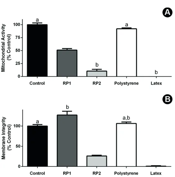
After exposure to the conditioned media, deposition of material was observed over cells on the powder group, but not on putty PLGA/HA. Both putty and powder configurations were considered cytotoxic by the XTT reduction test, with viability ranging below 70% of the control, as established on ISO 7405:2008 (Fig. 2A). The Neutral Red Uptake assay identified powder PLGA/HA as non-cytotoxic, while the putty PLGA/HA induced levels of cell survival significantly lower than control (untreated) group (p<0.05) (Fig. 2B). Putty PLGA/HA was significantly more toxic than the powder configuration (p<0.05), behaving similarly to the extremely toxic positive control.
Histological Analysis
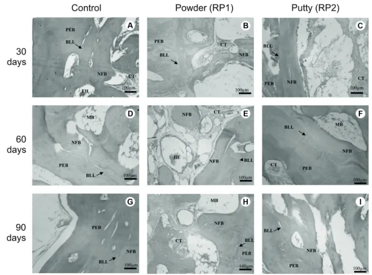
The implantation of both powder (RP1) and putty (RP2) of the PLGA/HA composite resulted in new bone formation
in vivo in the three experimental periods (Fig. 3). Thirty days after implantation, in control and RP1 groups, osteoblastic
paving and new bone formation from the periphery to the center of the defect were detected, as well as connective tissue mostly in the defect center (Figs. 3A and 3B). In RP2 sections, preexisting bone, a basophilic limit line, novel bone trabeculae and a considerable amount of connective tissue in the center of the defect were observed (Fig. 3C). Sixty days after implantation, new bone formation progressed, as more mature trabeculae from the periphery to the center of defects were detected in all groups sections (Figs. 3D, 3E, and 3F). Islands of newly formed bone circled by connective tissue were observed in RP1 sections too (Fig. 3E). By the 90th day, such increase on ossification was still noticed, and no inflammatory cells could be observed in all groups (Fig. 3G, 3H and 3I). Fibrillar areas in RP1 (Fig. 3H) and spaces in RP2 (Fig. 3I) were observed and both considered as effects of biomaterial residues.
Histomorphometric Analysis
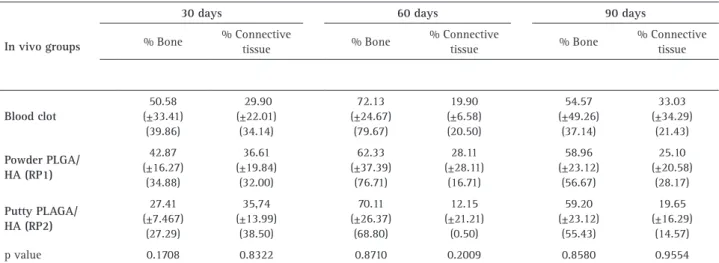
Table 1 summarizes the quantitative data of the
In vitro and in vivo biocompatibility of ReOss® hydro
xyapatite
histomorphometric analysis of control, RP1 and RP2 grafted defects during all experimental times. No significant differences were observed between the experimental groups, regarding newly formed bone volume and connective tissue (soft tissue) volume in the three experimental periods.
Post-Implantation SEM and EDS
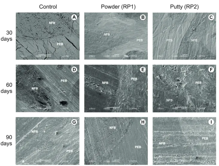
Preexisting bone and newly formed bone from the periphery to the center of the defects were detected in micrographics of control, RP1 and RP2 related to the 30, 60 and 90 experimental periods (Fig. 4). EDS revealed the presence of C, O, and Au (gold) in 30 days period for both RP1 and RP2 (Fig. 5A and 5B). The presence of C, O, and
Au was observed in RP2 and also Ca (calcium) in RP1, in 60 days spectrums (Fig. 5C and 5D). The 90 days period EDS analysis showed the presence of Ca, C, O, Al (aluminum) and Au in control; Ca, C, O, N (nitrogen) and Au in RP2; C, O, Ca and Au in RP1 (Figs. 5E and 5F).
Discussion
The PLGA/HA ReOss® was approved by FDA and is commercially available in both powder and putty configurations. Although the powder configuration had been previously evaluated (13), there was no data on literature concerning the possible differences in the biological responses between the powder and the putty configurations. The present work assessed this issue
A. V
. B
. Pintor et al.
through microscopic morphology, in vitro cytotoxicity, biocompatibility and in vivo performance as bone substitutes.
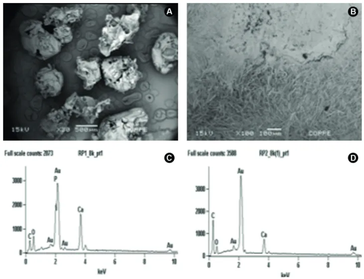
The manufacturer of ReOss (Intra-Lock Int.) describes the product composition as a usual 50/50% combination of HA and PLGA, indicating no chemical difference between the putty and powder presentations. Indeed, the chemical microanalysis performed in this work by Energy Dispersive Spectroscopy (EDS) (Fig. 1) found very similar results for both configurations. On the other hand, the morphological assessment was able to identify a source of potential differences in the biological effects of these materials. For the powder form (RP1), SEM analysis revealed the predominance of porous particles with variable granulometry (Fig. 1). These results were very similar to those presented by Kim et al. (3), in the original description of this material in the scientific literature.
On the other hand, putty PLGA/HA (RP2) presented a very different morphology, where its composite nature is more evidenced by two phases: the first, very similar to the porous agglomerates of RP2, and a second fibrous phase (Fig. 1), which closely resembled the nanofibrous composite of PLGA and nanoscaled hydroxyapatite obtained by electrospinning process described by Jose et al.(14). The morphological differences found between the putty and powder configurations could suggest different contributions to the outcome of bone tissue regeneration. The homogenous and porous design of the powder form could favor rapid diffusion or flow of nutrients and cell-material interactions.(4) On the other hand, the fibrous and HA agglomerate-containing morphology of the putty configuration could emulate collagenous fibers, while at the same time disposing calcium ions for novel bone formation (15).
In vitro and in vivo biocompatibility of ReOss® hydro
xyapatite
In order to investigate if such structural differences impacted on the viability of bone cells exposed to these materials, an in vitro cytotoxicity assessment was performed with Murine MC3T3-E1 pre-osteoblasts, a well-known model for biocompatibility studies of biomaterials for bone therapy. The results revealed that the putty configuration is strongly cytotoxic (Fig. 2), according to international parameters (ISO 7405:2008), as assessed by two different cell viability tests (XTT and NRU). The powder configuration, curiously, induced different effects on the cell viability parameters assessed in the present study (Fig. 2). The XTT assay, employed as a measure of cell metabolism, identified a significant reduction for RP1 when compared to control (p<0.05). Indeed, the previous report by Coimbra et al. (13) has described a similar effect for the total extracts of powder ReOss® over murine cells, even though this impact on metabolism presented a dose-response and could be reverted by dilution to 75% of the extract. It indicates
that this acute toxicity would probably disappear in the in vivo model, where homeostasis and the rapid flow/dilution of toxicants would occur. Nevertheless, the present work, by employing a second cell viability parameter, the NRU assay, a well-recognized endpoint for cell integrity (OECD Guide 129) (Fig. 2), identified that, even as metabolism was reduced in RP-1 exposed cells, cell survival remained unaffected. The use of sequentially combined cell viability parameters during a cytotoxicity assay increases the chances of the detection of adverse effects and contributes to the refinement of the understanding of the mechanisms involved on toxicity (16). In this context, other studies of PLGA/HA composites found similar good cell viability by employing other non-metabolic assays, such as the LDH test with human hFOb 1.19 osteoblast-like cells (17), or through fluorescence-based LIVE-DEAD tests with RAW 264.7 murine macrophages (18). Moreover, the in vitro evaluation of biocompatibility of the PLGA/HA composite scaffolds
A. V
. B
. Pintor et al.
performed by Kim et al. (3) has indicated good adhesion and proliferation of seeded rat calvaria osteoblasts.
Usually, during the development of novel biomaterials,
in vitro results pointing to strong cytotoxic effects, such
as those identified for the ReOss putty configuration (Fig. 2), would discourage the performance of in vivo tests with animals, for ethical and technical issues. However, since both configurations are already commercially
In vitro and in vivo biocompatibility of ReOss® hydro
xyapatite
available (i.e., human exposure to these materials already occurs), a complementary in vivo evaluation was performed, as recommended by ISO7405:2008, in order to better understand their biocompatibility by direct interactions with host tissues, and contribute to predict their clinical outcomes in bone therapy. Therefore, the implantation test was chosen, using White New Zealand rabbit’s calvaria as experimental surgery model (19). This animal model enables evaluation of the biological response of biomaterials implanted both in critical (≥15 mm) and non-critical (<15 mm) defects. Furthermore, the surgical model allows easy access and, since the site is not related to the masticatory or locomotor animals systems, there are fewer complications risks. In addition, calvaria’s intramembranous bone formation, similar to that of maxillae and mandible, turned it a good site for evaluation of biomaterials designed for filling and elevation of intraoral or maxilla-facial defects, such as the PLGA/HA biomaterial studied.
In the literature, favorable in vivo results were reported for the biological response for PLGA:HA composites as bone substitutes (13,17,20), as authors observed osteogenesis, and no inflammatory persistent reactions or osteolysis in implant sites. A PLGA/HA nanostructured composite, manufactured by baryosynthesis route, the same route described for the ReOss®, resulted in a composite with enhanced biocompatibility and osteogenic properties compared to a similar composite fabricated by the solvent casting/particulate leaching process (3). Coimbra et al. (13) observed newly bone formation after implantation of ReOss® powder form in Wistar rat calvaria, in the center and periphery of non-critical defects, whereas newly bone
formation was observed after PLGA/HA implantation on non-critical defects in rabbits (17). Recently, novel graft compositions have been developed blending different proportions of HA, tricalcium phosphate (TCP), PLGA and collagen (20), with higher amounts of bone substituted area and lower levels of soft tissue infiltration observed. The present in vivo results showed new bone formation related to both ReOss® configurations, without inflammatory or osteolytic reactions, evidencing the good biocompatibility and interaction with host tissues. Our results confirm the safety for clinical use of ReOss® in both configurations, due to the absence of significant adverse effects, even though both were considered cytotoxic by the XTT reduction test. It is important to notice that some reports on the literature have described the existence of discrepancies between in vitro standardized test results and the clinical outcomes of dental materials (21). In this sense, novel methods are nowadays being developed and validated, aiming to provide more consistency between in vitro and in vivo settings (22), according to the paradigms of the 21st-century toxicology.
The bone healing process involves an initial inflammation, with subsequent new bone formation and ensuing remodeling process that reshapes the bone tissue by removing and replacing and re-organizing the bone matrix (23). Since rabbits show fast skeletal changes and bone turn over (19), the histomorphometric data (Table 1) at the 30, 60 and 90 days experimental times may have evidenced such phases, even though without statistical significance, most probably due the high standard deviation presented by the model. In this manner, the present study could not identify an increase in new bone
Table 1. Results of the histomorphometric analysis showing the % of novel bone and % connective tissue (Mean ± SD) (Median) in the three experimental periods (n=5 animals for each group/experimental periods). P value provided for a Wilcoxon test comparing the in vivo groups in each experimental time (apha=0.05)
In vivo groups
30 days 60 days 90 days
% Bone % Connective
tissue % Bone
% Connective
tissue % Bone
A. V
. B
. Pintor et al.
formation when compared to control, through the in vivo
model employed. Nevertheless, this result should not discourage the use of these biomaterials for bone therapy. ReOss® showed to be osteoconductive, biocompatible and presented clear evidence of bioresorption and partial replacement with bone tissue at the interface biomaterial/ bone, similar to results obtained previously for Bio-Oss (24), a bovine xenograft widely used as a bone substitute. The clinical management of the putty PLGA:HA (RP-2), a dense spherical mass that was easily handled and positioned in the surgical sites, although a little brittle, could be promising on the guiding of novel bone tissue on vertical bone defects. This biomaterial would be useful in clinical situations in which bone contour and bone volume control are desired, since augmenting and correcting the bone defects contribute to the final bone structure thickness and shape aiming to better functional and esthetic results (25). Otherwise, the powder PLGA: HA (RP-1), that consisted of dried granules, presented a more difficult clinical handling, as the granules roll one upon another, although they were well fitted for horizontal defects such as those of our study. In the present study, we have shown that, regardless of their morphological/structural differences, or the different impact on the cytocompatibility on a murine in vitro model, both powder and putty configurations of ReOss® were considered biocompatible and presented osteoconductive activity, even though without significant differences in the amount of novel bone observed, as compared with control group. The hypothesis of this study was rejected since the final in vivo biological response was similar between the two groups tested. However, the main difference between these configurations might indeed rest on their applicability, since the main physical characteristics of each configuration might lend them suitable for different handlings and clinical situations.
In conclusion, the configuration of ReOss® affected the morphological characteristics and the in vitro
cytocompatibility, but did not impact on the in vivo
biological response, as measured by the present model.
Resumo
Este estudo avaliou comparativamente duas configurações (pó e massa) de um biomaterial composto com base de PLGA
(Poli(láctico-co-glicólico)/hidroxiapatita em nanoescala (ReOss®, Intra-Lock
International) através da morfologia microscópica, citotoxicidade in
vitro, biocompatibilidade e resposta in vivo como substituto ósseo. MEV e EDS caracterizaram os biomateriais antes/após o enxerto. A citocompatibilidade foi avaliada em pré-osteoblastos murinos. A osteocondutividade e a biocompatibilidade foram avaliadas em coelhos Branco da Nova Zelândia. Ambas as configurações foram implantadas na calvária de dezoito animais em defeitos não-críticos, com coágulo sanguíneo como grupo controle. Após 30, 60 e 90 dias, os animais foram eutanasiados e os fragmentos contendo os biomateriais e controles coletados. Blocos ósseos foram embebidos em parafina (n=15) destinados às análises histológica e histomorfométrica, e em resina (n=3) destinadas
à MEV e EDS. Antes da implantação, a configuração massa mostrou ambas fases morfológicas porosa e fibrosa. O pó revelou partículas porosas com granulometria variável. EDS mostrou cálcio, carbono e oxigênio na configuração massa, enquanto o pó mostrou também fósforo. Após a implantação a EDS revelou cálcio, carbono e oxigênio em ambas configurações. Os materiais foram considerados citotóxicos pelo teste XTT. A análise histológica mostrou nova formação óssea e nenhuma reação inflamatória nos sítios de implante. Entretanto, a análise histomorfométrica indicou que a quantidade de osso neoformado não foi estatisticamente diferente entre os grupos experimentais.
Embora ambos os materiais tenham apresentado citotoxicidade in
vitro, foram biocompatíveis e osteocondutores. A configuração do
ReOss® afetou as características morfológicas e a citocompatibilidade
in vitro, porém não impactou a resposta biológica in vivo, como medido pelo presente modelo.
References
1. Lichte P, Pape HC, Pufe T, Kobbe P, Fischer H. Scaffolds for bone healing : Concepts , materials and evidence. Injury 2011;42:569-573. 2. Hutmacher DW. Scaffolds in tissue engineering bone and cartilage.
Biomaterials 2000;21:2529-2543.
3. Kim S-S, Sun Park M, Jeon O, Choi CY, Kim B-S. Poly(lactide-co-glycolide)/hydroxyapatite composite scaffolds for bone tissue engineering. Biomaterials 2006;27:1399-1409.
4. Karageorgiou V, Kaplan D. Porosity of 3D biomaterial scaffolds and osteogenesis. Biomaterials 2005;26:5474-5491.
5. Rezwan K, Chen QZ, Blaker JJ, Boccaccini R. Biodegradable and bioactive porous polymer / inorganic composite scaffolds for bone tissue engineering. Biomaterials 2006;27:3413-3431.
6. Dorozhkin S V. Biomaterials Bioceramics of calcium orthophosphates. Biomaterials 2010;31:1465-1485.
7. Okada S, Ito H, Nagai A, Kotomori J, Imai H. Adhesion of osteoblast-like cells on nanostructured hydroxyapatite. Acta Biomater 2010;6:591-597.
8. Ge Z, Jin Z, Cao T. Manufacture of degradable polymeric scaffolds for bone regeneration. Biomed Mater 2008;3:022001.
9. Hong Z, Zhang P, He C, Qiu X, Liu A, Chen L, et al. Nano-composite of poly(L-lactide) and surface grafted hydroxyapatite: Mechanical properties and biocompatibility. Biomaterials 2005;26:6296-6304. 10. Higashi S, Yamamuro T, Nakamura T, Ikada Y, Hyon S-H, Jamshidi K.
Polymer-hydroxyapatite composites for biodegradable bone fillers. Biomaterials 1986;7:183-187.
11. Liu H, Webster TJ. Mechanical properties of dispersed ceramic nanoparticles in polymer composites for orthopedic applications. Int J Nanomedicine 2010;5:299-313
12. International I. http://intra-lock.com/index.php?option=com_ content&task=view&id=151 Internet. Available from: http://intra-lock.com/index.php?option=com_content&task=view&id=151 13. Coimbra ME, Salles MB, Yoshimoto M, Allegrini Jr. S, Fancio E, Higa
O, et al. Physico / Chemical Characterization , In Vitro , and In Vivo Evaluation of Hydroxyapatite / PLGA Composite and Tricalcium Phosphate Particulate Grafting Materials. Titanium 2009;1:16-28. 14. Jose M V, Thomas V, Johnson KT, Dean DR, Nyairo E. Aligned PLGA/HA
nanofibrous nanocomposite scaffolds for bone tissue engineering. Acta Biomater Internet. Acta Biomaterialia 2009;5:305-315. 15. Li X, Wang L, Fan Y, Feng Q, Cui FZ, Watari F. Nanostructured
scaffolds for bone tissue engineering. J Biomed Mater Res - Part A 2013;101A:2424-2435.
16. De-Deus G, Canabarro A, Alves G, Linhares A, Senne MI, Granjeiro JM. Optimal Cytocompatibility of a Bioceramic Nanoparticulate Cement in Primary Human Mesenchymal Cells. J Endod 2009;35:1387-1390. 17. Cieslik M, Mertas A, Morawska-Chochól A, Sabat D, Orlicki R,
Owezarek A et al. The evaluation of the possibilities of using PLGA co-polymer and its composites with carbon fibers or hydroxyapatite in the bone tissue regeneration process - in vitro and in vivo examinations. Int J Mol Sci 2009;10:3224-3234.
In vitro and in vivo biocompatibility of ReOss® hydro
xyapatite
-glycolide ) in vitro. J Biomed Mater Res A 2006;79:582-590. 19. Li Y,Chen S-K, Li L, Qin L, Wang X-L,Lai Y-X. Bone defect animal
models for testing efficacy of bone substitute biomaterials. J Orthop Transl 2015;3:95-104.
20. Khan R, Witek L, Breit M, Colon D, Tovar N, Janal MN et al. Bone Regenerative Potential of Modified Biphasic Graft Materials. Implant Dent Internet. 2015;1. Available from: http:// content.wkhealth.com/linkback/openurl?sid=WKPTLP:landingpage &an=00008505-900000000-99625
21. Scelza MZ, Linhares AB, da Silva LE, Granjeiro JM, Alves GG. A multiparametric assay to compare the cytotoxicity of endodontic sealers with primary human osteoblasts. Int Endod J 2012;45:12-8. 22. Restle L, Costa-Silva D, Lourenço ES, Bachinsky RF, Batista AC,
Linhares ABR, et al.. A 3D osteoblast in vitro model for the evaluation of biomedical materials. Advances in Mater Sci Eng 2015, Article ID 268930, 8 pages.
23. Lin C-Y, Chang Y-H, Sung L-Y, Chen C-L, Lin S-Y, Li K-C, et al. Long-term trackin of segmental bone healing mediated by genetically engineered adipose-derived stem cells: focuses on bone remodeling and potential side effects. Tissue Eng 2014;20:1392-1402. 24. Calasans-Maia MD, Ascoli FO, Novellino ATNA, Rossi AM, Granjeiro
JM. Comparative histological evaluation of tibial bone repair in rabbits treated with xenografts. Acta Ortop Bras 2009;17:340-343. 25. Liu J, Kerns DG. Mechanisms of guided bone regeneration: a review.
Open Dent J 2014;8:56-65.