http://dx.doi.org/10.1590/s2175-97902018000217438
Article
*Correspondence: T. S. O. Capote. Departamento de Morfologia, Faculda-de Faculda-de Odontologia Faculda-de Araraquara, UniversidaFaculda-de Estadual Paulista, UNESP. Rua Humaitá, 1680. CEP: 14801-903. Araraquara – SP. Brazil. E-mail: [email protected]
BONEFILL
®block as alternative for bone substitute: a toxicological
evaluation
Karine Melchior
1, Sybele Saska
2, Fernanda Coelho
2, Raquel Mantuaneli Scarel-Caminaga
2,
Ticiana Sidorenko de Oliveira Capote
2*1São Paulo State University (UNESP), School of Pharmaceutical Sciences, Araraquara, São Paulo, Brazil, 2São Paulo State University (UNESP), School of Dentistry, Araraquara, São Paulo, Brazil
Bone substitutes based on hydroxyapatite (HA) and Bonefill® (BO - inorganic bovine bone) associated with poly(lactic-co-glycolic acid) (PLGA) (HA/PLGA and BO/PLGA) were evaluated concerning cytotoxicity, genotoxicity and mutagenicity as potential candidates for bone repair. The materials were developed and provided by Bionnovation Biomedical Products Ltda. Eluates from these bone substitutes were prepared for toxicity evaluations using eukaryotic cell cultures. HA/PLGA was used as a comparison
for Bonefill®. Cell viability was evaluated by XTT assay and surviving fraction was calculated for clonogenic survival. Additionally, tail moment was used to assess genotoxicity (comet assay). The frequencies of binucleated cells with micronucleus (FBMN), micronucleus (FMN), nucleoplasmic bridges (NPBs), and nuclear buds (NBUDs) were analysed by cytokinesis-block micronucleus assay (CBMN
assay). Results showed no statistical difference in cell viability compared with negative control (NC)
The eluates did not promote delayed cytotoxicity whereas the surviving fraction rate for cultured cells
was similar to NC. Furthermore, no genotoxicity or mutagenicity effects were observed for cultured cells with the Bonefill/PLGA and HA/PLGA eluates. In conclusion, the negative cytotoxicity, genotoxicity
and mutagenicity results indicate that these bone substitutes presented interesting preliminary results as potential biomaterials for bone repair.
Keywords: BONEFILL®/toxicity. Biomaterials/evaluation. Bone regeneration. Hydroxyapatite.
INTRODUCTION
Over the years, therapeutic approaches and alloplastic materials have been developed which improve bone repair/regeneration mainly in tissue engineering. However, bone grafts are still used as an alternative to support bone repair in traumatic or non-traumatic injuries (Suchanek, Yoshimura, 1998; Kaveh et al., 2010; Dimitriou et al., 2011). The gold standard for bone reconstruction is autograft, due to the low rejection factor and its osteogenic properties. However, the disadvantages of this graft include additional surgery and limited donor quantities (Costantino et al., 1991; Suchanek, Yoshimura, 1998; Kannana et al., 2014). In
addition, allografts and xenografts have been used as alternatives for bone repair, although they may promote rejection, and diseases can be transmitted when not properly chemically processed (Suchanek, Yoshimura, 1998). For these reasons, alternative bone substitutes auto, allo and xenografts to replace auto, allo, and xeno bone grafts (Saska et al., 2015) which contain materials with similar characteristics to bone tissue, such as osteoconductive, osteoinductive and osteogenic properties for decreasing immunogenic and compatibility problems (Saska et al., 2015).
properties of pure HA ceramics (Suchanek, Yoshimura, 1998). Due to their bioactivity and reabsorption features, they promote direct binding to living tissue and their own slow and gradual degradation which is replaced by the tissues in which they are implanted (Wang et al., 2005). However, metallic, ceramic and polymer materials have been combined with HA to improve its mechanical and biological properties (Suchanek, Yoshimura, 1998; Shi
et al., 2015).
Other alloplastic materials, such as synthetic polymers, have been used to improve bone replacement. One of those, poly(lactic-co-glycolic acid) (PLGA) is a biodegradable aliphatic polyester polymer obtained from hydroxyl acids (De Lima etal., 2011); it is used in several biomedical applications, such as tissue and genetic
engineering and drug delivery (Cieślik et al., 2009). This polymer has the capacity to modulate the mechanical properties of others compounds and provide versatility in their structure (Saska et al., 2015), and it can be used as supporting or stabilizing elements by creating composites
based on them (Cieślik et al., 2009). The association with other compounds provides biocompatibility, bioactivity, satisfactory mechanical properties, and osteoconduction, which makes them potential materials in regenerative medicine therapies (Saska et al., 2015).
A l t h o u g h x e n o g r a f t s m a y i n d u c e s o m e immunogenic and inflammatory reactions depending on the host, bovine grafts have been a good option for bone regeneration in humans. As their inorganic phases are similar to human bone, and if bovine grafts are correctly processed, they are a reliable and high available source (Galia et al., 2008; Galia et al., 2009). Moreover, xenografts have good biocompatibility without unfavourable immunologic responses (Rios et al., 1996; Araújo et al., 2009).
Engineered nanomaterials have become prevalent in our everyday life, raising awareness of nanotoxicology to accelerate our understanding of the ill effects that
different nanomaterials can bring to biological systems
(Setyawati et al., 2013). In this context, this study aimed
to evaluate toxicological effects of alloplastic materials
based on hydroxyapatite and PLGA (HA/PLGA) and inorganic bovine bone and PLGA, named Bonefill®
(Bionnovation Biomedical Products Ltda, Brazil). We therefore evaluated the cytotoxicity, genotoxicity and mutagenicity of the bone substitutes in block, HA/PLGA
and Bonefill®, as the biologic safety of medical devices
form part of the risk management and analysis process, and this information also plays an important role in the safe use of the biomaterial.
MATERIALS AND METHODS
Materials
The HA/PLGA and Bonefill® block materials were
provided by Bionnovation Biomedical Products Ltda (Brazil), and sterilized by gamma irradiation at dose of 25 kGy.
Cell culture experiments
Chinese hamster ovary cells (CHO-K1) were grown in 1:1 Ham-F10+D-MEM (Sigma®, St. Louis, MO) culture medium supplemented with 10 % (v/v) fetal bovine serum (FBS) (fetal bovine serum-Cultilab, Campinas, Brazil) and kanamycin (1%) (Gibco, Carlsbad, CA) at 37 ºC in a 5% CO2 atmosphere. Cells were used between the 3rd and 8th passages. CHO cell line has been
widely used for studies that assess cytotoxicity and genotoxicity (Yalkinoglu, Schlehofer, Hausen, 1990) and are recommended by The Organization for Economic Cooperation and Development (2004) (OECD, 2014) for genotoxicity screening.
Eluates from each HA/PLGA and Bonefill®
(Bonefill/PLGA) block were prepared considering weight
(0.2 g mL). The materials were immersed in 1:1 Ham-F10+D-MEM medium (Sigma®) without fetal bovine
serum (FBS) at 37 ºC for 72 h (Iso, 2008), shaking at 180
×g in an incubator (New Brunswick Scientific – Excella
E24 Incubator Shaker Series).
Cytotoxicity tests
XTT assay. These experiments used CHO-K1 cells, a CellProliferation Kit II (Roche Applied Science), and 24 h of seeding. CHO-K1 cells (2×10⁴ cells seeded)
were treated with Bonefill® or HA/PLGA eluates at 100% concentration for 24 h in 24-well plates. Each well containing the respective eluate was supplemented with 10% FBS. Negative controls (NC) were wells containing culture medium supplemented with 10% FBS without any eluate (untreated controls), while positive controls (PC) were wells containing CHO-K1 cells, treated with doxorubicin (3 µg.mL−1) for 24 h (all treatments performed in triplicate). After treatment, cultures were washed with PBS and fresh medium was added. Subsequently, cultures were washed with PBS and 500 µL DMEM without phenol red was immediately added, followed by 60 µL of XTT/
electron solution (50:1) (Cell Proliferation Kit II – Roche
measured by a Microplate Reader (VersaMax, Molecular Devices, Sunnyvale,CA) at 492 and 690 nm. Absorbance is directly proportional to the number of metabolically active cells (viable cells) in each treatment after 24 h of exposure. Cell viability was calculated from the absorbance. Three independent experiments were conducted.
Clonogenic assay. Clonogenic assay or colony formation assay is an in vitro cellsurvival assay based on the ability of a single cell to grow into a colony. The colony
is defined as consisting of at least 50 cells (Franken et al., 2006). After 24 h of seeding, CHO-K1 cells (5×10⁴ cells
seeded) were exposed to Bonefill® or HA/PLGA eluates
at 100% concentration for 24 h in 24-well plates. Each well containing the respective eluate was supplemented with 10% FBS. NC were wells containing culture medium supplemented with 10% FBS without any eluate, and PC were wells containing CHO-K1 cells, treated with hydrogen peroxide (80 µmol.L−1) for 10 min. After exposure, the cultures were washed with PBS and fresh medium added. Exponentially growing cells were seeded after treatment at 150 cells per 25 cm2 flasks in duplicate
for each treatment. The flasks were incubated at 37 ºC, 5%
CO2, for 7 days without medium exchange. Colonies were
fixed with methanol:acetic acid:water (1:1:8, v/v/v) and
stained with 5% Giemsa. The number of colonies counted in the negative control group was considered 100%. From this, survival fraction (FS) calculations were performed: FS = number of colonies counted in each treatment × 100/Number of colonies observed in the negative control group. Three independent experiments were conducted.
Genotoxicity and mutagenicity assays
Comet assay. The alkaline version of the comet assay was used according to a previously described method (Singh et al., 1988). CHO-K1 cells were seeded (5×10⁴ cells seeded) and after 24 h exposed to Bonefill®
or HA/PLGA eluates at 100% concentration for 24 h in 24-well plates. Each well containing an eluate was supplemented with 10% FBS. NC were wells with culture medium supplemented with 10% FBS without any eluate and PC were wells containing CHO-K1 cells treated with hydrogen peroxide (80 µmol.L−1) for 10 min (all treatments were carried out in duplicate). After exposure, cultures were washed with PBS and harvested with trypsin. Five hundred microliters of cells in suspension were obtained, kept on ice, and protected from light. After centrifugation, the pellet was re-suspended in 200 µL of 0.5% (w/v) low melting point agarose and the mixture spread onto two microscope slides (Knittel, Germany) pre-coated with 1.5% (w/v) normal melting point agarose (Gibco).
Coverslips were placed over the gel. When the gels had
solidified, the coverslips were gently removed and the slides
immersed in cold (4 ºC) lysis solution (1% Triton X-100, 10% DMSO, 2.5 mmol.L−1 NaCl, 100 mmol.L−1 Na2EDTA, 100 mmol.L−1 Tris, pH 10) for 24 h. Immediately after this step, slides were placed in a horizontal electrophoresis unit containing freshly prepared electrophoresis buffer (1 mmol.L−1 Na2EDTA, 300 mmol L−1 NaOH, pH > 13). The DNA was allowed to unwind for 20 min; electrophoresis was then performed at 43V, 308 mA for 25 min. The
slides were then gently immersed in neutralization buffer
(0.4 mol.L−1 Tris– HCl, pH 7.5) for 15 min and then fixed with ethanol. All steps of the comet assay were conducted under subdued light. Three independent experiments were conducted. DNA damage was determined blinded regarding treatment in 100 nucleoids per slide. Slides were prepared in triplicate, stained with ethidium bromide, and screened
with a fluorescent microscope (ZEISS®, Jena,Thuringia, DEU) equipped with a 515–560 nm excitation filter, a 590 nm barrier filter, and a 40X objective. The level of DNA
damage was assessed by an image analysis system (TriTek CometScore®1.5, 2006, Sumerduck, VA, USA), and the percentage of DNA in the tail and Tail Moment were obtained for each treatment.
Cytokinesis-blocked micronucleus assay (CBMN).
CBMN assay formutagenicity evaluation was performed according to a reliable study (Fenech, 2000) with minor
modifications. CHO-K1 cells (37×104 cells/culture flask)
were seeded in 25 cm2 culture flasks at 37 ºC, 5% CO 2.
After 24 h of seeding, cells were exposed for 24 h to
Bonefill® or HA/PLGA eluates at 100% concentration. Each culture flask containing an eluate was supplemented with 10% FBS. NC were culture flasks containing culture
medium supplemented with 10% FBS without any eluate
(untreated controls), and PC were culture flasks containing
CHO-K1 cells treated with doxorubicin (0.3 µg.mL−1) for 4 h. Cytochalasin-B (CytB) was added to the CHO-K1
cultures at a final concentration of 5 µg.mL−1 and left for
24 h. After treatments, the cultures were washed with PBS, trypsinized and centrifuged for 5 min at 406×g. The pellet was then resuspended in cold hypotonic solution
(0.3% KCl, w/v) for 3 min. Cells were fixed twice with
methanol:glacial acetic acid (3:1, v/v) and with four drops of formaldehyde, and then carefully homogenized with a Pasteur pipette. The cell suspensions were dripped on to
a slide with a film of distilled water at 4 ºC. Slides were
stained with 5% Giemsa solution diluted in phosphate
buffer (Na2HPO
4 0.06 mol.L−1, KH 2PO
4 0.06 mol.L−1 –
pH 6.8) for 7 min, washed with distilled water, air dried,
and examined by light microscopy (400× magnification).
Five hundred (500) viable cells were scored to determine the frequency of cells with 1, 2, 3, or 4 nuclei.
The nuclear division index (NDI) was calculated using the formula: [NDI = M1 + 2(M2) + 3(M3) + 4 (M4)/N],
where M1–M4 represents the number of cells with 1–4 nuclei, respectively, and N is the total number of viable cells scored (Eastmond, Tucker, 1989; Fenech, 2000). The frequency of binucleated cells with micronuclei (MNBCF), total frequency of micronuclei (MF), frequency of nucleoplasmic bridges (FNPBs) and the frequency of nuclear buds (NBUDs) were scored in 1000 binucleated cells for each treatment. The criteria used for identifying micronuclei were based on Fenech (2000).
Statistical analysis
At least 3 experiments were conducted for each analysed parameter. The experimental results were expressed as mean and standard error (SE). The Shapiro-Wilk test was used to assess data normality and Levene’s
test for data homogeneity. In view of the results, parametric
tests were utilized. For XTT, Clonogenic Survival, Comet, and CBMN assays, one-way ANOVA test followed by
Tukey’s test were applied to the data. In addition, data
from treated groups were compared with negative controls (Dunnett’s test). The non-parametric Kruskal-Wallis test
was applied for NDI (CBMN), followed by Dunn’s test.
Graphpad Prism 5.01 was used to perform the statistical
tests. Differences were considered statistically significant
when p<0.05.
RESULTS AND DISCUSSION
Although there is some published research on the toxicity of PLGA, HA and xenograft from bovine
bone, inasmuch as a chemical modification takes place,
toxicity potential regarding these materials requires investigation. Hence, toxicity data obtained by these studies may determine whether the material is safe for medical implants (Galia et al., 2008). Safety assessments of medical materials can be conducted by toxicological
guidelines recommended by the International Organization of Standardization (ISO 10993-1/EN 30993-1). Depending
on the type and extent of contact of a material with the patient, a standardized battery of biological safety tests
are suggested by the ISO (Scarel-Caminaga et al., 2014). Considering that the materials investigated in this study are potential candidates for bone substitute as alloplastic block grafts requiring long-term contact with
host fluids and tissues, some cytotoxicity and genotoxicity assessments are required under ISO guidance
(Scarel-Caminaga etal., 2014).
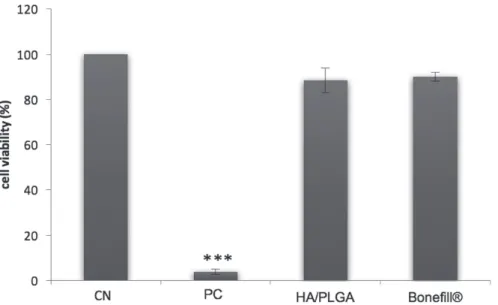
Cytotoxicity results are shown in Figure 1 (XTT cell viability assay). Cell viability is related to absorbance. Negative control corresponded to 100% cell viability.
The results obtained for NC and HA/PLGA and Bonefill®
eluates did not significantly differ (p>0.05, Dunnett’s test), indicating that the materials did not affect cell viability.
Literature describes several assays that can be used to determine toxicity in polymeric nanostructured systems, including assays involving cell viability analyses
FIGURE 1 - XTT assay in CHO-K1 (Cell viability). Cells treated with 100%concentration HA/PLGA and Bonefill® eluates. NC:
of mammalian cells, such as the tetrazolium salt reduction
assay (XTT), MTT assay and genotoxicity assays (Cieślik
et al., 2009). Therefore, the XTT assay was used to evaluate cell viability after exposure to eluates from HA/PLGA and
Bonefill® materials. This assay is based on cell metabolic
activity where the reduction of yellow tetrazolium salt to orange formazan dye only occurs in viable cells by mitochondrial dehydrogenases; this can be measured
by absorbance. It is important to bear in mind that the
XTT assay aims to demonstrate immediate cytotoxic
effect on cultured cells, whereas the clonogenic survival
assay shows whether other damage has occurred to cells that interfere with or stop their proliferative capacity at a later time (Sumantran et al., 2007). The results of this
study showed the absence of cytotoxic effects from HA/ PLGA and Bonefill® eluates, which are in accordance with
literature demonstrating HA, bovine bone, and PLGA as biocompatible materials (Galia et al., 2008;Cieślik et al., 2009; Trif et al., 2015). PLGA nanoparticles associated withpolyethylene glycol (PEG) and poly L-lysine (PLL) (PEG-PLL-PLGA) have demonstrated low cytotoxicity by MTT assay (Guo et al., 2015).
Furthermore, different concentrations of PLGA
nanoparticles have shown no cytotoxicity to fibroblasts
(3T3) by MTT assay (De Lima et al., 2011). In contrast,
high concentrations (5,000 µg.mL-1) of PLGA nanoparticles
induced moderate cytotoxicity in Madin–Darby bovine kidney (MDBK) cells (Trif et al., 2015). However, the same concentration did not induce a cytotoxic effect inhuman colorectal adenocarcinoma (Colo 205) cells. Also, no cytotoxicity was seen from PLGA and HA/ PLGA composites by the lactate dehydrogenase (LDH)
test (Cieślik et al., 2009). Additionally, lyophilized
bovine bone prepared on a semi-industrial scale showed
no cytotoxicity potential by the agar diffusion test (Galia
et al., 2008). Studies have verified that even with a high
particle surface area, particles concentrations between 5.4 and 540 µg/mL are incapable of inducing cell toxicity (Semete et al., 2010; De Lima et al., 2011), agreeing with previously presented explanations.
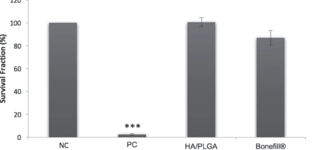
The surviving fraction obtained by clonogenic
survival assay revealed no statistical difference with NC
(p>0.05, Dunnett’s test; Figure 2).
The clonogenic assay evaluated whether the material interferes in mitotic replication of CHO-K1 cells, since only mitotically viable cells can produce progenitor cells.
We verified that HA/PLGA and Bonefill® eluate did not
interfere in cell survival or the proliferation capacity of a
single cell. Increasing eluate HA concentration was shown
to increase cytotoxic effects by inhibiting cell-colony
formation (Jantová et al., 2008).
The general cytotoxicity results revealed that HA/ PLGA and Bonefill® were not cytotoxic, showing no
disturbance in mitotic cell replication.
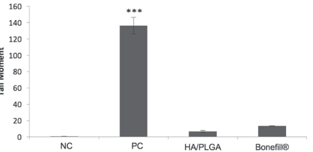
Tail moment evaluation verified that neither HA/ PLGA or Bonefill® promoted genotoxic effects (p>0.05,
Dunnett’s test; Figure 3).
The alkaline version of Comet assay can detect DNA double-strand breaks, alkali-labile sites, and single-strand breaks associated with incomplete excision repair sites (Tice et al., 2000; Araújo et al., 2009). Tail moment evaluation verified that HA/PLGA and Bonefill® bone
substitutes did not promote genotoxic effects. The concept
of tail moment (calculated as the product of tail length and total DNA fraction in the tail) is a measurement of DNA migration. This method incorporates relative
FIGURE 2 - Clonogenic survival assay in CHO-K1. Cells treated with 100%concentration HA/PLGA and Bonefill® eluates. NC:
measurements of both the smallest detectable size of migrating DNA (reflected by comet tail length) and the number of broken pieces of DNA (represented by the staining intensity of DNA in the tail) (Liao,
Mcnutt, Zhu, 2009). The study of DNA damage at the
chromosome level is an essential part of genetic toxicology whereas chromosomal mutation is an important event in carcinogenesis. Studies investigating HA toxicity have
shown that it does not induce genotoxic effects (Kannana
et al., 2014), however comet assay showed increasing HA concentrations (10% to 100%; v/v) induced DNA damage (between 13.1 and 14.2%), which was dose-dependent
(Jantová et al., 2008).
Table I shows mutagenicity assay results of: nuclear division index (NDI), the frequency of binucleated cells
with micronuclei (BCMN), and micronucleus (MN), nucleoplasmic bridge (NPB), nuclear bud (NBUD) frequencies.
NDI was similar between groups (p>0.05; Kruskal Wallis’s), except for PC and Bonefill® (p<0.05; Kruskal
Wallis’s). Nuclear Division Index (NDI) is a marker of
cell proliferation in cultures and is considered a measure of general cytotoxicity (Eastmond, Tucker, 1989; Fenech,
2000; Ionescu et al., 2011). There may be an induction of mitotic delay which, by not allowing the repair of genotoxic lesions, will modify the number of cells entering mitosis and modify the proportion of mono-/bi-/tri- and
tetranucleated cells (Ionescu et al., 2011). Thus, lower
NDI can signify fewer cell divisions. There is also the
hypothesis of a clastogenic effect from mutagens with an aneugenic action, inducing some degree of cell cycle
blockade. Therefore, more cells will not divide and NDI
will again be low. There was no significant difference
in NDI between NC and both Bonefill® or HA/PLGA
materials, indicating that these materials did not induce
CHO-K1 nuclear division arrest. Therefore, Bonefill® and
HA/PLGA did not decreased cell division.
HA/PLGA and Bonefill® were not mutagenic, as
only PC was statistically higher than NC for FBCMN,
FMN, and FNBUD. In FNPB, only Bonefill® was
statistically lower than NC emphasizing that this material did not induce nucleoplasmic bridges.
TABLE I - CBMN assay in CHO-K1. Mean and standard error of nuclear division index (NDI), frequency of binucleated cells with
micronucleus (FBCMN), frequency of micronuclei (FMN), frequency of nucleoplasmic bridges (FNPBs) and frequency of nuclear
buds (NBUDs), in 1000 binucleated cell for each treatment. Cells treated with 100% concentration of HA/PLGA and Boneffil®
eluates. NC: negative control; PC: positive control; *p<0.05 compared to NC group (Dunnett’s test); a,b = p<0.05 (Kruskal-Wallis’s
test).
TREATMENT NDI
Mean ± SE
FBCMN Mean ± SE
FMN Mean ± SE
FNPBs Mean ± SE
FNBUDs Mean ± SE
NC 1.914± 0.002 16.33 ±0.298 21.67±1.075 5.33±0.789 16.57±2.024
PC 1.754±0.005a 91.67±1.814* 143.67±5.873* 40.6±1.300* 101.29±7.928*
HA/PLGA 1.977±0.006 15.33±1.193 20.33 ± 1.578 4.33±0.298 11.86±2.436
Bonefill® 2.032±0.013b 11.67±1.660 12.67±1.193 2.33±0.298* 13.71±1.723
FIGURE 3 - Comet assay in CHO-K1. Cells treat with 100% concentration HA/PLGA and Bonefill® eluates. NC: negative control;
The micronucleus assay (or cytokinesis-blocked micronucleus) has emerged as one of the preferred methods for assessing chromosome damage because it enables both chromosome loss and chromosome breakage to be reliably measured (Fenech, 2000). Although using a different method, the Salmonella Typhimurium bacterial test,
Jantová et al., (2008) also observed no mutagenic effect in
100%HA-concentration eluates. Therefore, HA was not
mutagenic, even at high concentration, by two different
reliable tests (CBMN and bacterial mutagenicity test). Our study showed that 100% HA/PLGA eluate concentration (v/v) did not promote cytotoxic, genotoxic,
or mutagenic effects. This is in agreement with Cieślik et al. (2009) who demonstrated that an HA/PLGA composite was fully biocompatible and the bone defects were fully repaired after 48 weeks. They concluded that this
composite did not induce toxic effects on bone-forming
cells.
Cyto- and genotoxicity effects on materials prepared
with PLGA nanoparticles require further study, even though this polymer is one of the most widely used in the preparation of polymer nanoparticles, mainly for pharmaceutical and medical processes. Studies concerning the impact of these nanostructures on living organisms and the environment are therefore needed so that the safety of these nanosystems can be assessed before they become even more widely commercialized (De Lima et al., 2011). For example, the aforementioned PEG-PLL-PLGA showed no blood toxicity and mutagenicity by MN (Guo et al., 2015). Positively charged PLGA nanoparticles led to chromosomal aberrations without primary DNA damage in human bronchial epithelial cells (Platel et al., 2016). Human peripheral blood mononuclear cell cultures treated with PLGA-PEO nanoparticles revealed no increase in the number of micronucleated binucleated cells (Tulinska et al., 2015). On the other hand,
PLGA-PEO lead to a weak but significant increase in the level
of MN in TK6 human B-lymphoblastoid cells, which did not induce DNA strand-breaks (detected by comet assay), nor was it cytotoxic (measured by relative cell growth
activity and cytokinesis-block proliferation index (CBPI))
(Kazimirova et al., 2012).
Previous studies have shown that HA/PLGA composites not only promote adequate bone regeneration (Galia et al., 2008), but also do not induce cytotoxicity (Galia et al., 2008; Cieślik et al., 2009; Kannana et al., 2014), mutagenicity (Kazimirova et al., 2012; Kannana et al., 2014), or genotoxicity (Cieślik et al., 2009; Kannana
et al., 2014), which corroborate with the results of this
study. In this way, HA/PLGA results were used as a bench mark to evaluate whether Bonefill® could have the similar
toxicity profile to that of HA/PLGA. Both materials demonstrated similar results - not inducing cytotoxic,
genotoxic, or mutagenic effects in CHO-K1 cells.
CONCLUSION
Considering the negative cytotoxicity, genotoxicity and mutagenicity results found for HA/PLGA and
Bonefill®, it was concluded that these bone substitutes
presented interesting preliminary results as potential biomaterials for bone repair.
DECLARATION OF INTERESTS
The authors certify that they have no commercial or
associate interests that represent a conflict of interest in
connection with the manuscript.
ACKNOWLEDGMENTS
The authors are grateful to Bionnovation Biomedical Products Ltda. Brazil, for providing the materials (HA/
PLGA and Bonefill® Block).
REFERENCES
Araújo JMSD, Quintans TC, Santos SDD, Sousa CD, Queiroga AS, Limeira Júnior F. Enxerto ósseo bovino como alternativa
para cirurgias de levantamento de assoalho de seio maxilar. Rev
Cir Traumatol Buco-maxilo-fac. 2009;9(3):89-96.
Cieślik M, Mertas A, Morawska-Chochół A, Sabat D, Orlicki R, Owczarek A, Król W, Cieślik T. The evaluation of the
possibilities of using PLGA co-polymer and its composites with
carbon fibers or hydroxyapatite in the bone tissue regeneration process - in vitro and in vivo examinations. Int J Mol Sci.
2009;10(7):3224-3234
Costantino PD, Friedman CD, Jones K, Chow LC, Pelzer HJ, Sisson, G.A. Hydroxyapatite cement: I. Basic chemistry
and histologic properties. Arch Otolaryngol Head Neck Sur. 1991;117(4):379-384.
De Lima R, Santo Pereira ADE, Porto R M, Fraceto LF. Evaluation of cyto- and genotoxicity of
Poly(lactide-co-glycolide) nanoparticles J Polym Environ. 2011;19(1):196-202.
Dimitriou R, Jones E, Mcgonagle D, Giannoudis PV. Bone
Eastmond DA, Tucker JD. Identification of aneuploidy‐ inducing agents using cytokinesis‐blocked human lymphocytes
and an antikinetochore antibody. Environ Mol Mutagen. 1989;13(1):34-43
Fenech M. The in vitro micronucleus technique. Mutat. Res.
2000;455(1):81-95.
Franken NA, Rodermond HM., Stap J, Haveman J, Van Bree C.
Clonogenic assay of cells in vitro. Nat Protoc. 2006;1(5):2315-2319.
Galia CR, Macedo CA, Rosito R, Mello TMD, Camargo LMAQ,
Moreira LF. In vitro and in vivo evaluation of lyophilized bovine
bone biocompatibility. Clinics. 2008;63(6):801-806.
Galia CR, Macedo CADS, Rosito R, Mello TMD, Diesel C, Moreira LF. Physicochemical characterization of bovine and human freeze-dried bones. Rev Col Bras Cir. 2009;36(2):157-160.
G u o L , C h e n B , L i u R , L i u P, X i a G , Wa n g Y, e t a l . Biocompatibility assessment of polyethylene glycol-poly l-lysine-poly lactic-co-glycolic acid nanoparticles in vitro and
in vivo. J Nanosci Nanotechnol. 2015;15(5):3710-3719.
Ionescu ME, Ciocirlan M, Becheanu G, Nicolaie T, Ditescu
C, Teiusanu AG, et al. Nuclear division index may predict neoplastic colorectal lesions. Maedica. 2011;6(3):173.
International Standard. ISO 10993-12: 2008 - Biological
evaluation of medical devices- Part 12: Sample preparation and
reference materials. German version: DIN EN ISO,
2008;10993-10912.
Jantová S, Theiszová M, Letašiová S, Birošová L, Palou
TM. In vitro effects of fluor-hydroxyapatite, fluorapatite and hydroxyapatite on colony formation, DNA damage and mutagenicity. Mutat Res. 2008;652(2):139-144.
Kannana TP, Lahc NASN, Ahmada A, Fatimah S. Genotoxic evaluation of synthetic hydroxyapatite using mammalian
bone marrow chromosome aberration test. Arch Orofac Sci.
2014;9(1):1-7.
Kaveh K, Ibrahim R, Bakar MZA, Ibrahim TA. Bone grafting and bone graft substitutes. J Anim Vet Adv. 2010;9(6):1055-1067.
Kazimirova A, Magdolenova Z, Barancokova M, Staruchova
M, Volkovova K, Dusinska M. Genotoxicity testing of PLGA–PEO nanoparticles in TK6 cells by the comet assay and the cytokinesis-block micronucleus assay. Mutat Res. 2012;748(1):42-47.
Liao W, McNutt MA, Zhu WG. The comet assay: a sensitive
method for detecting DNA damage in individual cells. Methods. 2009;48(1):46-53.
Organisation for Economic Co-operation and Development. OECD. Guidelines for Testing of Chemicals Section for Health
Effects. Test No. 487: In vitro mammalian cell micronucleus
test. Paris: OECD; 2014. p. 1-26.
Platel A, Carpentier R, Becart E, Mordacq G, Betbeder
D, Nesslany F. Influence of the surface charge of PLGA
nanoparticles on their in vitro genotoxicity, cytotoxicity, ROS production and endocytosis. Appl Toxicol. 2016;36(3):434-444.
Rios ALBB, Barbosa CEDM, Rached A, Georges RS, Gabrielli
MFR, Okamoto T. Comportamento biológico de implantes de
osso bovino anorgânico em arco zigomático de ratos: estudo
histológico. Rev Odontol UNESP. 1996;25(esp):87-101.
Saska S, Mendes LS, Gaspar AMM, Capote TSDO. Bone
substitute materials in implant dentistry. Implant Dent.
2015;2:158-167.
Scarel-Caminaga RM, Saska S, Franchi LP, Santos RA, Gaspar AMM, Capote TSDO, et al. Nanocomposites based on bacterial cellulose in combination with osteogenic growth peptide for
bone repair: cytotoxic, genotoxic and mutagenic evaluations. J
App Biol Biotech. 2014;2(1):001-008.
Semete B, Booysen L, Lemmer Y, Kalombo L, Katata L,
Verschoor J, Swai HS. In vivo evaluation of the biodistribution
and safety of PLGA nanoparticles as drug delivery systems. Nanomedicine. 2010;6(5):662-671.
Setyawati MI, Khoo PKS, Eng BH, Xiong S, Zhao X, Das GK,
et al. Cytotoxic and genotoxic characterization of titanium
dioxide, gadolinium oxide, and poly (lactic‐co‐glycolic acid) nanoparticles in human fibroblasts. J Biomed Mater Res A.
2013;101(3):633-640.
Shi C, Gao J, Wang M, Fu J, Wang D, Zhu Y. Ultra-trace
BONEFILL® block as alternative for bone substitute: a toxicological evaluation
Singh NP, McCoy MT, Tice RR, Schneider EL. A simple technique for quantitation of low levels of DNA damage in individual cells. Exp Cell Res. 1988;175(1):184-191.
Suchanek W, Yoshimura M. Processing and properties of hydroxyapatite-based biomaterials for use as hard tissue
replacement implants. J Mater Res. 1998;13(1):94-117.
Sumantran VN, Boddul S, Koppikar SJ, Dalvi M, Wele A, Gaire V, Wagh UV. Differential growth inhibitory effects of W. somnifera root and E. officinalis fruits on CHO cells. Phytother
Res. 2007;21(5):496-499.
Tice RR, Agurell E, Anderson D, Burlinson B, Hartmann A, Kobayashi H, et al. Single cell gel/comet assay: guidelines for in vitro and in vivo genetic toxicology testing. Environ Mol Mutagen. 2000;35(3):206-221.
Trif M, Florian PE, Roseanu A, Moisei M, Craciunescu O, Astete CE, Sabliov CM. Cytotoxicity and intracellular
fate of PLGA and chitosan‐coated PLGA nanoparticles in
Madin–Darby bovine kidney (MDBK) and human colorectal
adenocarcinoma (Colo 205) cells. J Biomed Mater Res A.
2015;103(11):3599-3611.
Tulinska J, Kazimirova A, Kuricova M, Barancokova M,
Liskova A, Neubauerova E, et al. Immunotoxicity and
genotoxicity testing of PLGA-PEO nanoparticles in human
blood cell model. Nanotoxicology. 2015;9(Supp1):33-43.
Wang YW, Wu Q, Chen J, Chen GQ. Evaluation of three-dimensional scaffolds made of blends of hydroxyapatite and
poly (3-hydroxybutyrate-co-3-hydroxyhexanoate) for bone reconstruction. Biomaterials. 2005;26(8):899-904.
Yalkinoglu AO, Schlehofer JRR, Hausen HZ. Inhibition of N-methyl-N”-nitro-‐n‐nitrosoguanidine‐induced methotrexate and adriamycin resistance in CHO cells by adeno‐associated virus type 2. Int J Cancer. 1990;45(6):1195-1203.
Received for publication on 28th July 2017