ABSTRACT
Objective: To determine the frequency of active smoking among patients with asthma and individuals without asthma by self-report and urinary cotinine measurement. Methods: This was a cross-sectional study conducted in the city of Salvador, Brazil, and involving 1,341 individuals: 498 patients with severe asthma, 417 patients with mild-to-moderate asthma, and 426 individuals without asthma. Smoking status was determined by self-report (with the use of standardized questionnaires) and urinary cotinine measurement. The study variables were compared with the chi-square test and the Kruskal-Wallis test. Results: Of the sample as a whole, 55 (4.1%) reported being current smokers. Of those, 5 had severe asthma, 17 had mild-to-moderate asthma, and 33 had no asthma diagnosis. Of the 55 smokers, 32 (58.2%) were daily smokers and 23 (41.8%) were occasional smokers. Urinary cotinine levels were found to be high in self-reported nonsmokers and former smokers, especially among severe asthma patients, a finding that suggests patient nondisclosure of smoking status. Among smokers, a longer smoking history was found in patients with severe asthma when compared with those with mild-to-moderate asthma. In addition, the proportion of former smokers was higher among patients with severe asthma than among those with mild-to-moderate asthma. Conclusions: Former smoking is associated with severe asthma. Current smoking is observed in patients with severe asthma, and patient nondisclosure of smoking status occurs in some cases. Patients with severe asthma should be thoroughly screened for smoking, and findings should be complemented by objective testing.
Keywords: Asthma; Smoking; Cotinine.
Self-reported smoking status and urinary
cotinine levels in patients with asthma
Gabriela Pimentel Pinheiro1,2,a, Carolina de Souza-Machado1,3,b, Andréia Guedes Oliva Fernandes4,c, Raquel Cristina Lins Mota5,d, Liranei Limoeiro Lima2,e, Diego da Silva Vasconcellos6,f,Ives Pereira da Luz Júnior7,g, Yvonbergues Ramon dos Santos Silva7,h, Valmar Bião Lima1,4,i, Sérgio Telles de Oliva8,j, Luane Marques de Mello9,k, Ricardo David Couto10,l, José Miguel Chatkin11,m,
Constança Margarida Sampaio Cruz12,13,n, Álvaro Augusto Cruz1,14,o
Correspondence to:
Gabriela Pimentel Pinheiro. Programa para o Controle da Asma na Bahia (ProAR), Centro de Saúde Carlos Gomes, Rua Carlos Gomes, 270, 7º andar, CEP 40060-330, Salvador, BA, Brasil.
Tel.: 55 71 3321-8467. E-mail: [email protected]
Financial support: This study received financial support from the Brazilian Conselho Nacional de Desenvolvimento Científico e Tecnológico/Programa de Apoio a Núcleos de Excelência (CNPq/PRONEX, National Council for Scientific and Technological Development/Program for the Support of Centers of Excellence; Grant no. 020/2009), the Fundação de Amparo à Pesquisa do Estado da Bahia (FAPESB, Foundation for the Support of Research in the State of Bahia; Grant no. 6353 PNX 0018/2009), and GlaxoSmithKline’s Trust in Science program investigator-initiated grant (2012-2015).
a. http://orcid.org/0000-0002-6088-2805; b. http://orcid.org/0000-0001-7328-9608; c. http://orcid.org//0000-0001-5584-5658; d. http://orcid.org/0000-0001-9723-8717; e. http://orcid.org/0000-0002-6129-8221; f. http://orcid.org/0000-0003-2247-4883; g. http://orcid.org/0000-0002-9640-0193; h. http://orcid.org/0000-0003-0297-5226; i. http://orcid.org/0000-0001-8479-3666; j. http://orcid.org/0000-0002-8874-7736; k. http://orcid.org/0000-0002-4462-8364; l. http://orcid.org/0000-0003-2119-437X; m. http://orcid.org/0000-0002-4343-025X; n. http://orcid.org/0000-0002-3885-4314; o. http://orcid.org/0000-0002-7403-3871
INTRODUCTION
Smoking is recognized worldwide as a chronic disease resulting from nicotine dependence and as a risk factor for the development and worsening of chronic
respiratory diseases such as asthma and COPD.(1) Smoking is a major cause of
preventable death and is associated with increased health care costs, morbidity, and mortality, accounting for more than 6 million deaths per year.(2)
Asthma is a chronic disease that has a high worldwide prevalence (i.e., 1-16%).(3,4)
In the city of Salvador, Brazil, 13.4% of all adolescents and 5.1% of all adults have asthma.(5,6) Smoking is directly related to uncontrolled asthma and increased asthma
severity, increasing the risk of exacerbations, decreased lung function, persistent dyspnea,(7) and limited response to treatment with corticosteroids.(8) Nevertheless,
smoking remains prevalent among patients with asthma. In a study conducted in the city of São Paulo, Brazil, the prevalence of self-reported smoking among asthma
patients was 3%, and the prevalence of self-reported former smoking was 33%.(9)
1. Programa para o Controle da Asma na Bahia – ProAR – Universidade Federal da Bahia – UFBA – Salvador (BA) Brasil. 2. Programa de Pós-Graduação em
Ciências da Saúde, Universidade Federal da Bahia – UFBA – Salvador (BA) Brasil.
3. Escola de Enfermagem, Universidade Federal da Bahia – UFBA – Salvador (BA) Brasil. 4. Programa de Pós-Graduação em
Medicina em Saúde, Universidade Federal da Bahia – UFBA – Salvador (BA) Brasil.
5. Hospital Heliópolis, São Paulo (SP) Brasil. 6. Programa de Pós-Graduação em
Química, Universidade Federal da Bahia – UFBA – Salvador (BA) Brasil. 7. Laboratório de Química Analítica e
Ambiental, Instituto de Química, Universidade Federal da Bahia – UFBA – Salvador (BA) Brasil.
8. Departamento de Química Analítica,
Instituto de Química, Universidade Federal da Bahia – UFBA – Salvador (BA) Brasil.
9. Faculdade de Medicina de Ribeirão
Preto, Universidade de São Paulo, Ribeirão Preto (SP) Brasil. 10. Laboratório de Bioquímica Clínica,
Departamento de Análises Clínicas e Toxicológicas, Faculdade de Farmácia, Universidade Federal da Bahia – UFBA – Salvador (BA) Brasil.
11. Escola de Medicina, Pontifícia Universidade Católica do Rio Grande do Sul, Porto Alegre (RS) Brasil. 12. Programa de Pós-Graduação em
Medicina e Saúde Humana, Escola Bahiana de Medicina e Saúde Pública, Salvador (BA) Brasil.
13. Obras Sociais Irmã Dulce, Hospital Santo Antônio, Salvador (BA) Brasil. 14. Faculdade de Medicina da Bahia,
Universidade Federal da Bahia – UFBA – Salvador (BA) Brasil.
Submitted: 4 September 2017.
Self-reported smoking status and urinary cotinine levels in patients with asthma
Exposure to smoking can be determined by patient self-report and measurement of biological markers such as exhaled carbon monoxide and carboxyhemoglobin, as well as thiocyanate, nicotine, and cotinine levels, which can be measured in saliva, plasma, and urine. (10)
Patient self-report is commonly used because it is an easy-to-use and inexpensive method for assessing smoking; however, inaccurate self-reporting (patient nondisclosure of smoking status) constitutes a disadvantage.(11)
Cotinine is a byproduct of nicotine metabolism, and measurement of cotinine levels is the most widely recommended method for quantifying exposure to
tobacco smoking because it is not influenced by other
exposures.(12) The technique used in order to measure
cotinine levels is reliable(12-16) and allows detection
of smoking occurring 19-40 h prior to urine sample collection.(17-19) This is due to the fact that the renal
excretion of cotinine is low, meaning that cotinine can be easily detected by laboratory monitoring and has
the prerequisites of specificity that make it the analyte
of choice for quantifying exposures.(20)
The objective of the present study was to determine the frequency of active smoking among patients with varying degrees of asthma severity and individuals without asthma in the city of Salvador by self-report (with the use of standardized questionnaires) and urinary cotinine measurement.
METHODS
Study design
This was a cross-sectional study of patients diagnosed with asthma. The study was conducted between 2013 and 2015 at the Federal University of Bahia Center of Excellence in Asthma, which is located in the city of
Salvador and is a research center affiliated with the
Programa para o Controle da Asma na Bahia (ProAR, Bahia State Program for the Control of Asthma and Allergic Rhinitis).
The present study is part of a larger study entitled “Fatores de risco, biomarcadores e endofenótipos da asma grave” (Severe asthma: risk factors, biomarkers, and endophenotypes), which is a case-control study investigating patients with severe asthma and involving two control groups: participants with mild-to-moderate asthma and participants without asthma.
Selection and sampling
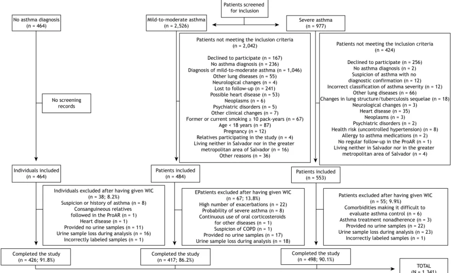
A total of 1,341 individuals were studied. Of those, 915 had been diagnosed with asthma. Of those, 417 had mild-to-moderate asthma and 498 had severe asthma (and were followed in the ProAR). The study also included 426 individuals without asthma.
The severe asthma patients participating in the study had not been under regular treatment prior to admission to the ProAR, when they were diagnosed with severe asthma (having been followed ever since). The study participants with mild-to-moderate asthma
and those without asthma were recruited through advertisements in the media, in public transportation, and in public places, as well as through peer referral. Asthma severity was determined on the basis of the 2012 Global Initiative for Asthma criteria.(21)
Individuals ≥ 18 years of age living in Salvador (or
in the greater metropolitan area of Salvador) and
treated via the Brazilian Unified Health Care System
were included in the study. All of the severe asthma patients included in the study had been under regular treatment for at least six months. Patients presenting
with comorbidities that made it difficult to evaluate
asthma control (including congestive heart failure, stroke, myopathies, advanced neoplasia, psychiatric disorders, and lung diseases other than asthma) were excluded, as were those with a smoking history of
more than 10 pack-years, because of the difficulty
in making a differential diagnosis between asthma and COPD. At the end of the study period, some of the participants were excluded for various reasons, including problems with the urine sample, treatment abandonment, and exacerbation of comorbidities that
made patient evaluation difficult (Figure 1).
Participants with severe asthma
Severe asthma was diagnosed in accordance with the Global Initiative for Asthma criteria(21) by two
specialists, who reviewed patient medical records during the selection phase. Disagreements between the two specialists regarding asthma diagnosis or severity were resolved by a third specialist. At the end of this phase, 949 patients meeting the inclusion criteria were contacted by telephone and invited to visit the Federal University of Bahia Center of Excellence in Asthma. Of those, only 553 visited the Federal University of Bahia Center of Excellence in Asthma, where they underwent clinical evaluation and spirometry. A total of 55 individuals were excluded, the total sample of patients with severe asthma therefore consisting of 498 individuals (Figure 1).
Participants with mild-to-moderate asthma or without asthma
A total of 2,526 patients with mild-to-moderate asthma and individuals without asthma were contacted for prescreening. Of those, 484 patients with
mild-to-moderate asthma were included in the study. However,
only 417 completed all tests. For comparison purposes, 464 individuals without asthma were included in the
study. However, only 426 completed all tests (Figure 1).
Study procedures and data collection
During appointment scheduling, participants were
instructed to collect first morning urine samples
following basic safety and hygiene procedures. After delivery, the samples were labeled and stored in a
freezer at −70°C. Patients were then referred for a clinical evaluation in order to confirm the diagnosis and
Pinheiro GP, Souza-Machado C, Fernandes AGO, Mota RCL, Lima LL, Vasconcellos DS, da Luz Júnior IP,
Silva YRS, Lima VB, Oliva ST, Mello LM, Couto RD, Chatkin JM, Cruz CMS, Cruz AA
No asthma diagnosis (n = 464)
Mild-to-moderate asthma (n = 2,526)
Severe asthma (n = 977)
Individuals included (n = 464)
Patients included
(n = 484) Patients included(n = 553)
Completed the study (n = 426; 91.8%)
Completed the study (n = 417; 86.2%)
Completed the study (n = 498; 90.1%)
TOTAL (N = 1,341) No screening
records
Individuals excluded after having given WIC (n = 38; 8.2%)
Suspicion or history of asthma (n = 8) Consanguineous relatives followed in the ProAR (n = 1)
Heart disease (n = 1) Provided no urine samples (n = 11) Urine sample loss during analysis (n = 16)
Incorrectly labeled samples (n = 1)
EPatients excluded after having given WIC (n = 67; 13.8%)
High number of exacerbations (n = 22) Probability of severe asthma (n = 8) Continuous use of oral corticosteroids
for other diseases (n = 1) Suspicion of COPD (n = 1) Provided no urine samples (n = 17) Urine sample loss during analysis (n = 18) Patients not meeting the inclusion criteria
(n = 2,042)
Declined to participate (n = 167) No asthma diagnosis (n = 236) Diagnosis of mild-to-moderate asthma (n = 1,046)
Other lung diseases (n = 55) Neurological changes (n = 4) Lost to follow-up (n = 241) Possible heart disease (n = 53)
Neoplasms (n = 6) Psychiatric disorders (n = 5) Other clinical changes (n = 7)
Former or current smoking ≥ 10 pack-years (n = 67)
Age < 18 years (n = 87) Pregnancy (n = 12)
Relatives participating in the study (n = 4) Living neither in Salvador nor in the greater
metropolitan area of Salvador (n = 16) Other reasons (n = 36)
Patients not meeting the inclusion criteria (n = 424)
Declined to participate (n = 256) No asthma diagnosis (n = 2) Suspicion of asthma with no diagnostic confirmation (n = 12) Incorrect classification of asthma severity (n = 12)
Other lung diseases (n = 66)
Changes in lung structure/tuberculosis sequelae (n = 18) Neurological changes (n = 3)
Heart disease (n = 35) Neoplasms (n = 3) Psychiatric disorders (n = 2) Health risk (uncontrolled hypertension) (n = 8)
Allergy to asthma medications (n = 2) No regular follow-up in the ProAR (n = 1) Living neither in Salvador nor in the greater
metropolitan area of Salvador (n = 4)
Patients excluded after having given WIC (n = 55; 9.9%)
Comorbidities making it difficult to evaluate asthma control (n = 6) Asthma treatment nonadherence (n = 3)
Provided no urine samples (n = 22) Urine sample loss during analysis (n = 23)
Incorrectly labeled samples (n = 1)
Figure 1. Flow chart of patient recruitment. ProAR: Programa para o Controle da Asma na Bahia (Bahia State Program for the Control of Asthma and Allergic Rhinitis); and WIC: written informed consent.
Self-reported smoking status and urinary cotinine levels in patients with asthma
and use of medications. None of the participants were on nicotine replacement therapy.
Self-reported smoking status
The participants who reported smoking cigarettes daily or occasionally were considered to be current smokers. The participants who reported being former smokers and having quit smoking at least six months before their interview were considered to be former smokers.
Data regarding exposure to smoking were collected by asking participants questions regarding smoking history (the questions being part of the Brazilian Telephone-based System for the Surveillance of Risk and Protective Factors for Chronic Noncommunicable Diseases questionnaire)(22) and exposure to secondhand
smoke at home, school, and work, as well as questions regarding exposure to smoking in public transportation and in public places (the questions being part of a questionnaire used by the Brazilian Institute of Geography and Statistics in the 2010 Census).(23)
Urinary cotinine measurement
Urinary cotinine was measured in accordance with the procedures described by Cattaneo et al.(24)
A high-performance liquid chromatograph (1290
Infinity; Agilent®, Santa Clara, CA, USA) equipped
with a Zorbax Eclipse XDB-C8 (4.6 mm × 150 mm ×
5 µm) column and a UV-Vis (λ = 260 nm) detector
(Agilent®) was used, with an injection volume of 20 µL and an isocratic mobile phase flow rate of 0.4
mL/min. The methodology was validated by using
the parameters set forth in Brazilian National Health
Oversight Agency Resolution no. 899.(25) Because of
its sensitivity and specificity, high-performance liquid
chromatography is recommended for measuring cotinine; in addition to being less expensive than other methods, high-performance liquid chromatography allows determination of low concentrations of cotinine. (26)
The limits of detection and quantification were 6.46
µg/L and 19.59 µg/L, respectively.
Urinary cotinine levels are directly related to biological
factors such as renal function, urine flow, and urine pH. For increased accuracy, urinary cotinine levels
were adjusted for urinary creatinine levels (urinary cotinine/creatinine ratio, in µg/g).(27)
Urinary creatinine was measured with a creatinine assay kit and a spectrophotometer with a thermostated
cuvette at 37°C (readings at 30 s and 90 s; wavelength, 510 ηm). An automated chemistry analyzer (BT 3000
PLUS; Wiener lab Group, Rosario, Argentina) was used.
Statistical analysis
All severe asthma patients followed in the ProAR until study initiation were included. Therefore, there was no sample size calculation. The numbers of participants with mild-to-moderate asthma and without asthma were established in order to guarantee the comparability of the groups.
The collected data were processed with the Statistical Package for the Social Sciences, version 17.0 (SPSS Inc., Chicago, IL, USA) and are presented as graphs and tables. The Shapiro-Wilk test and the Kolmogorov-Smirnov test were used in order to determine the nature of the distribution of the variables. Continuous variables were expressed as mean and standard deviation if distribution was Gaussian or as median and interquartile range (IR) if distribution was non-Gaussian. Categorical variables were expressed as absolute frequency and valid proportion. The chi-square test was used in order to compare proportions, and the Kruskal-Wallis test was used in order to compare continuous variables, given that most of the data had non-normal distribution.
Ethical considerations
The study was approved by the Research Ethics Committee of the Federal University of Bahia Climério
de Oliveira Maternity Hospital (Ruling no. 099/2009;
addendum no. 032/2014), as well as by the Brazilian
National Health Council (Ruling no. 450/10). All of
the study participants gave written informed consent.
RESULTS
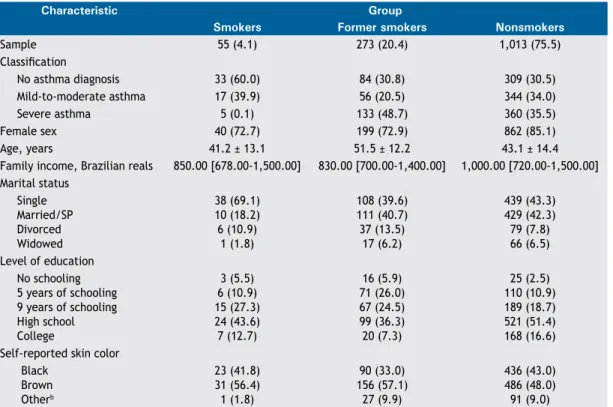
A total of 1,341 patients were evaluated. Of those, 55 (4.1%) reported being current smokers, 273 (20.4%) reported being former smokers, and 1,013 (75.5%) reported being nonsmokers. The characteristics of the study participants are described in Table 1. Participants were divided into three groups on the basis of asthma status and severity: severe asthma
(n = 498), mild-to-moderate asthma (n = 417), and no asthma diagnosis (n = 426).
Of the 55 participants who reported being active smokers, 32 (58.2%) reported smoking cigarettes daily and 23 (41.8%) reported smoking occasionally (Table 1). Table 2 provides detailed information on smoking in each study group. Among current smokers, smoking duration was longer in patients with severe asthma and individuals without asthma than in patients with mild-to-moderate asthma.
Smoking initiation was found to have occurred at an early age (i.e., during adolescence). Among smokers and former smokers, the mean age at smoking initiation
was significantly lower in the group of patients with
severe asthma (15.9 ± 5.3 years) than in that of patients with mild-to-moderate asthma (18.8 ± 5.7 years) and that of individuals without asthma (16.8
± 4.2 years; p = 0.02).
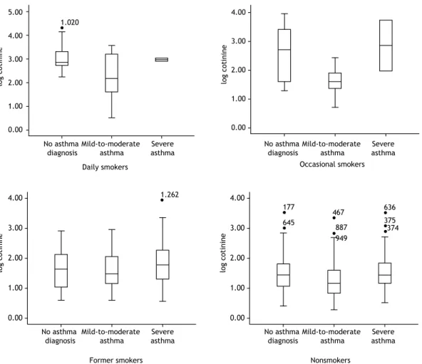
All of the study participants who reported smoking daily were positive for urinary cotinine. Of the study participants who reported smoking occasionally, 8 had urinary cotinine levels below the limit of detection. Median urinary cotinine levels were higher among daily smokers (758.2 µg/g; IR: 433.2-2,066.8) than among occasional smokers (97.1 µg/g; IR: 30.7-1.036.9; Table 3). Among daily and occasional smokers, urinary cotinine levels were highest in the group of patients with severe asthma.
Of the study participants who reported being
nonsmokers (n = 1,286), 273 (21.3%) were former
smokers. Median urinary cotinine levels were higher among former smokers (44.9 µg/g; IR: 17.4-147.9) than among individuals who reported never having smoked a cigarette (24.2 µg/g; IR: 10.9-58.5). Median urinary cotinine levels were higher among former smokers in the severe asthma group than among those in the remaining groups (Table 3). Figure 2 shows median urinary cotinine levels in smokers, former smokers, and nonsmokers, by asthma status.
Among former smokers, median urinary cotinine levels were highest in those with severe asthma. Among former smokers, median urinary cotinine levels were higher in those with severe asthma (62.5 µg/g; IR: 19.2-409.5) than in those with mild-to-moderate asthma (30.3 µg/g; IR: 13.0-110.3) and those without asthma (40.9 µg/g; IR: 9.9-129.1; p > 0.05).
Of the study participants who reported being nonsmokers, 440 (34.3%) reported having been exposed to secondhand smoke (at home, at work,
in public transportation, in public places, or any combination of the four) in the last 24 h. Of the nonsmokers who reported having been exposed to secondhand smoke in the last 24 h, 36.7% were patients with severe asthma, 34.6% were patients with mild-to-moderate asthma, and 30.8% were individuals without asthma.
DISCUSSION
In the present study, 4.1% of the participants reported being active smokers, a proportion that is lower than the mean proportion of smokers in the Brazilian population but similar to the proportion of smokers among adults in the city of Salvador.(5)
As expected, urinary cotinine levels were higher among daily smokers than among occasional smokers, former smokers, and nonsmokers. In addition, urinary cotinine levels were found to be higher in
former smokers than in nonsmokers, a finding that
suggests patient nondisclosure of smoking status. Urinary cotinine levels were higher in severe asthma patients who reported being former smokers than in former smokers with mild-to-moderate asthma and
no asthma diagnosis, a finding that suggests that the
issue of patient nondisclosure of smoking status is even more problematic in patients with severe asthma. The proportion of former smokers was highest among patients with severe asthma and lowest among patients with mild-to-moderate asthma.
The prevalence of current smoking was found to be higher in individuals without asthma than in patients
Table 1. Sociodemographic characteristics of the study sample, by self-reported smoking status.a
Characteristic Group
Smokers Former smokers Nonsmokers
Sample 55 (4.1) 273 (20.4) 1,013 (75.5)
Classification
No asthma diagnosis 33 (60.0) 84 (30.8) 309 (30.5)
Mild-to-moderate asthma 17 (39.9) 56 (20.5) 344 (34.0)
Severe asthma 5 (0.1) 133 (48.7) 360 (35.5)
Female sex 40 (72.7) 199 (72.9) 862 (85.1)
Age, years 41.2 ± 13.1 51.5 ± 12.2 43.1 ± 14.4
Family income, Brazilian reals 850.00 [678.00-1,500.00] 830.00 [700.00-1,400.00] 1,000.00 [720.00-1,500.00] Marital status
Single Married/SP Divorced Widowed
38 (69.1) 10 (18.2) 6 (10.9)
1 (1.8)
108 (39.6) 111 (40.7) 37 (13.5) 17 (6.2)
439 (43.3) 429 (42.3) 79 (7.8) 66 (6.5) Level of education
No schooling 5 years of schooling 9 years of schooling High school College
3 (5.5) 6 (10.9) 15 (27.3) 24 (43.6) 7 (12.7)
16 (5.9) 71 (26.0) 67 (24.5) 99 (36.3) 20 (7.3)
25 (2.5) 110 (10.9) 189 (18.7) 521 (51.4) 168 (16.6) Self-reported skin color
Black Brown Otherb
23 (41.8) 31 (56.4) 1 (1.8)
90 (33.0) 156 (57.1) 27 (9.9)
436 (43.0) 486 (48.0) 91 (9.0)
Self-reported smoking status and urinary cotinine levels in patients with asthma
with asthma, being lower in severe asthma patients than in mild-to-moderate asthma patients. The low rates of self-reported smoking among asthma patients in the present study are similar to those found in the literature(9) and might be due to the fact that
smoking has a negative impact on the clinical status and quality of life of asthma patients, who therefore avoid cigarettes. Because of their disease, patients with asthma are less likely to continue smoking. Another factor that can play an important role in reducing smoking among patients with asthma is being followed at health care clinics that provide education on the
harmful effects of smoking. However, the possibility
of patient nondisclosure of smoking status should be taken into account.(9,11)
The fact that the proportion of former smokers in the present study was highest among severe asthma patients suggests that smoking is a risk factor for the development of severe asthma in asthma patients who smoke despite feeling discomfort and despite warnings about the effects of smoking. In asthma patients with
an increased smoking history, increased asthma severity might be due to asthma-COPD overlap syndrome.
Among smokers and former smokers in the present study, smoking initiation was found to have occurred
during adolescence, a finding that is consistent with
those of Malcon et al.(28) and Abreu et al.(29) In the
present study, smoking initiation was found to have occurred earlier in the group of patients with severe asthma than in that of those with mild-to-moderate
asthma (15.9 years vs. 18.8 years), a finding that
is consistent with the possibility that exposure to smoking is a risk factor for the development of severe asthma.(30)
Among smokers and former smokers, the median duration of smoking was shorter in those with mild-to-moderate asthma than in those with severe asthma. This suggests that smoking is associated with asthma severity. In the present study, mild-to-moderate asthma patients smoked less than did severe asthma patients and individuals without asthma. It is possible that the discomfort associated with cigarette smoke inhalation
Table 2. Exposure to smokinga and creatinine-corrected urinary cotinine levels (in µg/g) in the study groups.b
Characteristic Group p*
Current smokers Former smokers
(n = 55) (n = 273)
Age at smoking initiation, years Severe asthma
Mild-to-moderate asthma No asthma diagnosis
20.0 [13.5-23.5] 18.0 [16.5-20.5] 17.0 [15.0-19.8]
15.0 [13.0-18.0] 18.0 [15.0-20.8] 16.0 [14.0-18.0]
0.20 0.25 0.20 Age at smoking cessation, years
Severe asthma
Mild-to-moderate asthma No asthma diagnosis
31.5 [23.0-40.0] 30.0 [24.0-59.0] 32.0 [25.0-40.0] Attempted to quit smoking
Severe asthma
Mild-to-moderate asthma No asthma diagnosis
3 (60.0) 6 (35.3) 11 (34.4) Duration of smoking, years
Severe asthma
Mild-to-moderate asthma No asthma diagnosis
33.0 [8.5-43.5] 10.0 [6.0-18.0] 27.5 [16.3-37.0]
15.0 [5.3-24.0] 11.3 [3.0-14.5] 10.2 [7.0-25.0]
0.14 0.07 < 0.01 Number of cigarettes/day
Severe asthma
Mild-to-moderate asthma No asthma diagnosis
2.0 [1.5-12.5] 2.0 [1.0-4.0] 5.0 [3.0-9.5]
6.0 [3.0-20.0] 5.0 [3.0-10.0] 10.0 [3.0-20.0]
0.18 < 0.01
0.03 Smoking history, pack-years
Severe asthma
Mild-to-moderate asthma No asthma diagnosis
25.5 [0.4-36.9] 1.3 [0.2-4.0] 7.7 [2.5-18.4]
4.4 [1.2-16.8] 1.2 [0.8-7.0] 8.0 [1.3-19.8]
0.52 0.25 0.89 Urinary cotinine, µg/gc
Severe asthma
Mild-to-moderate asthma No asthma diagnosis
807.8 [49.1-3.239.3] 41.1 [4.1-201.6] 598.3 [219.8-2.027.8]
62.5 [19.2-409.5] 30.3 [13.0-110.1] 40.9 [9.9-129.1]
0.03 0.27 < 0.01 Exposure to secondhand tobacco
smoke in the last 24 h Severe asthma
Mild-to-moderate asthma No asthma diagnosis
4 (80.0) 10 (58.8) 24 (72.7)
59 (44.4) 24 (42.9) 29 (34.5)
0.12 0.25 < 0.01
Figure 2. Creatinine-corrected urinary cotinine levels, by self-reported smoking status. Results expressed as (log) per µg/g of creatinine.
Table 3. Creatinine-corrected urinary cotinine (in µg/g) in the study participants (n = 1,341), by self-reported smoking status.a
Smoking status Number of participants Urinary cotinine,
µg/gb
p*
n/N %
Daily smoker Severe asthma
Mild-to-moderate asthma No asthma diagnosis
TOTAL
2/498 7/417 23/426 32/1,341
0.4 1.7 5.4 2.4
930.4 (807.8-1,053.1) 140.4 (11.9-2,189.7) 710.8 (499.1-2,357.7) 758.2 (433.2-2,066.8)
0.35
Occasional smoker Severe asthma
Mild-to-moderate asthma No asthma diagnosis
TOTAL
3/498 10/417 10/426 23/1,341
0.6 2.4 2.3 1.7
2,761.3 (97.1-5,425.5) 41.1 (16.2-129.1) 635.1 (32.3-3,945.0)
97.1 (30.7-1,036.9)
0.17
Former smoker Severe asthma
Mild-to-moderate asthma No asthma diagnosis
TOTAL
133/498 56/417 84/426 273/1,341
26.7 13.4 19.7 20.4
62.5 (19.2-409.5) 30.3 (13.0-110.3) 40.9 (9.9-129.0) 44.9 (17.4-147.9)
0.17
Nonsmoker Severe asthma
Mild-to-moderate asthma No asthma diagnosis
TOTAL
360/498 344/417 309/426 1,013/1,341
72.3 82.5 72.5 75.5
27.7 (14.3-69.5) 14.3 (6.8-39.9) 28.2 (11.4-67.3) 24.2 (10.9-58.5)
< 0.01
aValues expressed as median (interquartile range). bIndividuals presenting with results below the limit of detection are not included. *Kruskal-Wallis test.
5.00
4.00
3.00
2.00
1.00
0.00
1.020
1.262
177
645
467
887 949
636
374 375
log cotinine
4.00
3.00
2.00
1.00
0.00
log cotinine
4.00
3.00
2.00
1.00
0.00
log cotinine
No asthma diagnosis
Mild-to-moderate asthma
Severe asthma
4.00
3.00
2.00
1.00
0.00
log cotinine
Daily smokers Occasional smokers
Former smokers Nonsmokers
No asthma diagnosis
Mild-to-moderate asthma
Severe asthma
No asthma diagnosis
Mild-to-moderate asthma
Severe asthma
No asthma diagnosis
Mild-to-moderate asthma
Self-reported smoking status and urinary cotinine levels in patients with asthma
led mild-to-moderate asthma patients to quit smoking, whereas those who continued to smoke developed asthma that is more severe.
In the present study, patients with severe asthma were found to have a longer smoking history (in pack-years) than that of those with mild-to-moderate
asthma, a finding that suggests an association between
smoking and increased asthma severity.
In the present study, urinary cotinine levels varied among the groups, differences being found between urinary cotinine measurements and self-reported smoking status. Median urinary cotinine levels were found to be higher in self-reported daily smokers than in self-reported occasional smokers, except in the
group of patients with severe asthma, a finding that
suggests patient nondisclosure of smoking behavior. Cotinine levels are typically lower in individuals who do not smoke daily than in those who do, being
high in those who smoke more cigarettes daily,(31)
a single measurement of cotinine being sufficient to
show that.(32)
Other studies have shown discrepancies between self-reported smoking status and cotinine measurements,(33,34)
suggesting patient nondisclosure of smoking status. In a study conducted in the city of São Paulo, Brazil, urinary cotinine levels were found to be high in severe asthma patients who reported being former smokers,
a finding that alerts us to the possibility of inaccurate
self-reporting.(11)
Smoking is known to be associated with a poor asthma prognosis, reducing patient response to inhaled corticosteroids, increasing asthma symptoms, increasing the need for emergency room visits, increasing the need for hospitalization, and increasing treatment costs, as well as having a negative impact on quality of life. Cessation of smoking and smoke exposure can improve the clinical status of patients with asthma.(8,35)
Although our sample was large, the present study has limitations that should be taken into account. Urinary cotinine measurements might have been affected by passive exposure to tobacco smoke, ethnicity, and consumption of nicotine-containing foods, such as tomatoes, potatoes, and black tea.(36,37) However,
there were no differences in cotinine levels among nonsmokers or self-reported ethnicities exposed to secondhand smoke (exposure being expressed as
number of hours). The influence of dietary habits on
urinary cotinine levels was not investigated in the present study. The low frequency of current smokers in our sample reduced the power of subgroup analyses. During patient recruitment, asthma patients who reported a
smoking history ≥ 10 pack-years were excluded in
order to avoid mistaking COPD for asthma and ensure that the inclusion criteria were similar for patients with severe asthma and those with mild-to-moderate asthma. This might have introduced a bias in the comparison
with the individuals without asthma. However, the bias
would have favored a shorter smoking history among asthma patients; the fact that this was not observed in the severe asthma group reinforces the internal validity of our study. The proportion of former smokers was considerably higher in the severe asthma group (i.e., 27%) than in the remaining groups.
In conclusion, the prevalence of self-reported smoking was low among patients with varying degrees of asthma severity, being particularly low among those
with severe asthma. However, among patients with severe asthma, findings of an increased proportion of
self-reported former smokers, an increased smoking history, and increased urinary cotinine levels suggest patient nondisclosure of smoking status and an association between exposure to active smoking and severe asthma. Patients with severe asthma should be thoroughly screened for smoking via interviews and objective assessment.
REFERENCES
1. World Health Organization. WHO strategy for prevention and control of chronic respiratory diseases. Geneva: WHO; 2002.
2. World Health Organization. WHO global report on trends in prevalence of tobacco smoking. Geneva: WHO; 2015.
3. World Health Organization. WHO Report on the Global Tobacco Epidemic 2015: raising taxes on tobacco. Geneva: WHO; 2015.
4. Global Initiative for Asthma (GINA). Global Strategy for Asthma
Management and Prevention: online appendix. Bethesda: GINA; 2016.
5. Brasil. Ministério da Saúde. Secretaria de Vigilância em Saúde. Departamento de Vigilância de Doenças e Agravos não Transmissíveis e Promoção da Saúde. Vigitel Brasil 2016: vigilância de fatores de risco e proteção para doenças crônicas por inquérito telefônico: estimativas sobre frequência e distribuição sociodemográfica de fatores de risco e proteção para doenças crônicas nas capitais dos 26 estados brasileiros e no Distrito Federal em 2016. Brasília: Ministério da Saúde; 2017.
6. Barreto ML, Ribeiro-Silva RC, Malta DC, Oliveira-Campos M, Andreazzi MA, Cruz AA. Prevalence of asthma symptoms among adolescents in Brazil: National Adolescent School-based Health Survey (PeNSE 2012). Rev Bras Epidemiol. 2014;17 Suppl 1:106-15. https://doi.org/10.1590/1809-4503201400050009
7. Global Initiative for Asthma (GINA). Global Strategy for Asthma Management and Prevention. Bethesda: GINA; 2016.
8. Polosa R, Thomson NC. Smoking and asthma: dangerous liaisons. Eur Respir J. 2013;41(3):716-26. https://doi. org/10.1183/09031936.00073312
9. Dias-Júnior AS, Pinto RC, Angelini L, Fernandes FL, Cukier A,
Stelmach R. Prevalence of active and passive smoking in a population of patients with asthma. J Bras Pneumol. 2009;35(3):261-5. https:// doi.org/10.1590/S1806-37132009000300011
10. Haufroid V, Lison D. Urinary cotinine as a tobacco-smoke exposure index: a minireview. Int Arch Occup Environ Health. 1998;71(3):162-8. https://doi.org/10.1007/s004200050266
11. Stelmach R, Fernandes FL, Carvalho-Pinto RM, Athanazio RA,
Rached SZ, Prado GF, et al. Comparison between objective measures of smoking and self-reported smoking status in patients with asthma or COPD: are our patients telling us the truth? J Bras Pneumol. 2015;41(2):124-32. https://doi.org/10.1590/S1806-37132015000004526
12. Kim H, Lim Y, Lee S, Park S, Kim C, Hong C, et al. Relationship between environmental tobacco smoke and urinary cotinine levels in passive smokers at their residence. J Expo Anal Environ Epidemiol. 2004;14 Suppl 1:S65-70. https://doi.org/10.1038/sj.jea.7500360
13. Benowitz NL. Cotinine as a biomarker of environmental tobacco smoke exposure. Epidemiol Rev. 1996;18(2):188-204. https://doi. org/10.1093/oxfordjournals.epirev.a017925
Methods for quantification of exposure to cigarette smoking and environmental tobacco smoke: focus on developmental toxicology. Ther Drug Monit. 2009;31(1):14-30. https://doi.org/10.1097/ FTD.0b013e3181957a3b
15. Matsumoto M, Inaba Y, Yamaguchi I, Endo O, Hammond D, Uchiyama S, et al. Smoking topography and biomarkers of exposure among Japanese smokers: associations with cigarette emissions obtained using machine smoking protocols. Environ Health Prev Med. 2013;18(2):95-103. https://doi.org/10.1007/s12199-012-0293-7
16. Machado Jde B, Plínio Filho VM, Petersen GO, Chatkin JM. Quantitative effects of tobacco smoking exposure on the maternal-fetal circulation. BMC Pregnancy and Childbirth. 2011;11:24. https:// doi.org/10.1186/1471-2393-11-24
17. Benowitz NL, Kuyt F, Jacob P 3rd, Jones RT, Osman AL. Cotinine
disposition and effects. Clin Pharmacol Ther. 1983;34(5):604-11. https://doi.org/10.1038/clpt.1983.222
18. Etzel RA, Greenberg RA, Haley NJ, Loda FA. Urine cotinine excretion
in neonates exposed to tobacco smoke products in utero. J Pediatr. 1985;107(1):146-8. https://doi.org/10.1016/S0022-3476(85)80637-5
19. Jacob P 3rd, Benowitz NL, Shulgin AT. Recent studies of nicotine
metabolism in humans. Pharmacol Biochem Behav. 1988;30(1):249-53. https://doi.org/10.1016/0091-3057(88)90453-4
20. Malafatti L, Martins I. Analytical aspects of the continine’s
determination in biological matrices [Article in Portuguese]. Rev Bras Toxicol. 2009;22(1-2):9-20.
21. Global Initiative for Asthma (GINA). Global Strategy for Asthma
Management and Prevention. Bethesda: GINA; 2012.
22. Brasil. Ministério da Saúde. Departamento de Vigilância de Doenças
e Agravos não Transmissíveis e Promoção da Saúde. Vigitel Brasil 2010. Vigilância de fatores de risco e proteção de doenças crônicas por inquérito telefônico: estimativas sobre frequência e distribuição sociodemográfica de fatores de risco e proteção para doenças crônicas nas capitais dos 26 estados brasileiros e no distrito federal em 2010. Brasília: Ministério da Saúde; 2011.
23. Brasil. Ministério da Saúde. Instituto Nacional do Câncer; Organização Pan-Americana da Saúde. Pesquisa especial de tabagismo (PETab): relatório Brasil. Rio de Janeiro: Instituto Nacional do Câncer; 2011.
24. Cattaneo R, Alegretti AP, Sagebin FR, Abreu CM, Petersen GO, Chatkin JM, et al. Validação do método para determinação de cotinina em urina por cromatografia líquida de alta eficiência. Rev Bras Toxicol. 2006;19(1):25-31.
25. Brasil. Ministério da Saúde. Agência Nacional de Vigilância Sanitária
(ANVISA). Guia para validação de métodos analíticos e bioanalíticos. Resolução RE nº 899, de 29 de maio de 2003. Brasília: ANVISA; 2003.
26. Petersen GO, Leite CE, Chatkin JM, Thiesen FV. Cotinine as a
biomarker of tobacco exposure: development of a HPLC method and comparison of matrices. J Sep Sci. 2010;33(4-5):516-21. https://doi. org/10.1002/jssc.200900575
27. Watts RR, Langone JJ, Knight GJ, Lewtas J. Cotinine analytical
workshop report: consideration of analytical methods for determining cotinine in human body fluids as a measure of passive exposure to tobacco smoke. Environ Health Perspect. 1990;84:173-82. https:// doi.org/10.1289/ehp.9084173
28. Malcon MC, Menezes AB, Chatkin M. Prevalence and risk factors for smoking among adolescents [Article in Portuguese. Rev Saude Publica. 2003;37(1):1-7. https://doi.org/10.1590/S0034-89102003000100003
29. Abreu MN, Souza CF, Caiaffa WT. Smoking among adolescents and young adults in Belo Horizonte, Minas Gerais State, Brazil: the influence of family setting and social group [Article in Portuguese]. Cad Saude Publica. 2011;27(5):935-43. https://doi.org/10.1590/ S0102-311X2011000500011
30. Annesi-Maesano I, Oryszczyn MP, Raherison C, Kopferschmitt
C, Pauli G, Taytard A, et al. Increased prevalence of asthma and allied diseases among active adolescent tobacco smokers after controlling for passive smoking exposure. A cause for concern? Clin Exp Allergy. 2004;34(7):1017-23. https://doi.org/10.1111/j.1365-2222.2004.02002.x
31. Caraballo RS, Giovino GA, Pechacek TF, Mowery PD. Factors associated with discrepancies between self-reports on cigarette smoking and measured serum cotinine levels among persons aged 17 years or older: Third National Health and Nutrition Examination Survey, 1988-1994. Am J Epidemiol. 2001;153(8):807-14. https://doi. org/10.1093/aje/153.8.807
32. Lee K, Lim S, Bartell S, Hong YC. Interpersonal and temporal variability of urinary cotinine in elderly subjects. Int J Hyg Environ Health. 2011;215(1):46-50. https://doi.org/10.1016/j.ijheh.2011.07.007
33. Boyd NR, Windsor RA, Perkins LL, Lowe JB. Quality of measurement of smoking status by self-report and saliva cotinine among pregnant women. Matern Child Health J. 1998;2(2):77-83. https://doi. org/10.1023/A:1022936705438
34. Man CN, Fathelrahman AI, Harn GL, Lajis R, Samin AS, Omar M, et al. Correlation between urinary nicotine, cotinine and self-reported smoking status among educated young adults. Environ Toxicol Pharmacol. 2009;28(1):92-6. https://doi.org/10.1016/j. etap.2009.03.003
35. Thomson NC, Chaudhuri R, Livingston E. Asthma and cigarette
smoking. Eur Respir J. 2004;24(5):822-33. https://doi.org/10.1183/0 9031936.04.00039004
36. Bramer SL, Kallungal BA. Clinical considerations in study designs that use cotinine as a biomarker. Biomarkers. 2003;8(3-4):187-203. https://doi.org/10.1080/13547500310012545
37. Siegmund B, Leitner E, Pfannhauser W. Determination of the