Toxic Epidermal Necrolysis:
The Experience of Coimbra’s Burn Unit
Necrólise Epidérmica Tóxica:
A Experiência da Unidade de Queimados de Coimbra
1. Serviço de Cirurgia Plástica e Queimados. Centro Hospitalar e Universitário de Coimbra. Coimbra. Portugal. 2. Serviços Farmacêuticos. Centro Hospitalar e Universitário de Coimbra. Coimbra. Portugal.
Recebido: 14 de Outubro de 2012 - Aceite: 15 de Abril de 2013 | Copyright © Ordem dos Médicos 2013
Susana PINHEIRO1, Ricardo CARVALHO1, Sara RAMOS1, Carla DIOGO1, Marisa CAETANO2, Luís CABRAL1, Celso
CRUZEIRO1
Acta Med Port 2013 Jul-Aug;26(4):341-348
RESUMO
Introdução: A necrólise epidérmica tóxica é uma doença sistémica grave, potencialmente fatal, caracterizada por febre,
descola-mento dermoepidérmico extenso e erosão das mucosas. Em 95% dos casos, consiste numa reacção idiossincrática à administração de fármacos. A gravidade da doença é estratificada através da aplicação de uma escala de previsão da mortalidade, denominada SCORTEN. O tratamento obriga à suspensão imediata do fármaco suspeito e à referenciação do doente a uma Unidade de Queima-dos capaz de assegurar um tratamento específico.
Material e Métodos: Estudo retrospectivo de 21 doentes internados com SJS/ Necrólise Epidérmica Tóxica na Unidade de Queimados
dos Hospitais da Universidade de Coimbra, entre Janeiro de 1999 e Dezembro de 2010 com avaliação dos resultados e das opções terapêuticas. Comparação das taxas de mortalidade desses doentes com as previstas pelo SCORTEN, no sentido de avaliar a capa-cidade preditiva desta escala. Os dados foram analisados no programa SPSS 17.0®.
Resultados: Foram internados 13 doentes do sexo feminino (61,9%) e 8 do sexo masculino (38,1%), com média de idades de 55,6 ±
23,7 anos e com 51% ± 22,4% de superfície corporal atingida. A taxa de mortalidade dos doentes internados foi de 47,6% e a prevista pelo SCORTEN foi de 42,2%. %. O tratamento instituído centrou-se na remoção imediata do fármaco suspeito e na referenciação precoce do doente para a unidade.
Conclusão: A fisiopatologia da necrólise epidérmica tóxica não está completamente esclarecida, pelo que não existe actualmente
uma terapêutica específica, comprovadamente eficaz. A utilização do SCORTEN permite uma previsão adequada da taxa de mortali-dade nestes doentes.
Palavras-Chave: Necrolise Epidérmica Tóxica; Queimaduras; Unidades de Queimados; Índice de Gravidade de Doença. ABSTRACT
Introduction: Toxic Epidermal Necrolysis is a drug-induced life-threatening systemic disease, characterized by extensive
dermoepider-mal detachment and mucositis. At least 95% of cases are believed to be drug-induced. SCORTEN is a scoring system used to stratify severity and predict mortality. Treatment demands immediate withdrawal of the causative drug and early transfer to a burn centre for specific and intensive care.
Material and Methods: Authors have performed a retrospective study of 21 consecutive patients with SJS/ Toxic Epidermal Necrolysis
admitted in the Burn Centre of Coimbra’s University Hospital, between January 1999 and December 2010, and have compared the actual mortality rate with that predicted by SCORTEN, in order to assess the predictive capacity of SCORTEN. Analysis of results and treatment options were conducted. Data were analysed in SPSS 17.0®.
Results: Thirteen females (61.9%) and 8 males (38.1%) were treated, mean age 55.6 ± 23.7 years and with a mean of 51% ± 22.4%
epidermal detachment. The overall observed mortality rate was 47.6% and the one predicted by SCORTEN 42.2%. Immediate with-drawal of the causative drug and early transfer of the patient to our burn centre were the basis of treatment.
Conclusion: Toxic Epidermal Necrolysis pathophysiology remains to be clarified and no specific treatment has unequivocally proven
to be effective. SCORTEN seems to be an accurate scoring system for estimation of mortality rate.
Keywords: Burns; Burns Units; Epidermal Necrolysis, Toxic; Predictive Value of Tests; Severity of Illness Index.
ARTIGO ORIGINAL
INTRODUCTION
Toxic epidermal necrolysis (TEN) is a rare, life-threat-ening disease characterized by hyperthermia, painful and extensive dermoepidermal exfoliation and mucosal
ero-sions.1 Mucosal involvement can lead to the appearance of
ophthalmologic and genito-urinary complications, as well as to respiratory failure and gastro-intestinal bleeding.
In 95% of cases, it is recognized to be an idiosyncratic reaction to the administration of certain type of drugs, no-tably to anticonvulsivants, allopurinol, sulfonamides,
non-steroidal anti-inflammatory drugs, among others,2
compris-ing 1% of all hospital admissions related to adverse drug
reactions.
The yearly incidence of TEN reaches about 0.4 to 1.3
per million individuals in Western populations.3,4 Mortality
rate is estimated to be 30% to 50%, being mainly due to
sepsis and multiorgan failure.3
Studies seem to indicate that epidermolysis is due to keratinocyte apoptosis (mediated by Fas-FasL and perforin-granzyme B systems) triggered by themselves in the pres-ence of a specific drug or metabolite, which is amplified by
inflammatory cells (particularly CD8+ T lymphocytes) and
ne-crosis and massive epidermal destruction.5
Accordingly to Bastuji-Garin et al,6 Stevens-Johnson
syndrome (SJS) and TEN are considered to be on oppo-site sides of essentially the same disease, being differenti-ated only by the extent of total body surface area (TBSA) involved. In SJS, epidermal loss affects less than 10% of TBSA, in TEN the involvement is higher than 30%, being the rest of the cases classified as SJS/TEN overlap. Clini-cally, SJS and TEN are characterized initially by flu-like symptoms, which are followed in the next days by rapid and extensive dermoepidermal detachment and erosion of mucous membranes, the latest considered as an important clue to diagnosis. Skin involvement begins as painful purpu-ric macules with necrotic centers or atypical target lesions with centrifugal distribution that tend to coalesce to form flaccid blisters and evolve to diffuse epidermal detachment with a positive Nikolsky sign (easy removal of non-detached
areas by finger pressure).7
SCORTEN8 (severity-of-illness score for TEN) is a
prog-nostic scoring system used to predict disease severity and patient outcome from TEN, based on seven clinical and laboratorial variables present during the first 24 hours of ad-mission. Its use among patients admitted in Burn Centers has been questioned by many authors, since these burn units usually treat the most aggressive forms of the disease and SCORTEN was initially designed to be applied to the
whole spectrum of SJS-TEN patients.9
Since TEN pathophysiology remains to be clarified no specific treatment has unequivocally proven to be effective. It has been demonstrated that immediate suspension of the offending drug and all non-essential medications as well as early transfer of the patient to a Burn Centre are
associ-ated to lower mortality rates.7 Supportive therapy includes
fluid and electrolyte balance, infection prevention and ther-moregulation, similarly to the treatment of a burned patient, in order to promote rapid reepidermization of denuded ar-eas.
This study was conducted to review the SJS, SJS/TEN and TEN patients admitted to the Burn Unit of the Univer-sity Hospitals of Coimbra, Portugal, over a period of twelve years (1999 to 2010) as well as to analyse the results and treatment options in these patients. In addition, the authors propose to assess the predictive capacity of SCORTEN based on this sample.
MATERIAL AND METHODS Data Collection
During the period from January 1999 to December 2010 all patients admitted to the Burns Unit of the University Hos-pitals of Coimbra (HUC) with the diagnosis of SJS, SJS/ TEN or TEN were included in the study. Through a retro-spective chart, the authors reviewed clinical and laboratory data present in medical files of twenty-one patients. Cases were excluded if any of the variables used to determine SCORTEN in the first 24 hours of admission (age, presence of malignancy, % TBSA of detached epidermis, peak heart rate and serum blood urea nitrogen, glucose and
bicarbo-nate levels) were omitted in the medical files. In addition to these variables, collected data included gender, length of hospital stay, probable causative agent, time interval be-tween first symptoms and referral as well as time interval between beginning of the disease and withdrawal of the suspected drug. Relevant comorbidities, previous therapy with corticosteroids, mucosal involvement, acute compli-cations (namely, pneumoniae and sepsis), modality treat-ments and outcome were also registered. Based on labora-tory records, authors studied the microbiological profile of patients who sustained pneumoniae during hospitalization. TEN diagnosis was made through cutaneous biopsy, when-ever possible, or through clinical criteria. Sepsis was con-sidered whenever signs of SIRS (temperature greater than 38.5 °C or less than 35 °C, heart rate greater than 90 beats/
min, respiratory rate greater than 20 breaths/min or PaCO2
less than 32 mmHG, white blood count greater than 12000
cells/mm3 or less than 4000 cells/mm3 or relative count of
immature forms greater than 10%) were present associated with infection confirmed by positive cultures. In order to as-sess the accuracy of SCORTEN, mortality rate was calcu-lated among the population in study and compared with the mortality rate predicted by SCORTEN. Statistical analyses were performed using SPSS for Windows version 17.0
ap-plying unpaired two-tailed student’s t-tests and χ2 as
appro-priate. A p value < 0.05 was considered significant.
Treatment Protocol
After investigating recent exposure to drugs, all non-essential medications, probable causative drug as well as antibiotics which have been previously introduced were immediately suspended. Corticosteroids instituted before admission were either stopped acutely or tapered. Percent-age of TBSA of detached epidermis was estimated using Lund and Browder charts and fluid needs were calculated accordingly with Parkland’s formula and replaced with crys-talloids (Ringer’s lactate). Since fluid requirements in these patients are usually less than in burn patients, the initial in-fusion rate, despite of being estimated through Parkland’s formula, was carefully titrated based on urinary output and clinical response of the patient. Other intensive care at-titudes were used as needed, namely hemodynamic and respiratory support, enteric nutrition and supplementation with glutamine and other immunomodulatory nutrients. In order to prevent infection, patients were treated in isolation rooms with strict aseptic techniques. At admission, areas of loose epidermal patches and mucosal plaques were re-moved under sedation and disinfected with chlorhexidine solution, which was repeated daily until no more epidermal detachment was observed. Raw areas were dressed with
Omiderm® or nanocrystalline silver impregnated systems.
Analgesia was given regularly, mainly with opioids. Anti-biotic therapy was only initiated when first signs of sepsis occurred. Broad spectrum antibiotics were initially chosen and posteriorly adapted accordingly to the antibiogram. Since 2006, date of enforcement of the present protocol, intravenous high doses of N-acetylcysteine (2 g/d),
ARTIGO ORIGINAL venous immunoglobulin (1 g/Kg/d for 3 days) and
plasma-pheresis (usually 3 sessions) were initiated on admission in all patients. Daily observation by an ophthalmologist to prevent ophthalmological complications was the rule. Daily clinical analyses and sampling for bacteriological cultures (skin, blood, urine and bronchial aspirates/sputum) and an-tibiogram three times a week were performed in all critical ill patients.
RESULTS
Table 1 summarizes clinical and demographic charac-teristics and outcome of all patients included in the study. During the period in study, 21 patients were admitted for SJS/TEN or TEN in our Burns Center, 13 female (61.9%) and 8 male (38.1%) with a mean age of 55.6 ± 23.7 years (range 14 - 91 years) and a mean of 50.5 ± 22.5% TBSA epidermal detachment. In 4 patients (19%), SJS/TEN was the clinical diagnosis whereas TEN was present in 17 pa-tients (81%). None of the admissions in the study group cor-responded to SJS.
Mean time interval between first symptoms and refer-ral to our Burns Unit was 5.3 ± 4.9 days and time interval between beginning and withdrawal of the suspected drug was 9.2 ± 10.5 days (it was not possible to quantify this interval in 5 patients due to lack of information in medical files). Mean length of hospital stay among the survivors was 13.5 ± 7.2 days.
The most frequently encountered causative drugs were beta-lactam antibiotics (23.8%) followed by allopurinol (19%). In the rest of the patients, the suspected agents were anticonvulsants (phenytoin and lamotrigine), paracetamol, antibiotics (cotrimoxazol, meropenem and azithromycin) and NSAIDs (diclofenac). In 3 patients (14.3%) the offend-ing drug wasn’t identified.
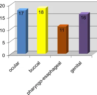
Mucosal involvement was present in all patients and affected most frequently buccal mucosa (n = 18). Fig. 1 shows distribution of mucosal involvement.
Relevant comorbidities were present in 17 patients. Eleven (52.4%) had medical history of cardiovascular dis-eases, namely hypertension and coronary disease, and metabolic disorders such as Diabetes Mellitus and dyslipid-emia were present in 6 patients (28.6%). Other comorbidi-ties identified included neurological diseases (n = 5); auto-immune disorders (n = 5) such psoriasis, myasthenia gravis and rheumatoid arthritis; malignancy (n = 3) namely breast cancer, lymphoma and cerebral tumor; chronic infectious diseases (n = 2); chronic renal failure (n = 2) and depres-sion syndrome (n = 1) – Fig. 2.
Data collection concerning previous treatment with cor-ticosteroids prescribed by general practitioners or in refer-ring hospitals show that this type of drugs was used in 13 patients (61.9%).
Pneumonia was the most prevalent acute complication (42.9%) during hospitalization obliging to ventilator support in 44.4% of cases. Microbiologic evaluation of sputum or bronchial aspirate samples showed MRSA (methicillin-re-sistant Staphylococcus aureus) in 33.3%, Candida albicans
in 11.1% and several Gram-negative species in the rest of the patients. Sepsis occurred in 19% of patients.
In terms of treatment modalities, 16 patients (76.2%) were treated with N-acetylcysteine and 11 patients (52.4%) with intravenous immunoglobulin (IVIG). Plasmapheresis was used in 10 patients (47.6%) as two or three sessions. Survival rate among the group of 5 patients who have not received any of these treatments was lower (40%) as com-pared with the group of 6 patients who have been treated with these three pharmacologic agents (66.7%), although without statistically significant difference (p = 0.567). With regard to long-term sequelae, 4 patients devel-oped long-term cutaneous complications, namely pigmen-tation abnormalities (n = 3), chronic pruritus (n = 2) and ungueal dystrophy (n = 1). Of the 17 patients with ocular involvement, one had symblepharon formation associated with photophobia and xerophthalmia and another one had epiphora and cilia depletion. One patient developed labia majora synechia.
The mean SCORTEN score was 3.2 ± 1.1 (range 2-5). Table 1 summarizes the number of patients and deaths for each SCORTEN level as well as the comparisons between real and predicted mortality rates. There was no statisti-cally significant difference between the overall mortality rate of 47.6% (10 patients) in the population in study and the overall mortality rate of 42.2% (8.9 patients) predicted by SCORTEN (P = 0.142) – Table 2.
Comparison between nonsurvivors and survivors con-cerning age, time interval between first symptoms and re-ferral to burn unit and duration of treatment with the sus-pected drug showed that the former group had a trend to-wards highest age (64.4 ± 23.6 years vs. 47.6 ± 21.7 years;
P = 0.104), longer time interval between first lesions and
referral to the Burns Center (6 ± 5.8 days vs. 4.7 ± 4.2 days;
p = 0.57) and towards wider treatment duration with
the causative drug (11 ± 10.8 days vs. 6.9 ± 10.4 days;
p = 0.45).
DISCUSSION
In Portugal, data concerning treatment of SJS and TEN patients in Burn Units are very scarce and the present retro-spective study corresponds to the largest series of patients admitted in a Burn Center due to these exfoliating diseases
in our country.10-12 Since SJS/TEN are rare conditions, our
sample size is limited but similar to other international
se-ries published in literature.13-15
TEN can affect people of all ages and all ethnicities, but is recognized to be more frequent at extreme ages. In our group of patients the mean age (55.6 years) was equivalent
or slightly higher to other studies,12-14,16 but is important to
state that, in opposite to other centers, admissions in our burn unit are restricted to patients older than 12 years old. We have observed a preponderance of cases in the female
gender, which is also in agreement with literature4,17 and
re-cent studies.13-15
In the majority of patients the final diagnosis was TEN (81%), reflecting the greater severity of patients who are
ARTIGO ORIGINAL
Table 1
- Patient characteristics
Patient
Age (years) Length of burns unit admission (days)
Days
between initial symptoms and referral Days of exposure to the probable offending drug
Probable causative drug
SCOR TEN TBSA (%) Plasma- pheresis IVIG Acetylcysteine Pneumonia Sepsis Outcome 1 79 4 7 ? ? 2 80 Survived 2 81 2 21 20 Paracetamol 5 44 Died 3 37 3 7 2 β-lactam 4 21 + + Survived 4 14 2 5 35 Lamotrigine 2 50 Died 5 63 10 4 4 β-lactam 3 80 Died 6 80 9 15 ? β-lactam 4 93 + + + Survived 7 67 1 2 5 Allopurinol 4 100 + + + Died 8 84 10 7 7 Allopurinol 3 50 + + + Died 9 34 8 1 3 Meropenem 2 17.5 + + + Survived 10 91 9 8 ? ? 4 25 + + + Died 11 50 3 6 16 Cotrimoxazol 5 38 + + + Died 12 64 23 2 ? ? 4 45 + + Survived 13 16 25 2 4 Paracetamol 2 46 + + Survived 14 15 16 6 ? Azitromycin 2 45 + + Survived 15 64 4 1 3 β-lactam 5 24 + + + Died 16 51 14 1 1 Phenytoin 4 35 + + + Survived 17 53 14 6 1 Allopurinol 3 60 + + + Survived 18 44 4 5 2 Diclofenac 2 37 + + + Died 19 86 8 1 7 β-lactam 4 45 + + + + + Died 20 49 18 3 7 Phenytoin 2 61 + + + + Survived 21 45 14 2 30 Allopurinol 2 30 + + + Survived
ARTIGO ORIGINAL
usually admitted in burn centers, being the less severe forms of these spectrum of diseases treated in other hos-pital departments (like Dermatology departments). Com-paring with recent studies, we have a much higher propor-tion of patients with TEN which is also consonant with high TBSA epidermal detachment values (50.5%) observed in our patients. In fact, a review of 61 patients at a Canadian
burn unit16 found 54% of TEN as the final diagnosis, almost
30% lower than in our study.
Most researchers consider TEN as an idiosyncratic drug
reaction in 95% of cases.2 In our study, we found that 18
cases (86%) could be attributed to a single drug based on clinical and chronological criteria, in agreement with litera-ture. Antibiotics, mainly beta-lactams, were the agents most frequently involved in this series, followed by allopurinol, as also noted by two other study groups from Birmingham’s
University Hospital14 and Sydney’s General Hospital.18
However the EuroSCAR study,19 a prospective case-control
study conducted in six countries (Austria, France, Germany, Israel, Italy and Netherlands), has identified allopurinol as the most common cause of SJS and TEN, particularly when administered in high doses.
Admission delay in our burn unit was 5.3 days (range
1-21), almost twice than in other studies,13,14 corresponding
to a very late referral and thus a delayed start of optimal supportive and therapeutic care, even though most of them have been transferred from another hospital department besides the Emergency Room. In addition, suspension of the causative drug was also belated (9.2 days; range 1-35). Together, these two points can reflect a delayed diagnosis due to lack of awareness among physicians for this type of pathologies, attending to their rarity and unspecific initial clinical features, turning educational campaigns of greatest
importance. In literature,7 besides high SCORTEN values,
late transfer to a specialized center as well as delayed iden-tification and withdrawal of the provocative agent have been associated with poor outcomes and higher mortality rates.
It has also been reported that early elimination of the
caus-ative drug may reduce the risk of death by 30% per day.7
Indeed, when analyzing our results concerning time interval between onset of symptoms and referral to our burn unit and duration of treatment with the suspected drug, nonsur-vivors had a trend towards wider time intervals (p = 0.57;
p = 0.45 respectively).
Clinically, mucosal involvement was present in all pa-tients with simultaneous commitment of different mucosal membranes in the majority of cases. In fact, it is well known that mucosal involvement occurs in more than 90% of pa-tients, usually preceding skin lesions in 1 to 3 days, display-ing a special tropism for stratified squamous epithelium and
affecting most often buccal, ocular and genital mucosa.4
Association of typical skin lesions and mucosal erosions are an important clue to early diagnosis, particularly in pres-ence of ocular involvement.
Sepsis and multiorgan failure are the two major causes of death in TEN, the former being responsible for 50% of
deaths in the acute phase of the disease.3 This high
inci-dence of sepsis is related to loss of skin barrier with con-sequent tissue invasion by endogenous flora as well as by exogenous microorganisms, which multiply exponentially in the exudate and necrotic tissues. It is also dependent of the very frequent use of central venous catheters in these patients. Sepsis can also result from pneumonia. Indeed, respiratory mucosa involvement is observed in 27% of pa-tients, with intubation and mechanical ventilation support
in 10 to 20% of cases.20 Increased alveolocapillary
perme-ability with secondary pulmonary edema, polypnea due to pain and aspiration of debris from oropharyngeal and tra-cheobronchial mucosa detachment can lead to atelecta-sias, bronchial pneumonia and, in more severe cases, to ARDS (adult respiratory distress syndrome). In agreement
with these data and with other studies,12-14 infectious
com-plications, like pneumonia and sepsis, were frequent in our group of patients. Pneumonia was a major cause of morbid-Table 2 - Number of patients and comparison between real and predicted mortality rates for each SCORTEN level
SCORTEN of patientsNumber Predicted mortality (No. of deaths) (No of deaths)Real mortality
0 0 1.2% _ 1 0 3.9% _ 2 8 12.2% (1) 25% (2) 3 3 32.4% (1) 66.7% (2) 4 7 62.2% (4.3) 42.9% (3) 5 3 85% (2.6) 100% (3) 6 0 95.1% _ 7 0 98.5% _ Total 21 42.2% (8.9) 47.6% (10)
10
5
15
0
20
genital
pharyngo-esaphageal
buccal
ocular
16
11
18
17
Figure 1 - Distribution of mucosal involvement in the study group.
4 2 6 0 12 10 8 infectious psychiatric auto-immune neurological malignancy renal metabolic cardiovascular
Figure 2 - Comorbidities present in the study group
ity during hospitalization and MRSA appeared as the prin-cipal microorganism cultured, probably related to the high incidence of this pathogen in skin flora.
As we described previously, corticosteroids instituted before admission were either stopped acutely or tapered (n = 13 patients). The role of corticosteroids in TEN remains
controversial. Several retrospective studies21-23 have shown
enhanced mortality (up to 12.5 times), delayed skin lesions healing, prolonged hospital stay and higher infectious rates (up to 5 times) in patients treated with systemic corticoste-roids. In addition, case reports of patients who developed TEN while being on treatment with corticosteroids for other comorbidities have been described, suggesting that corti-costeroids do not prevent development of disease.
Nev-ertheless, van der Meer et al24 and Kardaun et al25 have
suggested that high doses of corticosteroids administered as pulses and in an early phase of disease reduce signifi-cantly death risk. In conclusion, recent data seems to
indi-cate that corticosteroids could be useful in the erytrodermic stage, before dermoepidermal detachment. Administration beyond the initial 48 hours of disease is associated with
higher morbidity and mortality rates.26 Since TEN diagnosis
in non-bullous phase is rare and difficult, early treatment with corticosteroids rarely takes place. According to these data and as all patients presented already epidermal de-tachment at admission in our burn unit, our policy was to suspend corticosteroids.
Plasmapheresis has been used in many centers with promising results as concluded by some retrospective
re-ports, showing survival rates between 77% and 100%.27-29
Its proponents believe that it removes the causative drug or its metabolites and soluble inflammatory mediators from the bloodstream. The opponents argue that it may also elimi-nate anti-inflammatory mediators and, attending to the short half-life of cytokines, it will probably be instituted too late, limiting its value.
Several meta-analyses, retrospective and prospective studies have been published in the last years in order to understand the role of IVIG in the treatment of SJS/TEN patients, with contradictory results concerning morbidity and mortality, probably related to wide variation in treatment protocols and in anti-Fas activity among different batch-es.30-34 Despite these contradictions and although controlled
and randomized studies have not been published yet, the majority seems to indicate a benefit in mortality rates with early administration of high IVIG doses (1g/Kg/day, during 4 days, in continuous infusion).
Other immunomodulatory drugs like N-acetylcysteine, cyclosporin A, pentoxifylline, granulocyte colony-stimulating factor, anti-TNF-α antibodies, cyclophosphamide and ulina-statin are being used in multiple units, in spite of lack of clear evidence of a beneficial role in the management of
these patients.5
Since none of the mentioned specific treatments have proven to be completely effective or even superior to sup-portive care, immediate withdrawal of the causative drug and early transfer to a Burn centre in order to prevent
ARTIGO ORIGINAL tion, hydroelectrolytic changes and to promote rapid
reepi-dermization of denuded areas are the mainstream attitudes with confirmed benefits and impact in SJS/TEN morbidity and mortality. Burn centers are therefore recognized to be best suited to care SJS/TEN patients, attending to their ex-pertise in treating large denuded skin areas in critically ill
patients.35
SCORTEN is a scoring system developed by Bastuji-Garin et al which is used to stratify gravity and estimate
mortality.8 Its application in TEN patients treated in burn
units has been called into question by some authors who argue that SCORTEN overestimates mortality, since it was initially endorsed from a group with less severe forms of
the disease.9 In our sample we found a good correlation
between predicted and actual mortality which supports the accuracy of SCORTEN in predicting SJS/TEN mortality, as
already established in other studies.12,14,16
The mortality rate in our group of patients was high
(47.6%), but still consistent with literature.3,12,36 Several
fea-tures present in our sample may contribute to this high mor-tality rate namely higher proportion of patients with TEN, high mean TBSA involvement and mean age, high percent-age of comorbidities and late transfer of the patient to our unit as well as late withdrawal of the culprit drug.
Our study was designed as a retrospective analysis, with all the limitations inherent to this type of study, namely
in data collection. Besides, it was not possible to compare the real impact of specific therapeutic modalities in survival rates and to prove or disprove newer treatment protocols for TEN.
Since it is a rare disease, sample size was small which also restricts statistical data analyses and conclusions in some way.
CONCLUSION
Since SJS/TEN’s pathophysiology is still not completely clarified, a specific and efficient treatment hasn’t been yet identified. Nowadays, early diagnosis, immediate referral to a burn unit in order to provide a specific and intensive treat-ment and suspension of the causative drug are the pillars of treatment of these exfoliating disorders. Therefore, edu-cational campaigns assume greatest importance in order to alert physicians to these diseases.
Finally, SCORTEN seems to be an accurate method to predict mortality rate in SJS/TEN patients.
CONFLICT OF INTERESTS
None stated.
FUNDING SOURCES
None stated
REFERENCES
1. Lyell A. Toxic epidermal necrolysis: an eruption resembling scalding of the skin. Br J Dermatol. 1956;68:355-61.
2. Mockenhaupt M, Viboud C, Dunant A, Naldi L, Halevy S, et al. Stevens-Johnson Syndrome and Toxic Epidermal Necrolysis: assessment of medications risks with emphasis on recently marketed drugs. The Euro-SCAR study. J Invest Dermatol. 2008;128:35-44.
3. Abood GJ, Nickoloff BJ, Gamelli RL. Treatment strategies in toxic epidermal necrolysis syndrome: where are we at? J Burn Care Res. 2008;29:269-76.
4. Borchers AT, Lee JL, Naguwa SM, Cheema GS, Gershwin ME. Stevens Johnson syndrome and toxic epidermal necrolysis. Autoimmunity Rev. 2008;7;598-605.
5. Paquet P, Piérard GE. New insights in Toxic Epidermal Necrolysis (Ly-ell’s Syndrome). Clinical considerations, pathobiology and targeted treatments revisited. Drug Saf. 2010;33:189-212.
6. Bastuji-Garin S, Rzany B, Stern RS, Shear NH, Naldi L, Roujeau JC. Clinical classification of cases of toxic epidermal necrolysis, Ste-vens-Johnson syndrome, and erythema multiforme. Arch Dermatol. 1993;129:92-6.
7. Mukasa Y, Craven N. Management of toxic epidermal necrolysis and related syndromes. Postgrad Med J. 2008;84:60-5.
8. Bastuji-Garin S, Fouchard N, Bertocchi M, Roujeau JC, Revuz J, Wolk-enstein P. SCORTEN: a severity of illness score for toxic epidermal necrolysis. J Invest Dermatol. 2000;115:149-53.
9. Imahara SD, Holmes JH 4th, Heimbach DM, Engrav LE, Honari S, Klein MB, et al. SCORTEN overestimates mortality in the setting of a stan-dardized treatment protocol. J Burn Care Res. 2006;27:270-5. 10. Cabral L, Diogo C, Riobom F, Teles L, Cruzeiro C. Necrolise epidermica
toxica (Sindrome de Lyell). Uma Patologia para as Unidades de Quei-mados.Acta Med Port. 2004;17:129-40.
11. Cabral L, Riobom F, Diogo C, Teles L, Cruzeiro C. Toxic Epidermal Ne-crolysis – Lyell’s Syndrome. Ann Burns Fire Disasters. 2004; XVII. 12. Moniz P, Casal D, Mavioso C, Castro JV, Almeida MA. Síndroma de
Stevens-Johnson e necrolise tóxica epidérmica: um estudo retrospecti-vo de 15 anos. Acta Med Port. 2011;24:59-70.
13. Gerdts B, Vloemans AF, Kreis RW. Toxic epidermal necrolysis: 15 years’ experience in a Dutch burns center. J Eur Acad Dermatol Venereol. 2007;21:781-8.
14. Rajaratnam R, Mann C, Balasubramaniam P, Marsden JR, Taibjee SM, Shah F, et al. Toxic epidermal necrolysis: retrospective analysis of 21 consecutive cases managed at a tertiary center. CED. 2010;35:853-62. 15. Ugburo AO, Temiye EO, Ilombu A. A 12-year retrospective study of non-burn skin loss (non-burn-like syndromes) at a tertiary non-burns unit in a develop-ing country. Burns. 2008;34:637-43.
16. Cartotto R, Mayich M, Nickerson D, Gomez M. SCORTEN Accurately Predicts Mortality Among Toxic Epidermal Necrolysis Patients Treated in a Burn Center. J Burn Care Res. 2008;29:141-6.
17. Roujeau JC, Guillaume JC, Fabre JP, Penso D, Fléchet ML, Girre JP. Toxic epidermal necrolysis (Lyell syndrome), Incidence and drug aetiol-ogy in France, 1981-1985. Arch Dermatol. 1990;126:103-4.
18. Wong KC, Kennedy PJ, Lee S. Clinical manifestations and outcomes in 17 cases of Stevens-Johnson and toxic epidermal necrolysis. Aust J Dermatol. 2006;7:397-8.
19. Halevy S, Ghislain PD, Mockenhaupt M, Fagot JP, Bouwes Bavinck JN, Sidoroff A, et al. Allopurinol is the most common cause of Stevens-John-son syndrome and toxic epidermal necrolysis in Europe and Israel. J Am Acad Dematol. 2000;115:149-53.
20. Lebargy F, Wolkenstein P, Gisselbrecht M, Lange F, Fleury-Feith J, Del-claux C, et al. Pulmonary complications in toxic epidermal necrolysis; a prospective clinical study. Intensive Care Med. 1997;23:1237-44. 21. Engelhardt SL, Schurr MJ, Helgerson RB. Toxic epidermal necrolysis:
an analysis of referral patterns and steroid usage. J Burn Care Rehabil. 1997;18:520-4.
22. Halebian PH, Corder VJ, Madden MR, Finklestein JL, Shires GT. Im-proved burn center survival of patients with toxic epidermal necrolysis managed without corticosteroids. Ann Surg. 1986;204:503-12. 23. Kelemen JJ 3rd, Cioffi WG, McManus WF, Mason AD Jr, Pruitt BA Jr.
Burn center care for patients with toxic epidermal necrolysis. J Am Coll Surg. 1995;180:273-8.
24. van der Meer JB, Schuttelaar ML, Toth GG, Kardaun SH, Beerthuizen G, de Jong MC, et al. Successful dexamethasone pulse therapy in a toxic epidermal necrolysis (TEN) patient featuring recurrent TEN to ox-azepam. Clin Exp Dermatol. 2001;26:654-6.
25. Kardaun S, Jonkman M. Dexamethasone pulse therapy for Steven-Johnson syndrome/toxic epidermal necrolysis. Acta Dermatol Venereol. 2007;87:144-8.
26. Herndon D. Toxic epidermal necrolysis; a systemic and dermatologic disorder best treated with standard treatment protocols in burn intensive care units without the prolonged use of corticosteroids. J Am Coll Surg. 1995;180:340-2.
27. Chaidemenos GC, Chrysomallis F, Sombolos K, Mourellou O, Ioannides D, Papakonstantinou M. Plasmapheresis in toxic epidermal necrolysis. Int J Dermatol. 1997;36:218-21.
28. Egan CA, Grant WJ, Morris SE, Saffle JR, Zone JJ. Plasmapheresis as an adjunct treatment in toxic epidermal necrolysis. J Am Acad Dermatol. 1999;40:458-61.
29. Lissia M, Figus A, Rubino C. Intravenous immunoglobulins and plasma-pheresis combined treatments in patients with severe toxic epidermal necrolysis: preliminary report. Br J Plast Surg. 2005;58:504-10. 30. Metry DW, Jung P, Levy ML. Use of intravenous immunoglobulin in
chil-dren with Stevens-Johnson syndrome and toxic epidermal necrolysis: seven cases and review of literature. Pediatrics. 2003;112:1430-6. 31. Prins C, Kerdel FA, Padilla RS, Hunziker T, Chimenti S, Viard I, et al.
Treatment of toxic epidermal necrolysis with high-dose of intravenous
immunoglobulins: multicenter retrospective analysis of 48 consecutive cases. Arch Dermatol. 2003;139:26-32.
32. Trent JT, Kirsner RS, Romanelli P, Kerdel FA. Analysis of intravenous immunoglobulin for the treatment of toxic epidermal necrolysis us-ing SCORTEN: the University of Miami experience. Arch Dermatol. 2003;139:39-43.
33. Bachot N, Revuz J, Roujeau JC. Intravenous immunoglobulin treatment for Stevens-Johnson syndrome and toxic epidermal necrolysis. Arch Dermatol. 2003;139:33-6.
34. Brown KM, Silver GM, Halerz M, Walaszek P, Sandroni A, Gamelli RL. Toxic epidermal necrolysis: does immunoglobulin make a difference? J Burn Care Rehabil. 2004;25:81-8.
35. Palmieri TL, Greenhalgh DG, Saffle JR, Spence RJ, Peck MD, Jeng JC, et al. A multicenter review of toxic epidermal necrolysis treated in U.S. burn centers at the end of the twentieth century. J Burn Care Rehabil. 2002;23:87-96.
36. Lissia M, Mulas P, Bulla A, Rubino C. Toxic epidermal necrolysis (Lyell’s disease). Burns. 2010;36:152-63.