Universidade Nova de Lisboa
Faculdade de Ciências e Tecnologia
Departamento de Conservação e Restauro
Spectroscopy Studies on Conservation Issues
in Modern and Contemporary Paintings
Joana Andreia Lameiras Domingues
Dissertation presented to the Faculty of Sciences and Technology of New University of Lisbon in fulfilment of the requirements for the
Master’s degree in Conservation and Restoration
Specialization in easel painting
Supervisor:Prof. Dr. Costanza Miliani (CNR-ISTM)
Co-Supervisor:Dr. Francesca Rosi (SMAArt)
Acknowledgements
I am deeply thankful to my supervisor, Prof. Costanza Miliani, whose encouragement and guidance had been essential for the development of this thesis.
It was an honour for me to do my training stage with the SMAArt working group, an opportunity given by Prof. Bruno Brunetti, to whom I owe my deepest gratitude.
I would also like to show my sincere gratitude to Francesca and Brenda for all the help on the bench and portable equipments, and also those colleagues from SMAArt and CNR-ISTM that supported me from the beginning.
Finally, I would like to thank the kind collaboration from Vivi Pouli, (FORTH-IESL) and Grazia De Cesare (ISCR).
Sumário
A pintura de arte contemporânea é um dos grandes desafios da conservação-restauro de bens culturais. Algumas destas pinturas contêm repintes de composição química semelhante à das camadas pictóricas originais, colocando em causa a reversibilidade dos mesmos.
Neste trabalho a microscopia óptica e as espectroscopias de FTIR e Raman foram utilizadas para avaliar a eficácia e o dano associados às limpezas química e a laser para a remoção de repintes, tendo sido preparadas maquetes representativas com tintas comerciais. A limpeza a laser advém de um estudo interdisciplinar que pretende avaliar as limitações do método utilizando parâmetros de limpeza bastante agressivos.
O uso combinado das espectroscopias de FTIR e Raman permitiu identificar os compostos das tintas modernas e, contemporaneamente, controlar o seu comportamento durante a limpeza. A microscopia óptica permitiu, por outro lado, uma avaliação da morfologia da superfície. Equipamentos portáteis do MOLAB foram também utilizados devido à importância das análises in situ.
A remoção dos repintes revelou-se difícil com a limpeza química, enquanto com a limpeza a laser foi mais eficaz, no entanto, provocando degradação de ligante, com formação de carbono, e alteração do branco de titânio.
O protocolo espectroscópico proposto foi considerado útil para o controlo de diferentes métodos de limpeza em pinturas de arte contemporânea.
Abstract
Modern and contemporary paintings are one of today’s grand challenges in conservation of cultural heritage. Particularly, these paintings have often been retouched using materials rather similar to originals, thus, compromising the reversibility of the overpainting.
In this work, FTIR and Raman spectroscopies, assisted by optical microscopy, were used to evaluate the effectiveness and harmfulness of chemical and laser cleaning methods for the removal of overpaints. Representative mock-ups prepared with commercial paint formulations were used. The laser cleaning experiment was part of an interdisciplinary study which aims the evaluation of method’s limitations by using the most aggressive cleaning parameters.
The combined use of FTIR and Raman spectroscopies could identify constituent materials of modern paints, controlling their behaviour under cleaning, while optical microscopy allowed the evaluation on surface morphology. In addition, equivalent portable equipments from MOLAB were covered as a preparation for in situanalysis.
Several problems in the selective removal of overpaints were found with chemical cleaning. The laser cleaning showed better efficiency in removing them, although, some alterations occurred upon laser irradiation, for instance, binder degradation with carbon formation and titanium white alteration.
The proposed spectroscopic protocol was considered useful for controlling different cleaning methods in modern and contemporary paintings.
List of Abbreviations
Er:YAG – Erbium-doped: yttrium aluminium garnet FTIR – Fourier Transformed Infrared spectroscopy
IESL-FORTH – Foundation for Research and Technology-Hellas, Institute of Electronic Structure & Laser
ISCR – Istituto Superiore per la Conservazione ed il Restauro MOLAB – Mobile laboratory
Nd:YAG – Neodymium-doped: yttrium aluminium garnet OM – Optical Microscopy
PEA-MMA – Poly(ethyl acrylate-methyl methacrylate) PVA – Poly(vinyl acetate)
SMAArt – Scientific Methodologies Applied to Archaeology and Art Tg– Glass Transition Temperature
CONTENTS
1. Introduction ... 8
1.1. Modern paints... 8
1.2. Conservation issues on modern and contemporary paintings ... 11
1.3. Fundamentals of laser cleaning in conservation ... 11
1.4. Thesis aim ... 13
1.5. Optical and spectroscopic tools for cleaning control ... 13
2. Results and discussion ... 15
2.1. Mock-ups. ... 15
2.2. Chemical cleaning control ... 16
2.3. Laser cleaning control ... 19
3. Conclusions... 28
4. References... 30
APPENDICES
Appendix 1 - Materials and methods... A1
Appendix 2 – Photocatalytic properties of titanium dioxide... A9
Appendix 3 – General results on laser cleaning control... A10
LIST OF FIGURES
Figure 1 –poly(ethyl acrylate) and poly(methyl methacrylate) ... 8
Figure 3 –Poly vinyl acetate chemical structure [4]. ... 9
Figure 2 –poly styrene chemical structure [4]... 9
Figure 4 –Alkyd structure containing phthalic anhydride, glycerol and linoleic acid [4]. ... 10
Figure 5 –Cellulose nitrate structure [6]. ... 11
Figure 6 –Mock-up #1 after laser cleaning. Cotton canvas on stretcher with 29 x 25 cm2. ... 15
Figure 7 –OM images obtained from diverse points where the chemical cleaning method was performed. ... 16
Figure 8 –Reflection micro-FTIR spectra of ground layer and blue overpaint (n8 Liquitex®), for comparison with the one obtained from the area where cleaning was performed with acetone. ... 17
Figure 9 –Reflection micro-FTIR spectra of white overpaint (n5 Fly Color®), ... 18
Figure 10 –Reflection UV-Vis-NIR spectra obtained with the fibre-optics portable equipment, ... 19
Figure 11 –Optical microscopy images obtained from diverse points where the laser cleaning was tested... 20
Figure 13 –Fibre-optics video microscopy images obtained from the same points presented on figure 11. ... 21
Figure 12 –Fibre-optics video microscopy using an objective of 50x that works in contact mode with the surface of the mock-up. ... 21
Figure 14 –Reflection micro-FTIR spectrum of a cleaned area in n6 paint on mock-up#1 using the laser Nd:YAG 1064 nm and F = 3,02 J·cm-2, 5 pulses, compared with the white ground and canvas spectra. ... 22
Figure 15 –Reflection micro-FTIR spectrum of a cleaned area in n4 paint on mock-up #1 using laser Nd:YAG 355 nm (tp= 10ns) and F = 2,06 J·cm-2, 10 pulses, compared with the n4 overpaint and white ground spectra. .. 22
Figure 16 –Reflection micro-FTIR spectrum of a cleaned area in n3 paint on mock-up#4 using the excimer KrF laser at λ = 248 nm (tp= 30 ns, 3.06 J·cm-2, 30 pulses) compared with the n3 overpaint and white ground spectra. ... 23
Figure 17 –Reflection micro-FTIR spectrum of a cleaned area in n11 paint using the excimer KrF laser at λ = 248 nm (tp= 30 ns, 3.06 J·cm-2, 30 pulses) compared with standard spectra of n11 overpaint and white ground. ... 24
Figure 18 –Fibre-optics FTIR spectroscopy portable equipment. Photo courtesy of SMAArt (Perugia). ... 24
Figure 19 –Reflection FTIR spectra, showing examples of aggressive and incomplete cleaning, by comparison with n3 overpaint, white ground and canvas standard spectra. ... 25
Figure 20 –Ablation in paint n4 using the Nd:YAG 355 nm laser, tp= 10 ns, F = 2.06 J·cm-2and 10 pulses, a) the fibre-optics portable video microscopy image from cleaned area, b) and c) optical microscopy images showing the micro black particles, d) micro-Raman spectrum (=532 nm) of this area with carbon assignment bands. ... 25
Figure 22 –Micro-Raman spectra (=532 nm) of phthalocyanine paint n11, performed in the remaining blue spots on the ablation areas, which showed notable fluorescence around 667 nm and 617 nm. ... 26
Figure 21 –Raman spectra (=532 nm) of a cleaned area on n7 paint and the standard for white ground (rutile) for comparison (normalized [0,1]) [41]. ... 26
Figure 23 –Fibre-optics Raman spectroscopy portable equipment. Photo courtesy of SMAArt (Perugia) ... 27
LIST OF TABLES
Table 1 –General information about the three most important synthetic resin classes [3,4]... 10
Table 2 –Commercial paint formulations covered in the study. ... 15
Table 3 –General results for the cleaning experiment on mock-up #6 using solvents and chemical systems. The n6 is the considered original layer, except in its specific row, where the original layer is considered the canvas. . 19
1. Introduction
Up until the end of the nineteenth century there were mainly two typologies of easel paintings, oil and egg tempera, the first being, without doubt, the most important and diffused since Jan van Eyck (fifteenth century) [1].
However, at the beginning of the twentieth century the first synthetic materials started to emerge, as a result from the exponential growth in the paint and coatings industry. Hence, the first synthetic polymers appeared and started to be applied in paints as binding media. Not much time was needed for these materials to catch modern artists’ attention. Consequently, nowadays there is a vast collection of synthetic resin based paintings, which offers new problems for painting conservation [2].
1.1. Modern paints
A synthetic resin can be present in paint formulations as a solution, that is, it is dissolved in an organic solvent, or as emulsion, which means that it is dispersed in water due to surfactants and additives [3]. Moreover, there are three important classes of synthetic resins to consider in the study of modern and contemporary paintings, namely acrylic, polyvinyl acetate, and alkyd.
Acrylic resinswere the most diffused paint material in the artists’ paint market. The wide variety of synthetic acrylic resins formulations is useful not only as binding medium, but also as varnishes, additives (plasticizers) which, for example, can be used in traditional painting and as restoration materials [4]. This synthetic resin appeared first in 1936 as acrylic resin for the coatings industry, but soon in 1947 the first line of artists’ acrylic paints in turpentine solution was introduced by the paint maker Leonard Bocour with Sam Golden, and was commercially known by Magna®. However, it was finally in 1954 that an acrylic emulsion line for artist’s use was developed, called Liquitex®, by Henry Levison from Permanent Pigments Co. After this, many other lines of artists’ acrylic paints were developed by paint makers, mainly after 1964 [3].
The term “acrylic” covers a wide range of high molecular weight polymers based on acrylates and methacrylates. The only acrylic homopolymers that have the right properties to be used as acrylic solutions are the polybutyl methacrylates, namely the isomer pnBMA, which is used in acrylic solution paints, and piBMA, more appropriate for acrylic varnishes, because of their Tg’s [3]. The first was commercial available by the line Magna® and was used by important twentieth century artists, like Roy Lichenstein, Morris Louis and Kenneth Noland, although it was discontinued due the success of emulsion paints, since these offered better painting properties [3,4]. The most common synthetic resin formulations which compose emulsion paints are the copolymers composed by poly(ethyl acrylate) and poly(methyl methacrylate), which structures are showed in figure 1.
pEA pMMA
The first acrylic emulsion grade of paints was Liquitex®, as stated before, however, other posterior grades from other producers are important to consider, as well. Examples of such are Rohm and Haas Primal® (22, 34, 234, AC-634) and Rohm Plextol® B-500 [4].
With the aim of decreasing the overall cost of synthetic acrylic resin based paint formulations, styrene monomers are often added (structure in figure 2), shifting the final properties of the painting material, and, as a result, these are often present in
cost household paints [3,4]. Nevertheless, it is commonly found that artists experiment these low-cost paints either by philosophy or economic reasons. Thus, the eventual analysis of these materials is important to consider in modern and contemporary paintings. An example of a commercial paint containing this compound is the Brera® paint from Maimeri, which has a terpolymer p(n BMA-2EHA-styrene) [3].
Polyvinyl acetate resins. The group of vinyl resins include a wide range of polymeric materials, however, in the field of plastic and coatings’ industries is commonly accepted to restrict this group only to vinyl chloride polymers, vinyl acetate polymers, their copolymers, modified polymers and derivate products. The most important is the poly(vinyl acetate) resin (PVA) whose structure is shown in figure 3. The first commercial developments of PVA started in the mid 1920s as an adhesive [4]. The PVA paints are mainly waterborne emulsions. On the other hand, it usually requires the addition of plasticizers since PVA homopolymers are too brittle to form a continuous film [3].
Figure 3 –Poly vinyl acetate chemical structure [4].
The most common plasticizers, sometimes in quantities up to 20% (wt%), are the phthalates, for example, the dibutyl phthalate (DBP). These external plasticizers tend to migrate to the paint surface, and therefore the paint becomes brittle and the surface more tacky and likely to have dirt pick-up [3]. In 1928 a copolymerization process was tried involving vinyl acetate and vinyl chloride, producing an emulsion paint with better properties than pure polymers or alone monomers. In the mid 1940s, vinyl acetate emulsions were introduced by DuPont Co. and others [4]. By the 1960s a copolymerization of PVA with softer monomers was accomplished, mainly with the highly branched C9 and C10 vinyl esters, commercially known as vinyl versatates, or VeoVa, giving a PVA/VeoVa copolymer with improved hydrophobicity and UV resistance. However, almost every commercial PVA artist paint formulations were discontinued, except, for example, the artist grade called Flashe®, marketed by Lefranc & Bourgeois [3,5].
The PVA emulsion paints, comparing to the acrylic’s, are generally considered to be slightly inferior in what respects the toughness, binding power, and resistance to weathering. On the other hand, they are less expensive and suitable for interior use [3].
Alkyd resins are oil-modified polyesters, resulting from a reaction of a polybasic acid (or polyol), a polyhydric alcohol (or poly acid) and a source of fatty acids, giving the term “alkyd” (“al” from alcohol
Figure 2 –poly styrene chemical
and “kyd” from acid). The combined product is a very flexible polymer which was introduced in the early 1930s. It had magnitude in the paint technology because of its reduced drying time and superior durability and hardness when comparing with other sources of oils. Still, alkyd resins were more famous in the household paint technology, since only Winsor & Newton produced a full colour range for artists’ use, called Griffin®, introduced in 1970. In fact, alkyd paints, as household formulations, were used by renamed artists such as Pablo Picasso and Jackson Pollock [3,4]. The most important polyols used in alkyd resins are glycerol (propan-1,2,3-triol) and pentaerythritol (2,2-bis(hydroxymethyl) 1,3-propanediol).
As for the polybasic acids the most common is the dibasic acid phthalic anhydride (1,2-benzenedicarboxylic acid). The oil feature appears by the addition of a source of monobasic fatty acids, being the most common a drying oil, namely linseed and soy oils [3]. Example of an alkyd structure containing phthalic anhydride, glycerol and linoleic acid is shown in figure 4.
Some modifications are made to alkyd paints to improve certain properties. The most common are the addition of styrene, vinyl toluene, isocyanates, acrylic, epoxy, or silicone compounds [3]. Below a table with a summary on the three most important synthetic resin paint formulations is presented.
Table 1 –General information about the three most important synthetic resin classes [3,4].
Synthetic resin class
Chemical composition
Date of first appearance
Name of grade/
manufacturer Artists that used it
Acrylic
pnBMA
(solution) 1947
Magna®/ Leonard Bocour and Sam Golden
Roy Lichenstein Morris Louis Kenneth Noland
p(EA-MMA)
(emulsion) 1954
Liquitex®/ Henry Levison (Permanent Pigments Co.)
Helen Frankenthaler David Hockney Robert Motherwell Andy Warhol PVA PVA (emulsion) 1954
Flashe®/ Lefranc & Bourgeois
Sydney Nolan Kenneth Noland Bridget Riley PVA/VeoVa
(emulsion) 1966 VeoVa®/ Shell Chemicals
-Alkyd Polyol/polybasic
acid/drying oil 1970
Griffin®/ Winsor & Newton Gillian Ayres Peter Blake Patrick Caulfield Sydney Nolan Francis Picabia Pablo Picasso Jackson Pollock Pierre Soulages Frank Stella
Nitrocellulose. Other synthetic resins are also important to consider since they are included in some paint formulations that may be present in artworks, mainly in household paints. An example is nitrocellulose, which is the term applied to paints and lacquers that contain mixtures of cellulose nitrate polymer (chemical structure in figure 5) with a second resin (to improve gloss, adhesion and hardness) and plasticizers. This kind of paint only became important in the paint market after the 1920s, being
formulated as solution paint, and usually dissolved in esters, alcohols, ketones, or glycol ethers. Normally, alkyd resins are the second resin, mainly after the 1940s. As for the plasticizers, the phthalates are the most recently employed (e.g. dibutyl phthalate, dioctyl phthalate). After the 1950s, when alkyd paints started to dominate the household paint market, the nitrocellulose paints started to be sold as low-cost sprays. It was in this
form that Richard Hamilton used it, and it is documented that Pollock used the solution form in his drip paintings [3].
1.2. Conservation issues on modern and contemporary paintings
Modern and contemporary paints present new concerns in painting conservation, since they have some different properties from the traditional oil or egg tempera painting. In general, synthetic resin based paints, mainly the acrylic emulsions, are very sensible to organic solvents. This is a problem for the cleaning of artworks which contain such materials without disturbing the surface texture, colour or gloss. But, not only organic solvents cause damage to synthetic resin surfaces, also water or water-based cleaning systems can damage the paint surface, mainly because of swelling. Other main problem is the affinity for dirt pick-up when environmental conditions change, which can also be responsible for other problematic situations, for example, the irreversible mark of fingerprints and the attachment of exterior materials when they are in prolonged contact with the surface [7].
One of the most concerning problems in restoration of modern and contemporary paintings is the difficulty on the selective removal of the overpaints that are quite similar in composition to the original layers. Furthermore, it is not said that traditional restoration approaches offer a harmfulness treatment. As a matter of fact, the chemical or mechanical techniques, apart from being difficult to control, in many cases damage pigments or binders in the cleaning process. Adding to this, mechanical methods can damage the painting texture and chemicals can penetrate into the inner paint layers, ground and support, causing alterations on them. On the other hand, the actual laser cleaning method may offer a viable solution since it permits a layer by layer micro-removal [8-11]. Some experiments for the application of this method on modern and contemporary paintings have been recently tried [12-14]. However, it is still a method that has to be fine-tuned and far away to be used conventionally, due to many reasons, among which, the insufficient knowledge on laser interaction mechanisms, method expensiveness, and, the few developed diagnostic devices to provide qualitative and quantitative analysis during the laser cleaning intervention [10,15,16].
1.3. Fundamentals of laser cleaning in conservation
The emitted light from a laser is due to the energy loss of photons that are provided at one specific wavelength and one direction, being equal in this wavelength and direction. Thus, it emits a monochromatic light. For this to succeed, the majority of atoms, ions or molecules must be in the excited energy state rather than in the low-energy state (population inversion), and therefore a strong source of excitation is needed to maintain this situation, such as gaseous active materials (e.g. mixtures of noble/halogen gases in the excimer lasers) or solid active materials (e.g. the Nd+3 in the Yttrium Aluminium Garnet matrix) [17]. Usually, the laser ablation processes in conservation of cultural heritage aim to achieve a selective removal of material layers and, for that, conservators in general use a pulsed laser. This means that light is delivered in short energy discharges, as opposed to a Figure 5 –Cellulose nitrate
continuous wave laser. This pulsed operation allows more control, since each pulse consists of a measurable and repeatable amount of energy, which can be applied in cases with different materials and aggregation states [8,16,18,19].
Laser cleaning method is relatively recent in the area of conservation of cultural heritage, and, thus, in each case-study the project must include collaboration between conservators, conservation scientists and scientists well familiar with the physics of laser systems [18]. From 1993, the IESL-FORTH, expertise in the laser technology applied to cultural heritage, has been conducting research on laser developments for application in the cleaning of artworks. These applications comprise the laser cleaning, diagnostic analyses, imaging and authentication techniques. In 1995, for the first time a workshop on lasers applied to conservation was organized, named LACONA (Lasers in the Conservation of Artworks), which continues nowadays as an international biannual conference. In recognition to that, conservators became more familiar to laser technology and several research groups have been formed to develop its application in artworks, as is the example of the European research project “advanced workstation for controlled laser cleaning of artworks” [8,9,20].
Laser cleaning of paintings is very controversial, mainly because many studies are still being developed on the short and long term irradiation effects on pigments and binding media, which can be thermal, photochemical or photomechanical [9,21-23]. Thermal effects depend on the thermal diffusivity parameters of the material, the laser system’s pulse length (tp) and wavelength. Photochemical alterations are expected to be the main source of cleaning procedure side effects, mainly because of the radicals and ions produced during the irradiation, which can react and form photo-oxidation products. As for the photomechanical effects, it has been demonstrated the development of stress waves, which can be significant, especially when high laser fluences are employed [24]. There are mainly three kinds of laser systems that are currently being applied for the conservation of cultural heritage: excimer, Nd:YAG and Er:YAG lasers.
Excimer lasers (short name for excited dimmer) are based on rare gas-halide mixtures, emitting mainly in the UV region: ArF = 193 nm; KrF = 248 nm; XeCl = 308 nm; XeF = 351 nm. These laser systems are useful for treating paintings since the materials traditionally employed in paintings (varnishes and overpaints) absorb strongly in the UV, and therefore cause minimal light penetration to inner layers, although it is a more energetic regime. Because this effect depends on the nature and specific properties of the material substrates, it requires the optimization of laser parameters (energy density (F), pulse length (tp), number of pulses) to the particular needs of the application [9,14,18,19,24,25].
Nd:YAG lasershave been studied since the 1970s, firstly for the cleaning of stone surfaces. It is a neodymium-doped: yttrium aluminium garnet (Nd: Y3Al5O12) emitting generally in the near infrared region (NIR) at λ = 1064 nm (ω). However, it can also emit other radiations by multiplying the fundamental frequency: λ = 532 nm (2ω); λ = 355 nm (3ω); λ = 266 nm (4ω); and λ = 213 nm (5ω). It has been explored, only recently, in what concerns its application on the cleaning of paintings, because it offers better results in breaking bonds of inorganic materials. However, it must be carefully controlled because of its deep irradiation penetration [9,14,19,26].
cases where the materials are not rich in OH bonds, hydroxylated liquids can be added to control the radiation penetration and limit the heat effect [9,27].
An important aspect is the ablation threshold, the point at which absorbed laser energy is sufficient to break the bonds between molecules of a material. Hence, the careful determination of the laser fluence values at which substrate damage threshold occurs, is important for a safe cleaning [10].
1.4. Thesis aim
For good practice in the conservation of modern and contemporary artworks the characterisation of constituting materials is necessary in order to apply correct methodologies that can contribute to the painting’s longevity. Apart from such, information concerning the material composition can give important information to art historians, for instance, on artist’s creative process or about the originality in employing new materials not yet available in artists’ market. Even if today’s ethical values and international recommendations defend a much more preservation/conservation practice than a curative one, some critical cases must be re-thought, e.g. a monochromatic painting with posterior overpaints which interfere with the original colour homogeneity intended by the artist, or a painting in direct contact with storage materials (e.g. polyethylene) that can easily attach to the painted surface. In both cases the removal of non original materials may be considered. However, taking into account the difficulty in removing this kind of layers in modern and contemporary paintings, a study has necessarily to be overcome by scientific investigative assistance.
Considering the statements above, the aim of this thesis was to apply diverse spectroscopic tools for the qualitative analysis of the elemental and chemical composition of painted surfaces, in order to evaluate and control different chemical cleanings, whilst, contemporarily assessing the laser cleaning of modern and contemporary paintings. Since the cleaning of this kind of artworks is still a very delicate subject considered border-line between an efficient and a too evasive cleaning, this thesis aims to offer a spectroscopic protocol that permits a liable evaluation on the effectiveness and harmfulness of the cleaning method applied.
One of the aims of this work was also to evaluate the results equivalence between portable equipments from MOLAB1and the ones obtained from bench equipments. This is important since the latter are more adequate for mock-ups analysis in laboratory, while portable equipments are important for in situanalysis in case studies.
1.5. Optical and spectroscopic tools for cleaning control
The spectroscopic tools that were considered relevant were FTIR and Raman spectroscopies. Other efficient analytic techniques have been proposed by authors for the characterisation of modern paints [3,7], e.g., Pyrolysis-Gas Chromatography-Mass Spectrometry (Py-GC-MS), Pyrolysis-Gas Chromatography (Py-GC), and Direct Temperature-Resolved Mass Spectrometry (DTMS), which are destructive techniques and therefore not appropriate for controlling cleaning procedures. FTIR and Raman spectroscopies are not only non-invasive techniques but also suitable to be applied in situ, due to the fibre-optics portable equipments.
- FTIR spectroscopy.FTIR spectroscopy is a technique that allows the identification of molecular bonds in a material by their resonance frequencies. The group frequency region (or functional group)
1
exists from 4000 to 1300 cm-1, where there is information about the chemical classes present in the sample, while from 1300 to 500 cm-1, is the called “fingerprint region”. Even if more difficult to interpret, it contains specific bands for each compound, which can be matched with standards in databases. For this reason, FTIR analytical method is ideal for painting medium characterisation. However, when dealing with the characterisation of modern artists’ paints, the interpretation becomes more complex, because relevant library of known standards are still being built. Besides that, paint formulations have different components (binder, pigment, extender, additives) and sometimes one particular element may dominate the entire FTIR spectrum, masking the other components characteristic bands [3].
Of relevance in cultural heritage analyses is the reflection FTIR spectroscopy, since it permits a non contact analysis and no sample preparation. However, each spectrum obtained depends on the area analysed morphology and its optical properties, giving large distortions in the spectra, both in band shape and position [4,10,28-33]. There are three modes of external reflection measurements in spectroscopy: transmission-reflection (or transflection), specular reflection and diffuse reflection. Reflection FTIR spectroscopy generally collects both specular and diffuse reflection with a ratio that depends in majority on the surface roughness [4,34]. Specular reflection is observed when the material has a very smooth and flat surface which reflects the incident irradiation. Diffuse reflection, on the contrary, results from materials with rather rough surfaces, which scatter irradiation over a wide range of angles [4].
Specular reflection is based on Fresnel’s equations, depending in both absorption coefficient (k) and refractive index (n) [34]. Fresnel reflection gives quite distorted spectra, depending on the band strength. One example is the reststrahleneffect, often seen in minerals, which means a reflectance maximum occurring for those fundamental bands that show k >> n corresponding to total inverted bands shape. For weaker bands, the dispersion of n origins bands in a first derivative shape of a normal absorption or reflection band [4,34].
Diffuse reflection is governed by the Kubelka–Munk model, depending on the parameters kand s, which are the absorption and scattering coefficients, respectively. The spectrum obtained from diffuse reflection is not related to peak intensity and composition. In fact, weak absorption bands show an intensity increase in respect to stronger absorption bands. Hence, it is usually observed that relative intensities of overtones and combination bands are larger in reflection spectra than in traditional transmission ones [4,34].
With the stated above one can understand the difficulty in interpreting reflection FTIR spectra from paintings, due to the co-existence of specular and diffuse components of reflection in the surface, which cannot be resolved individually using the mathematical algorithms above, and therefore, the spectra interpretation must be accomplished with such distortions [4].
- Raman spectroscopy.Raman spectroscopy is a light scattering technique and its spectra can provide information on molecular structures, which can be used to identify both organic and inorganic materials, making it an ideal analytical method for pigment analysis, along with its sensitivity and reliability. One of the disadvantages relies on the broadband fluorescence, which may overlay Raman signals [10,31].
not have yet equivalent portable equipment and the one of AFM is still too much sensible to exterior factors to be used routinelyin situ. A portable fibre-optics video microscopy was preferred as the OM equivalent equipment, since it is easy to use and it gives useful information on morphology and material distribution.
Other spectroscopic techniques are being used for the cleaning control such as laser-induced breakdown spectroscopy (LIBS) and laser-induced fluorescence (LIF), which, however, are not available at the MOLAB [13,22,35,36].
2. Results and discussion
2.1. Mock-ups.
It was intended that the mock-ups used for the development of this thesis would be representative of the materials that can usually be found in modern and contemporary paintings. Therefore, the emphasis was made on the diversity of synthetic binding media, since in this area much research is still in development for the adequate chemical characterisation of synthetic binder formulations in modern paints [3,4]. Thus, the study involved, mainly, the three classes of synthetic resins: acrylic, polyvinyl acetate, and alkyd. A mixture between nitrocellulose and alkyd was also covered. Different pigments were studied, which together with the binding media give the ready-mixed commercial paint
formulations used, presented in table 2 (for more information on the materials see appendix 1, page A1-A5). Figure 6 shows one of the prepared mock-ups.
Table 2 –Commercial paint formulations covered in the study.
White paints Blue paints
A
rt
ist
’s
u
se n1 – Polycolor®PVA/VeoVa, Titanium white
n2 – Liquitex® PEA-MMA, Titanium white n3 – Griffin® Alkyd, Titanium white
n7 – Polycolor® PVA/VeoVa,Ultramarine blue n8 – Liquitex® PEA-MMA,Ultramarine blue n9 – Griffin® Alkyd Ultramarine blue
H o u s e h o ld
n4 – Boero® Alkyd, Titanium white
n5 – Fly color® Styrene-acrylic spray, Titanium white
n6 – Belton Molotow® Nitrocellulose-alkyd spray, Titanium white with iron component
n10 – Boero® Alkyd, phthalocyanine blue n11 – Fly color® Styrene-acrylic spray,
phthalocyanine blue and dioxazine violet n12 – Belton Molotow® Nitrocellulose-alkyd spray, Dioxazine violet
Furthermore, the mock-ups offer the possibility to test the difficulty on removing overpaints rather similar to the original layer, either by composition or colour. For instance, the white paints over the white ground represent what most probably the restorer will have to face in an overpainted artwork.
well as the equivalent fibre-optics portable equipment. In the FTIR spectroscopy, analyses were made using the transmission mode (preparing KBr pellets), for paint materials characterisation, and in reflection mode, both in bench and portable equipment, for the cleaning control.
2.2. Chemical cleaning control
For this experiment eight solvents (covering ketones, alcohols, aromatics, esters and hydrocarbons) and chemical systems were chosen. The mock-up prepared for this chapter has a white ground (considered the original layer) prepared with paint n6 Belton®. The cleaning experimental work was performed by an experienced restorer in the conservation of modern and contemporary paintings.
In a first simple observation by naked eye it was possible to verify that the blue paints were more easily removed (more the ultramarine blue paints than the phthalocyanine or dioxazine paints). It was also confirmed what was already stated in the introduction about the action of solvents or water-based solutions in modern paints. In fact, it was observed that in some cases alterations on the surface occurred significantly, such as change of gloss, damage in inner layers, incorporation of overpaint into original layer, etc.
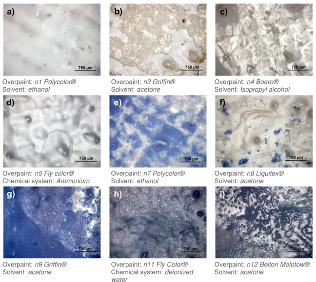
- Optical Microscopy (OM). The images obtained with optical microscopy (figure 7) confirmed some of the chemical cleaning problems explained previously.
Overpaint: n1 Polycolor® Solvent: ethanol
Overpaint: n3 Griffin® Solvent: acetone
Overpaint: n4 Boero® Solvent: Isopropyl alcohol
Overpaint: n5 Fly color® Chemical system: Ammonium
Overpaint: n7 Polycolor® Solvent:ethanol
Overpaint:n8 Liquitex® Solvent: acetone
Overpaint: n9 Griffin® Solvent: acetone
Overpaint: n11 Fly Color® Chemical system: deionized water
Overpaint: n12 Belton Molotow® Solvent: acetone
Figure 7 –OM images obtained from diverse points where the chemical cleaning method was performed.
a)
b)
c)
d)
e)
f)
For example in figure 7a) and g) it is possible to see an alteration on the surface gloss, which also indicates the exact area where the solvent was applied. Furthermore, this effect was seen in all blue paints containing ultramarine blue and cleaned with acetone. Other situations could be found, as for example the cleaning heterogeneity - figure 7b), e) and f) -, over cleaning - figure 7b) -, mechanical damage to the surface without removing the overpaint, possibly caused by the cotton swab - figure 7c) -, deposit marks from the left over solvent -figure 7d) -, swelling effect - figure 7h) -, and a mixture caused by the solvent, between the overpaint and the original layer - figure 7i) -, which are same binder based (nitrocellulose and alkyd), and therefore, it was already expected to occur.
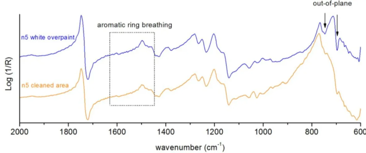
- FTIR spectroscopy. With the aim of assessing the chemical cleaning effectiveness and harmfulness a FTIR analysis was performed taking into account the binder behaviour under the cleaning in what respects: bands intensity, shifting of band position, relative increasing/decreasing of bands, and changes between derivative and positive maximum bands; comparing spectra with the standard ones obtained from the white ground (the considered original layer) and the overpaints [37,38].
As mentioned in the introduction, reflection spectroscopy has some practical implications in spectra interpretation. In this work, the mock-ups prepared have a quite rough surface and, therefore, rather being a disadvantage, it is an opportunity to prepare for the experience of interpreting reflection spectra when analysing an artworkin situ. For instance, in the example given in figure 8 it is possible to observe that the spectrum of the blue overpaint (ultramarine blue) has the silicate in-plane vibration assignment band in a reststrahlen inversion at about 1006 cm-1. The spectrum collected in the remaining blue overpaint of the cleaned area shows alterations in band shape. In fact, band has assumed a first derivative, possibly due to alteration to the optical properties of the surface or due to other bands interferences from ground layer.
The cleaning was not efficient because it could not completely remove the blue overpaint, showed by the ultramarine blue silicate presence in the spectrum of the cleaned area. Adding to this, the OM showed that cleaning was heterogeneous, leaving several residuals of blue overpaint.
Taking into account the analysis made on the paints containing phthalocyanine or dioxazine pigments, it had been observed that the binder is removed first than these organic dyes. In fact, in the spectra the bands associated to them become more clear and sharp. In addition, it was observed by OM that in the cleaned areas with acetone the blue pigment incorporated into the ground paint matrix.
About the white paints, another example of a partial cleaning is given in figure 9. In this example, xylene was the solvent used, which has an aromatic structure. With the principal “like dissolves like”, it is possible that this solvent had more interaction with the aromatic moiety styrene from the styrene-acrylic copolymer paint. In fact, a relative decreasing of the assignment bands of the aromatic C-H out-of-plane bending is visible (746 cm-1 and 698 cm-1). Also, the aromatic ring breathing area shows a slight relative decreasing. The same effect is visible in the corresponding blue paint (n11 Fly Color®).
Figure 9 –Reflection micro-FTIR spectra of white overpaint (n5 Fly Color®), and of the correspondent cleaned area using xylene.
In general, the efficiency in removing white paints was lower, which can be explain by the fact that the restorer had more difficulty in perceiving if it could go further or not with the cleaning.
Further below, a table is presented with the compiled results on the chemical cleaning effectiveness and harmfulness, obtained by interpretation of the results from OM and FTIR spectroscopy (table 3).
Table 3 –General results for the cleaning experiment on mock-up #6 using solvents and chemical systems. The n6 is the considered original layer, except in its specific row, where the original layer is considered the canvas.
Solvent Paint
Deionized
water Acetone Ethanol
Isopropyl
alcohol Xylene
Petroleum ether 80-100°C
Ethyl
lactate Ammonium
n1 –Polycolor® - * +++ - * + ++ - ++
-n2 – Liquitex® - ++ - * - ++ * - +++
-n3 – Griffin® - * ++ ! + * + * + * - +
-n4 – Boero® - +++ *! ++ * - * - * - - * - *
n5 – Fly color® - + * - * - + * - + - *
n6 – Belton® - * ++ + - - - -
-n7 – Polycolor® - * +++ * ++ ++ ++ - * ++ *
-n8 – Liquitex® - * +++ * ++ * ++ ++ - * +++ * + *
n9 – Griffin® - * +++ * ++ + * + * - * ++ * - *
n10 – Boero® - +++ *! ++ ++ * + - ++
-n11 – Fly color® - * +++ *! - * - * - * - + * - *
n12 – Belton® - * +++ ! - * - * - * - * - * - *
(-) no removal; (+) minor removal; (++) significant removal but with several overpaint residuals; (+++) total removal; (*) damage/alteration to the overpaint; (!) damage/alteration to the ground layer.
2.3. Laser cleaning control
Preliminary laser cleaning tests were performed mainly on a spot basis, in order to define the optimum parameters for efficient and safe overpaint removal and to establish the limitations of this method, by using the most aggressive cleaning parameters.
Hence, with that proposal four mock-ups were used, each one with a different white ground, namely, mock-up#1 – n1 Polycolor®; mock-up#2 – n2 Liquitex®; mock-up#3 – n3 Griffin®; mock-up#4 – n4 Boero®. However, only the mock-ups #1 e #4 were analysed in this work, since they covered all laser systems in study: mock-up #1 – laser Nd:YAG at λ= 1064 nm (tp= 10 ns), λ= 532 nm (tp= 10 ns) and λ= 355 nm (tp= 10 ns and 150 ps) and excimer KrF at λ= 248 nm (tp= 30 ns); mock-up #4 – Nd:YAG laser at λ= 355 nm (tp= 150 ps) and excimer KrF laser at λ= 248 nm (tp= 30 ns). To assist the interpretation of the results on pigment alteration upon laser irradiation, reflection UV-Vis-NIR spectra were obtained (figure 10), showing that titanium dioxide (TiO2) absorbs strongly in the UV zone. The n6 white paint, despite containing an iron component, which gives an ivory colour, has still a big absorption under 400 nm. As for blue paints, the ultramarine blue spectra show an absorption band centred at 590 nm, the phthalocyanine centred at 640 nm, and the dioxazine centred at 620 nm.
In all four canvases, the laser irradiation revealed in most cases a greyish discoloration in the white paints and a black-halo around some of their ablation areas. In what concerns the irradiation effect on blue paints, they seem to have been more easily removed and there is no visible alteration phase around their ablation areas.
The discoloration in paints is always a big problem in laser irradiation and in this specific case can be due to two situations: pigment or binder degradation/alteration. The greyish discoloration of paints containing titanium white is not new. In fact, authors have already observed this effect [21,39] defending that it can be caused by a chemical decomposition of the metal oxides or by physical effects (increase of particle size).
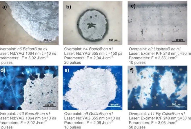
- Optical Microscopy. Optical microscopy observations clarified about cleaning homogeneity of the cleaned areas; discoloration on the white paints; presence of black-halos around ablation areas; aggressive cleanings, etc. Some examples are given in figure 11.
In fact, in figure 11a) and d) it is perfectly visible that the cleaning was aggressive, with canvas exposure. Further, several residuals of the blue overpaint on the d) were also observed, which cannot be a redeposition of the ablated material, since it was not observed around the crater, and it would have a darker appearance. The most reasonable is that it is related to the inhomogeneities of the laser beam. This last feature can also be correlated to the image from figure 11b) and f), all using different laser systems.
The aggressive cleaning seen with the laser Nd:YAG 1064 nm is possibly related to the wavelength, and thus, has to be still carefully studied before thinking its application on painted artworks.
Overpaint: n6 Belton® on n1 Laser: Nd:YAG 1064 nm tp=10 ns
Parameters: F = 3,02 J·cm-2 5 pulses
Overpaint: n4 Boero® on n1 Laser: Nd:YAG 355 nm tp=150 ps
Parameters: F = 2,04 J·cm-2 20 pulses
Overpaint: n2 Liquitex® on n1 Laser: Excimer KrF 248 nm tp=30 ns
Parameters: F = 2,33 J·cm-2 10 pulses
Overpaint: n10 Boero® on n1 Laser: Nd:YAG 1064 nm tp=10 ns
Parameters: F = 3,02 J·cm-2 3 pulses
Overpaint: n9 Griffin® on n1 Laser: Nd:YAG 355 nm tp=10 ns
Parameters: F = 2,06 J·cm-2 10 pulses
Overpaint: n11 Fly Color® on n1 Laser: Excimer KrF 248 nm tp=30 ns
Parameters: F = 3,06 J·cm-2 50 pulses
Figure 11 –Optical microscopy images obtained from diverse points where the laser cleaning was tested.
a)
b)
c)
In figure 11b) a black halo around the crater of the ablation area is visible, which can be explained by the fact that laser energy in the border is lower than at the centre, and thus, does not have enough energy to remove the material, leaving it damaged. In this figure, the already referred greyish discoloration is also visible. Furthermore, with a bigger magnification it is possible to observe presence of various micro black particles that can be responsible for the grey tone. On the contrary, in figure 11c) and e) examples are shown of what could be an approximation of a successful cleaning, i.e. with removal of overpaint and minimal damage to inner layers.
In general, in a first observation, the lasers in the UV regime (excimer KrF laser at λ = 248 nm and Nd:YAG laser atλ = 355 nm) have given the best
results. On the other hand, Nd:YAG at λ = 532 nm showed several defects on the beam profile. In fact, the few examples where the overpaint removal was complete a black halo on the limits of the ablation area was visible, although only on the white paints. Also the excimer KrF (248 nm) and the Nd:YAG (355 nm, tp = 150 ps) showed some defects in the beam profile.
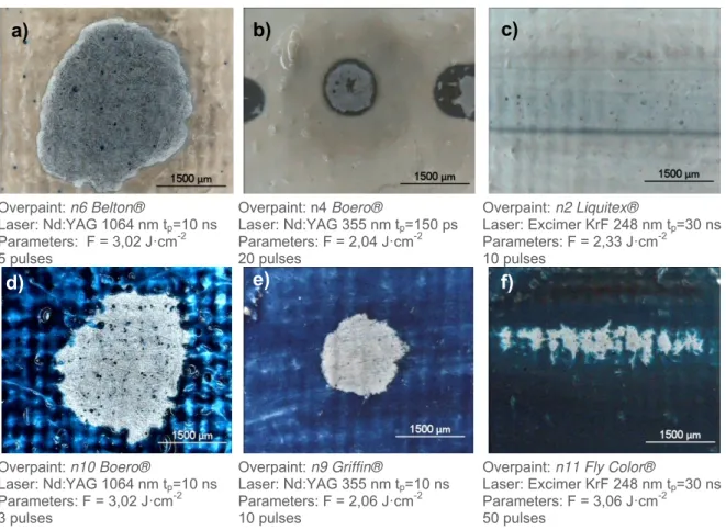
The equivalent portable equipment (figure 12)
allowed obtain very similar images to the ones acquired with OM, as it can be seen in figure 13. In fact, even if the objective magnification permitted by this equipment is lower than the one from OM, it is possible to make the same observations made before by analysing the images below, and therefore this portable equipment proves that it has the reliability to be used in situfor cleaning control.
Overpaint: n6 Belton®
Laser: Nd:YAG 1064 nm tp=10 ns
Parameters: F = 3,02 J·cm-2 5 pulses
Overpaint: n4 Boero®
Laser: Nd:YAG 355 nm tp=150 ps
Parameters: F = 2,04 J·cm-2 20 pulses
Overpaint: n2 Liquitex®
Laser: Excimer KrF 248 nm tp=30 ns
Parameters: F = 2,33 J·cm-2 10 pulses
Overpaint: n10 Boero®
Laser: Nd:YAG 1064 nm tp=10 ns
Parameters: F = 3,02 J·cm-2 3 pulses
Overpaint: n9 Griffin®
Laser: Nd:YAG 355 nm tp=10 ns
Parameters: F = 2,06 J·cm-2 10 pulses
Overpaint: n11 Fly Color®
Laser: Excimer KrF 248 nm tp=30 ns
Parameters: F = 3,06 J·cm-2 50 pulses
Figure 13 –Fibre-optics video microscopy images obtained from the same points presented on figure 11.
d)
e)
f)
Figure 12 – Fibre-optics video microscopy using an objective of 50x that works in contact mode with the surface of the mock-up.
- FTIR spectroscopy. This spectroscopic tool permitted to evaluate the cleaning effectiveness in a molecular level, especially in what concerns the binder behaviour under laser cleaning.
In what concerns first the analyses made on mock-up #1, it was confirmed that, in the majority of cases, cleaning with the Nd:YAG laser at λ = 1064 nm was injurious to the original paint, because when the laser could eliminate completely the overpaint, in almost every cases was damaging the ground layer and therefore the spectra show also bands corresponding to the canvas (see figure 14).
Figure 14 –Reflection micro-FTIR spectrum of a cleaned area in n6 paint on mock-up#1 using the laser Nd:YAG 1064 nm and F = 3,02 J·cm-2, 5 pulses, compared with the white ground and canvas spectra.
In fact, the spectrum from the ablated area is very similar to the one from canvas. However, some signals assigned to the ground are still visible, namely the bands at 1248 cm-1(first derivative shape) and 1749 cm-1, corresponding to the C-O-C symmetric stretching and C=O stretching of PVA, respectively [4].
In general, the laser irradiation with the Nd:YAG 532 nm offered less cleaning effectiveness because, in most cases, it did not reached the original layer, and when it reached there were still several residuals from the overpaint.
On the contrary, the results obtained with the laser Nd:YAG 355 nm, were the most satisfactory in removing overpaints, i.e. analysing the different spectra, it is possible to observe cleaning homogeneity and a progressive micro layer-by-layer removal. Adding to this, spectra of the most efficient cleaned areas showed the best similarity to the spectrum of white ground layer. An example of this is given in figure 15. Even if OM shows white ground discoloration, FTIR analysis demonstrated that the overpaint was completely removed, considering the difficulty in assessing visually an efficient removal of a white overpaint over a white ground. The white discoloration will be explained further by Raman spectroscopy.
The analysis made on the cleaned areas with the excimer KrF laser (λ = 248 nm) showed, as well, satisfactory results, even if problems like the darkish halo around ablated areas, discoloration of the white paints, and inhomogeneities of the laser beam, were still present.
About the analyses made on the mock-up #4, it was observed that the resulting spectra were quite “noisy”, due to the rougher surface of this mock-up, making it more difficult to interpret. This is one of the reasons why it is essential the acquisition of standard spectra of overpaints and the original layers (when possible) very near where the cleaning experiment is being tested.
The following example of ablation with the excimer KrF 248 nm (
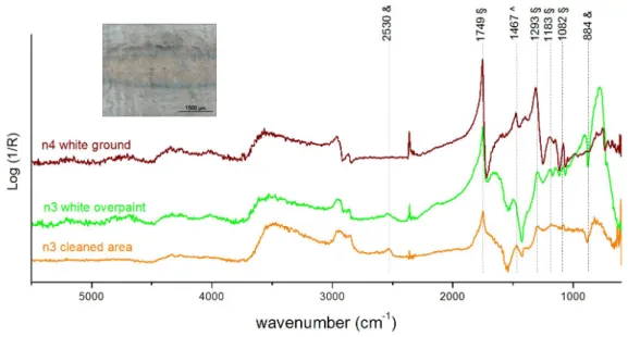
figure 16) is an incomplete cleaning, even if the OM image suggests an efficient overpaint removal.
Figure 16 –Reflection micro-FTIR spectrum of a cleaned area in n3 paint on mock-up#4 using the excimer KrF laser at λ = 248 nm (tp= 30 ns, 3.06 J·cm-2, 30 pulses) compared with the n3 overpaint and white ground spectra.
Although paints n3 and n4 are both alkyd based, the overall aspect of the spectra is different enough to affirm that the overpaint removal was not complete. For example the bands at 2530 cm-1 and 884 cm-1are definitely assigned to the n3 overpaint, which contains calcite, while the one at 1467 cm-1is assigned to the white ground.
Figure 17 –Reflection micro-FTIR spectrum of a cleaned area in n11 paint using the excimer KrF laser at λ = 248 nm (tp= 30 ns, 3.06 J·cm-2, 30 pulses) compared with standard spectra of n11 overpaint and white ground.
About the use of the Nd:YAG laser at λ = 355 nm and tp = 150 ps on this mock-up, the analyses show that the results were not so satisfactory as it were for the mock-up#1, since in most cases cleaning was incomplete and in cases where laser could remove the overpaint, the white ground was no longer visible, showing canvas exposure. This can
possibly be explained by the fact that the white ground layer is too much thin, and, adding to this, the laser at 355 nm is absorbed less by the material to be removed and, due to that, had a deeper penetration on the substrate.
The fibre-optics portable equipment (figure 18) has given acceptable results, in sense that the spectra obtained could be compared with the ones from bench equipment. The only disadvantages found are related to the analysed spot diameter, which is around 4 mm. I can be too big to analyse
ablated areas from Nd:YAG 355 nm and excimer KrF laser 248 nm. Other disadvantage is spectra range, which is in theory 7000-900 cm–1. However, in practice the background spectra obtained is not perfect. In fact, the fibres show a Se-H stretching absorption in the 2050-2250 cm−1range altering the signal to noise ratio in that region. Environmental moisture at 3100–3500 cm−1 and carbon dioxide at 2250 cm−1 can also cause interference, blinding the regions. Also, below 1000 cm-1the spectra are indiscernible. This feature can be a problem considering for example the characteristic silicate band of ultramarine blue, which in the reflection spectra has a reststrahlen inversion band at about 1000 cm-1, or, for example, the aromatic C-H out-of-plan bands characteristic of styrene acrylics at 746 cm-1and 698 cm-1.
In figure 19, reflection FTIR spectra from the portable equipment are presented, giving examples of aggressive cleaning and incomplete cleaning compared with overpaint, white ground and canvas standard spectra. In the example of incomplete cleaning an OM image of the cleaned area is shown to explain the difficulty in assessing visually the efficiency of overpaint removal. In fact, the image suggests that the overpaint was removed.
Figure 18 –Fibre-optics FTIR
Figure
-alterations most cases carbon (
However the greyish equipment, mea
of the dominant white zone
Figure
fibre-optics portable video microscopy micro black particles
Considering that this e same binders did
main role phenomena,
Figure 19 –Reflection
Raman spectroscopy.
alterations phases most cases, analysis carbon (figure 20), meaning
However, not all greyish discoloration equipment, meaning of the dominant white zone
Figure 20 –Ablation in
optics portable video microscopy micro black particles
Considering that this e same binders did
main role on this phenomena, which
a)
d)
Reflection FTIR spectra, showing examples of aggressive and with n3 overpaint, white ground
Raman spectroscopy.
phases formed and analysis made in
), meaning that probably not all ablation areas discoloration is visible. equipment, meaning that the black of the dominant white zone.
Ablation in paint n4 using the Nd:YAG 355 nm laser optics portable video microscopy
micro black particles, d) micro
Considering that this effect only binders did not shown sign
on this effect. In the which are originate
FTIR spectra, showing examples of aggressive and n3 overpaint, white ground
spectroscopy. This spectroscopic formed and some molecular
made in the micro that probably ablation areas showed discoloration is visible. This may
that the black particles
4 using the Nd:YAG 355 nm laser optics portable video microscopy image from cleaned area
micro-Raman spectrum
ffect only is visible signs of binder effect. In the literature,
originate from the
b)
FTIR spectra, showing examples of aggressive and n3 overpaint, white ground and canvas
spectroscopic technique some molecular alterations
black particles
that probably it is an alteration phase from the showed the presence
This may be explained particles are too little
4 using the Nd:YAG 355 nm laser image from cleaned area Raman spectrum (=532 nm)
is visible in the white paints (the corresponding blue binder degradation up
literature, TiO2 is characteris from the semiconductor
FTIR spectra, showing examples of aggressive and and canvas standard
technique permitted alterations on paint particles observed is an alteration phase from the
presence of carbon be explained by the
too little to be analys
4 using the Nd:YAG 355 nm laser, tp= 10 ns,
image from cleaned area, b) and c) optical microscopy =532 nm) of this area with
in the white paints (the corresponding blue radation upon irradiation),
is characterised semiconductor band gap,
FTIR spectra, showing examples of aggressive and incomplete standard spectra
technique permitted to observe on paint materials after observed with OM showed is an alteration phase from the binder
carbon on Raman explained by the sensitivity be analysed alone w
10 ns, F = 2.06 J , b) and c) optical microscopy
of this area with carbon
hite paints (the corresponding blue on irradiation), titanium dio
ed by the presence band gap, and can
c)
incomplete cleaning, by comparison spectra.
permitted to observe some materials after laser irradiation.
OM showed the presence binder degradation. Raman spectroscopy,
sensitivity of the spectroscopic alone without the interference
2.06 J·cm-2and 10 pulses , b) and c) optical microscopy images
carbon assignment bands.
hite paints (the corresponding blue paints with the itanium dioxide may play the presence of photoinduced and can lead to photocatalysis. cleaning, by comparison
observe some of the laser irradiation. In the presence of degradation.
spectroscopy, even if the spectroscopic ithout the interference
and 10 pulses, a) the images showing the assignment bands.
paints with the xide may play a of photoinduced to photocatalysis. the irradiation. In presence of
even if spectroscopic interference
the showing the
Thus, this phenomenon may be responsible for the photodegeneration of organic compounds [40] (for more information on the photocatalyst properties of TiO2see appendix 2, page A9).
Adding to this, a small but significant alteration of the relative intensity of the Raman bands of rutile has been observed upon laser interaction. The obtained spectra permitted to see a relative decreasing of the band assigned to vibrational group A1gin respect to the other signs of the rutile (see figure 21). Several analyses were made on the ablated areas to evaluate this decrease, and a systematic result was found, i.e. every laser system produced this effect on titanium white (rutile) pigment, when the white paint was found greyish. Further, the lasers with wavelengths 1064 nm and 355 nm showed in almost every case the more and less decrease of this vibrational band, respectively.
In this paper [41] the same effect was found on the same vibrational group upon laser irradiation. Since the mock-ups in this paper were TiO2 single crystals (40 mm in diameter and 1.5 mm thick) and the energy densities applied were much higher, at the end of the experiment the complete alteration from rutile structure to anatase was observed. However, in the results observed in the current work, because of the low titanium dioxide concentration (it is dispersed in the binder), in respect to the mock-up in that experience, the alteration process from rutile to anatase structure may not be complete.
In what concerns the blue pigments they did not show any significant alteration in Raman spectra that could be associated to a crystalline or molecular change. The observed changes in the spectra were only related to the absolute intensity. However, it must be reported that in the case of phthalocyanine blues an increase in the fluorescence background has been observed on the borders of the cleaned areas, probably suggesting the production of degradation products (see example in figure 22). This fluorescence is not equal in all phthalocyanine blues. In the n10 paint an accentuated fluorescence is observed very close to the laser irradiation line (532 nm) that possibly is deformed by the Raman notch. In the n11 paint there are two main relevant fluorescence bands that in wavelength correspond to absorptions around 667 nm and 617 nm. On the other hand, the analysis made on n12 paint, the dioxazine violet, did not show any significant alteration in the fluorescence.
Figure 22 –Micro-Raman spectra (=532 nm) of phthalocyanine paint n11, performed in the remaining blue spots on the ablation areas, which showed notable fluorescence around 667 nm and 617 nm. Figure 21 – Raman spectra (=532
In what concerns the ultramarine blues the relative intensity between the bands assigned to the chromophores S2-and S3-, namely the scattering at 581 cm-1and 549 cm-1, respectively, did not alter whatsoever. It was expected that chromophores would degrade first than the aluminosilicate matrix due to their removal from the sodalite cage. However, by analyses made with micro-Raman, when the chromophores were not visible anymore, also the assignment band of silicate in micro-FTIR was not visible. In fact, it has been demonstrated that this pigment is very stable to laser irradiation, suffering discoloration only at very high energy densities [23].
In respect of the fibre-optics Raman spectroscopy portable equipment (figure 23), it was not possible to identify the presence of carbon. In fact, the micro black particles are too little to be analysed without interference from the white paint matrix. However, the rutile alteration could be seen as well as the absolute decrease of the assignment bands of the phthalocyanine (figure 24) and dioxazine. In this case the use of the laser =785 nm in the analysis showed the best results.
Figure 23 –Fibre-optics Raman spectroscopy portable equipment. Photo courtesy of SMAArt (Perugia)
Figure 24 –Raman spectra (=785 nm) of two different ablation areas on paint n10 containing phthalocyanine blue, one successful, and other
incomplete, compared with the standard spectrum of the overpaint.
In the following table, the general results on the effectiveness and harmfulness of laser cleaning are presented, taking into account the analyses made with OM, FTIR and Raman spectroscopies. In each case a singular ablation area (with specific laser parameters) was chosen, considering it as the one where the cleaning was more efficient with that specific laser system. The same table, yet more detailed, is presented in the appendix 3 (page A10), with information added on laser parameters (fluence and number of pulses), fibre-optics video microscopy image, and observations.
Table 4 –General results for laser cleaning experiment on mock-up #1 and #4 using different laser systems. The n1 and the n4 are the original layers except in its specific row where the canvas is considered the original layer.
Canvas mock-up #1 #4
Laser system Paint Nd:YAG =1064 nm tp=10 ns Nd:YAG =532 nm tp=10 ns Nd:YAG =355 nm tp=10 ns Nd:YAG =355 nm tp=150 ps Excimer KrF =248 nm tp=30 ns Nd:YAG =355 nm tp=150 ps Excimer KrF =248 nm tp=30 ns
n1 - Polycolor® - * - * - * - * - * ++ *! + *
n2 - Liquitex® - * + !! +++ ! ++ *! - * ++ *! ++ *
n3 - Griffin® +++ !! + !! +++ *! ++ ! - * ++ *! ++ *
n4 - Boero® - !! ++ *!! +++ *! +++ ! + *! +++ *! n.t.
n5 - Fly color® - - * ++ ! +++ ! - * ++ *! ++ *
n6 - Belton® ++ ! + *! +++ *! ++ ! - + *!! + *
n7 - Polycolor® +++ !! ++ ! +++ +++ ! ++ ! ++ !! ++
n8 - Liquitex® +++ !! +++ ! +++ ! +++ ! ++ ! ++ !! +++
n9 - Griffin® +++ !! n.t. +++ ! ++ ! ++ ! ++ !! ++
n10 - Boero® +++ !! n.t. +++ ! ++ ++ ++ !! ++
n11 - Fly color® +++ !! n.t. +++ ++ +++ ++ !! +++
n12 - Belton® +++ !! n.t. +++ ++ +++ ++ !! ++ *
As it is possible to observe in the table, the lasers Nd:YAG 355 nm (tp= 10 ns and tp= 150 ps ) at rather high energy densities and several pulses (10 for the ns regime and 20 for the ps regime) showed the best results for the mock-up #1. On the contrary, the excimer KrF laser 248 nm (tp = 30 ns) showed to be the best choice in the mock-up #4, using energy densities around 3 J·cm-2and 30 pulses.
One possibility for the control of the most complicated side-effect found in this laser experiment, the greyish discoloration, can be the use of ultra-short laser systems, which were not tested in this work. Generally, the ns (nanosecond) cleaning regime gives photo-thermal phenomena, which are reduced upon ultra-short irradiation (i.e. picosencond - ps, femtosencond - fs), as confirmed from many studies [11,34,42-44]. In fact, these ultra-short lasers allow minimal thermal diffusion, heat accumulation after each pulse given, and the ablation morphology can be superior to that obtained with nanosecond laser pulses, mainly with the femtosecond lasers.
However, there are other conditions that influence the thermal, chemical or photo-mechanical damages of original layers, and govern on which of them will be more dominant. Firstly, it is important to choose the most appropriate wavelength, and whether is weakly or strongly absorbed by the paint material. In theory, the wavelengths more adapted for painted artworks are the ones ≤ 248 nm, to achieve a layer-by-layer removal. On the other hand, UV lasers are more capable to break the bonds of the polymer chains and will induce photo-chemical activity, while lasers with wavelengths of 1064 nm and 532 nm will mainly remove the material by explosive vaporization. It must also be appointed the importance of the original layer absorption in the electromagnetic spectrum, since this will influence substantially the cleaning process, mainly if the overpaint is very thin or does not absorb strongly.
3. Conclusions
The optical and spectroscopic analyses permitted the evaluation of the effectiveness and harmfulness of the different cleaning methods tested.
On the chemical cleaning control the analytical approach lead to the conclusion that acetone was the most efficient solvent, yet with strong action and deep penetration into substrate. Next in the line of efficacy is ethyl lactate, which seemed to allow more control in the cleaning, yet still leaving several overpaint residues behind. The considered medium-efficient solvents were ethanol, isopropyl alcohol, and xylene by giving partial cleaning or minor removal of the overpaints. The solvents and chemical systems that were not able to remove the overpaints were deionized water, ammonium and petroleum ether. To sum up, chemical cleaning has demonstrated to give several problems for the selective removal of overpaints.
Regarding the effectiveness of the laser cleaning method it is possible to highlight the following points. The Nd:YAG laser 1064 nm has proven to be a very aggressive irradiation regime within the parameters used. The hazard effects observed were the discoloration of the white paints, changes in the substrate morphology and the uncontrolled removal of overpaints, with canvas exposure in the majority of cases. The same laser at λ = 532 nm has shown to be the less efficient and, therefore, further research should be carried out by decreasing the energy density and increasing the number of
![Figure 4 – Alkyd structure containing phthalic anhydride, glycerol and linoleic acid [4].](https://thumb-eu.123doks.com/thumbv2/123dok_br/16624065.740332/11.892.145.796.651.1021/figure-alkyd-structure-containing-phthalic-anhydride-glycerol-linoleic.webp)