iii
Orientador: Doutora Filomena Macedo Dinis, Professor Auxiliar com Nomeação De-finitiva, DCR, FCT-UNL
Co-orientadores: Doutora Laura Rosado, Instituto Nacional de Saúde Doutor Ricardo Jorge I.P.
Doutora Valme Jurado, Instituto de Recursos Naturales y Agrobiología, CSIC
Júri
Presidente Prof. Doutor Fernando Pina
Arguentes Doutor António Manuel Santos Carriço Portugal, Professor Auxiliar
Vogais
Doutor Alan Phillips, Investigador
Doutora Maria Inês Durão de Carvalho Cordeiro, Directora da Biblioteca Nacional
Doutora Susana Marta Lopes Almeida, Investigadora Auxiliar
October, 2014
Ana Catarina Martiniano da Silva Pinheiro
Licenciada em Conservação e Restauro pela Universidade Nova de Lisboa
Licenciada em Ciências Farmacêuticas pela Universidade de Lisboa
Fungal Communities in Archives:
Assessment Strategies and Impact on Paper
Conservation and Human Health
Dissertação para obtenção do Grau de Doutor em
iii
Fungal Communities in Archives: Assessment Strategies and Impact on Paper Con-servation and Human Health Fungal
Copyright © Ana Catarina Martiniano da Silva Pinheiro, Faculdade de Ciências e Tecnologia, Universidade Nova de Lisboa.
v
vii
Acknowledgments
An endeavour such as a Philosophical Degree is never accomplished without the help and encouragement of a world of people. Because memory may fail me I would like to express my deep gratitude also to everyone I do not mention in the next lines.
This PhD was developed at the Department of Conservation and Restoration (FCT-UNL), the Mycology Laboratory at the National Institute of Health (URSZ, DDI,
INSA IP) and the Instituto de Recursos Naturales y Agrobiologia de Sevilla – Consejo
Superior de Investigaciones Cientificas (IRNAS-CSIC). It would not have been possible to gather together these three institutions without the support of the Fundação para a Ciência e Technology (SFRH/BD/36005/2007).
I would like to thank the Faculty of Science and Technology and the Conserva-tion and RestoraConserva-tion Department for stimulating the pursuit of higher knowledge and creating the conditions for the journey. Included are all the teachers and professionals in this institution. I would also like to thank Dr. Rui Silva (CENIMAT, FCT-UNL) for his time and help.
Everyone at the archives where this study was performed: Arquivo Histórico Ul-tramarino, Arquivo Distrital de Évora, Instituto de Habitação e Reabilitação Urbana and Torre do Tombo. Thank you for opening your doors and helping on the execution of this study.
A particular note of gratitude to Doutora Maria Filomena Macedo Dinis, Assis-tant Professor at DCR-FCT/UNL, who supervised this work improving it with her in-puts, valuable advices and guidance.
My gratitude goes also to Dr. Valme Jurado, PhD and Prof. Dr. Cesareo Saiz-Jimenez, PhD, for letting me in their team in Sevilla and for all the efforts in guiding me.
A very warmth thank you to everyone at the INSA’s Mycology Laboratory, esp
viii
To Catarina Silva, Sonia Pedro, Ana Cardoso and Luis Vieira for their help in the molecular biology field.
A special thank you to Carla Viegas and Susana Viegas (ESTESL) for all the ener-gy and expertise. You are truly inspirational.
Mathilda Larsson and Silvia Sequeira, for their inputs and shared concerns. You were a precious help! Maria João Pereira and Ana Zélia Miller for carving the way and being an inspiration and helpful hand.
To Phil Harrison from Transgenomic for the support while handling the DHPLC.
To all my friends for their support. Even from a distance.
Finally a very grateful thank you to my family for their encouragement and pres-ence. A special note of gratitude for my husband for his patience and reassurance
dur-ing the stressful moments and also a special note for my daughter…Hope this makes
ix
The faculty of art is to change events; the faculty of science is to foresee them.
— Henry Thomas Buckle 'The Influence of Women on the Progress of Knowledge,’ a discourse delivered at the Royal I
n-stitution (19 Mar 1858) reprinted from Fraser's Magazine (Apr 1858) in The Miscellaneous
xi
The main results presented in this PhD Dissertation have been published in interna-tional journals included in the Science Citation Index (SCI):
Pinheiro, A.C., Viegas, C., Viegas, S., Veríssimo, C., Brandão, J. & Macedo, M.F. (2014). Particulate Matter Distribution in Selected Portuguese archives: A Preliminary Study. International Journal of Conservation Science, 5 (2), 139-150.
Pinheiro, A.C., Viegas, C., Viegas, S., Veríssimo, C., Brandão, J. & Macedo, M.F. (2012). Indoor Air Quality in Portuguese archives: a Snapshot on Exposure Levels, Journal of Toxicology and Environmental Health, Part A: Current Issues, 75(22-23), 1359-70.
Pinheiro, A. C., Macedo, M. F., Jurado, V., Sáiz-Jiménez, C., Viegas, C., Brandão, J., & Rosado, L. (2011). Mould and yeast identification in archival settings: Preliminary re-sults on the use of traditional methods and molecular biology options in Portuguese archives. International Biodeterioration and Biodegradation, 65(4), 619-627
xiii
Abstract
Fungi are ubiquitous and, as such, are current contaminants in archives and librairies. Provided the adequate relative humidity, temperature and water activity, however, they can stop being innocent inhabitants and become dangerous threats to the organic materials that hold our written heritage. Their impact, however, is not re-stricted to the assets of an archive or library. New concerns arise from their incredible ability to thrive as human health can also be affected. In a contaminated environment, staff and attendees share the same space with fungal communities and the human body can suffer from this interaction in the most diverse ways. So, indoor air quality studies in archives and libraries should always comprehend not only the study of fun-gal communities but also their analysis under two perspectives: documents safekeep-ing and human health protection.
In this study it was important to determine the conditions provided by some of our heritage holder’s institutions and develop the best strategy to identify their fungal flora. This strategy encompassed both air and surfaces and both traditional culturing methods and molecular biology protocols. Knowing the environment in Portuguese institutions is essential for the development of guidelines and establishment of recom-mendations. Only by using this knowledge can a safer environment be created and the purpose of keeping our heritage while maintaining our health is fully attained.
xv
Resumo
Os fungos existem em todos os ambientes e, como tal, não surpreende a sua
presença em arquivos e bibliotecas. No entanto, quando existem as condições certas de
humidade relativa, temperatura e actividade da água, os fungos deixam de ser simples ocupantes e transformam-se numa perigosa ameaça ao nosso património. O seu impac-to, no entanimpac-to, não se limita ao acervo de um arquivo ou biblioteca. Da sua incrível te-nacidade nascem novas preocupações, desta vez relacionadas com a saúde humana. Num ambiente contaminado, tanto funcionários como visitantes podem sofrer com es-ta interacção. É por este motivo que os estudos de qualidade do ar interior neses-tas insti-tuições deverão sempre incluir não só o estudo das comunidades fúngicas como a sua análise sob ambas as perspectivas: a salvaguarda da nossa herança escrita e protecção da saúde humana.
Neste estudo foi importante conhecer as condições fornecidas por alguns dos nossos Arquivos e desenvolver a melhor estratégia para a identificação da microflora fúngica. Enquadradas nesta estratégia estiveram amostras de ar e de superficies tendo sido usados tanto os métodos tradicionais de cultura como protocolos de biologia mo-lecular. Conhecer o ambiente é essencial para o desenvolvimento de directrizes e recomendações. Só utilizando este conhecimento é que será possível criar ambientes mais seguros onde a salvaguarda do património pode caminhar lado a lado com a ma-nutenção da nossa saúde enquanto cuidadores.
xvii
Table of Contents
FIGURE LIST ... XXI TABLE LIST ... XXVII ABBREVIATIONS... XXIX
1. INTRODUCTION ... 17
FUNGI AND THE BIODETERIORATION OF PAPER ... 19
FUNGI AND HUMAN HEALTH ... 25
1.1RESEARCH PROBLEMS ... 30
1.2OBJECTIVES ... 31
1.3THESIS OUTLINE ... 32
2. FUNGI IN ARCHIVES.STATE OF THE ART ... 35
A REVIEW OF THE LITERATURE ... 37
2.1AIR STUDIES... 38
2.2SURFACE STUDIES ... 48
2.3CASE STUDIES IN DOCUMENTS ... 54
2.4FUNGAL CONTAMINATION AND PAPER CONSERVATION ... 62
2.5FUNGAL CONTAMINATION AND HUMAN HEALTH ... 71
2.6CONCLUSIONS AND FURTHER STUDIES: ... 74
3. METHODOLOGIES FOR THE EVALUATION OF FUNGAL COMMUNITIES IN ARCHIVES: STUDY DESIGN ... 77
3.1INTRODUCTION ... 79
3.2-SELECTED ARCHIVES AND SAMPLING LOCATIONS ... 80
3.3SAMPLING PROTOCOL ... 85
3.3.1 Air Samples ...85
3.3.2 Surface samples (Floor, Table, Shelves, Trays and DACs) ...86
3.3.3 Document samples ...87
3.4SAMPLE TREATMENT AND ANALYSIS ... 87
3.4.1. Conventional Culturing Methods ...87
3.4.1.1 Air samples ...88
xviii
3.4.2 Molecular Biology Protocols ... 88
3.5INDOOR AIR QUALITY STUDIES ... 102
4. AEROBIOLOGY OF FUNGI IN ARCHIVES ... 105
4.1INTRODUCTION ... 107
4.2RESULTS AND DISCUSSION ... 110
4.2.1 Air samples ... 110
4.2.2. Surface samples ... 132
4.2.3 Sampled Documents ... 179
4.2.4 AFCE Methodology Results ... 192
4.3CONCLUSIONS ... 194
5. INDOOR AIR QUALITY IN PORTUGUESE ARCHIVES: PRELIMINARY RESULTS . 201 5.1.INTRODUCTION ... 203
52.MATERIAL AND METHODS ... 207
5.3.RESULTS AND DISCUSSION ... 208
5.3.1 Carbon Dioxide ... 208
5.3.2 Carbon Monoxide ... 208
5.3.3 Volatile Organic Compounds (VOCs) ... 208
5.3.4 Formaldehyde ... 209
5.3.5 Particulate Matter ... 209
5.3.6 Ozone ... 212
5.3.7 Temperature and Relative Humidity ... 214
5.3.8 Fungi ... 215
5.4.CONCLUSIONS ... 221
6. WATER ACTIVITY AND FUNGAL PRESENCE ... 225
6.1INTRODUCTION ... 227
6.2METHODOLOGY ... 229
6.3RESULTS AND DISCUSSION ... 230
6.3.2 Water activity and Relative Humidity ... 232
6.3.3 Water activity measurements and fungal load ... 233
6.3.4 Risk of Condensation ... 234
6.3.5 Water activity and Document Samples ... 235
6.4CONCLUSIONS ... 238
7. FURTHER APPLICATIONS FOR THE DHPLC METHOD: FUNGI AND YEASTS IN CULTURAL HERITAGE AND CLINICAL STUDIES ... 239
7.1IDENTIFICATION OF A FUNGAL COMMUNITY ON GILDED WOOD CARVED HERITAGE... 243
7.1.1 Introduction ... 245
7.1.2 Materials and Methods ... 246
xix
7.1.4 Discussion ... 252
7.1.5. Conclusions ... 255
7.2DENATURING HIGH PERFORMANCE LIQUID CHROMATOGRAPHY AND AUTOMATED FLUORESCENT CAPILLARY ELECTROPHORESIS TO RESOLVE MIXED CANDIDA SP.CULTURES ... 257
7.2.1 Introduction ... 259
7.2.2 Methodology... 260
7.2.3 Results and Discussion ... 263
7.2.4 Conclusions ... 269
FINAL REMARKS AND FUTURE PERSPECTIVES ...271
xxi
Figure List
FIGURE 1.1-SPECIAL AREAS WERE SAMPLED FOR FUNGAL PRESENCE ANALYSIS.A STERILE COTTON SWAB WAS USED FOR THE EFFECT. ... 18
FIGURE 1.2–ALL DIFFERENT LIFE STAGES OF FUNGI CAN BE OBSERVED ON PAPER: SPORES, GERMINATED YOUNG HYPHAE OR ADULT MYCELIUM AND SPORE FORMING STRUCTURES.NONE OF THESE WERE VISIBLE TO THE NAKED EYE.IMAGE OBTAINED USING AN OPTICAL MICROSCOPE LEICA DMI5000M(50X TO 1000X), A COURTESY OF CENIMAT. ... 21 FIGURE 2.1-QUANTITATIVE AIR ANALYSIS STUDIES PERFORMED IN ARCHIVES AND THEIR RELATIONSHIP TO
PROPOSED LIMITS FOR CONSERVATION. ... 62
FIGURE 2.2–QUANTITATIVE AIR ANALYSIS RESULTS FROM THE AVAILABLE STUDIES. THESE RESULTS ARE
COMPARED TO PROPOSED LIMITS FOR GOOD AIR QUALITY. ... 72
FIGURE 3.1 -INDOOR AIR QUALITY STUDIES INCLUDE FUNGAL ASSESSMENT USING AIR SAMPLERS SUCH AS THIS M
AIR TESTER. ... 86 FIGURE 3.2-THE 10X10 CM METALLIC SQUARE USED FOR AREA DELIMITATION WAS DISINFECTED BETWEEN
SAMPLES.THE SURFACE INSIDE THE AREA WAS STREAKED HORIZONTALLY, VERTICALLY AND IN THE DIAGONAL. ... 86 FIGURE 3.3–SPECIAL AREAS WERE SAMPLED FOR FUNGAL PRESENCE ANALYSIS.A STERILE COTTON SWAB WAS USED FOR THE EFFECT. ... 87
FIGURE 3.4-SCHEMATIC REPRESENTATION OF DNA REGIONS IN THE FUNGAL GENOME ... 90
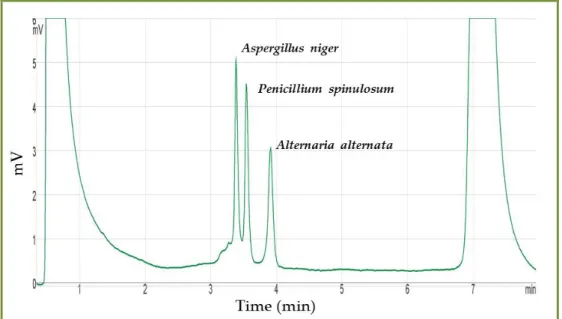
FIGURE 3.5-DHPLC CHROMATOGRAM OF A MIXTURE OF AMPLIFIED DNA FROM THREE DIFFERENT FUNGI
(ALTERNARIA ALTERNATA,ASPERGILLUS NIGER AND PENICILLIUM SPINULOSUM)... 97
FIGURE3.3.6-TOP:DHPLC CHROMATOGRAM OF A MIXTURE OF AMPLIFIED DNA FROM TWO DIFFERENT FUNGI
(CANDIDA PARAPSILOSIS ANDACREMONIUM SP.) USING PARTIALLY DENATURING TEMPERATURES (59°C, 55%B) AND A 0.5 ML/MIN FLOW RATE.BOTTOM:DHPLC CHROMATOGRAM OF A MIXTURE OF AMPLIFIED
DNA FROM THREE DIFFERENT FUNGI (CANDIDA PARAPSILOSIS AND ACREMONIUM SP. FROM ABOVE TO WHICH
ASPERGILLUS SP. WAS ADDED) USING THE SAME CONDITIONS AS ABOVE.IN THIS EXAMPLE, THE AMPLIFIED REGION IS THE ITS2(PINHEIRO ET AL,2010) ... 99
FIGURE 3.7 -DETECTION LIMITS FOR THE DHPLCDNASEP CARTRIDGE. ... 100
FIGURE 3.8 -PERFORMANCE OF A SINGLE SPECIES AT PARTIALLY DENATURING TEMPERATURES. ... 101
FIGURE 4.1-TOTAL VIABLE CFU/M3 IN THE AIR SAMPLES TAKEN FROM THE SELECTED LOCATIONS IN THE ADE ARCHIVE ... 110 FIGURE 4.2-TOTAL VIABLE CFU/M3 IN THE AIR SAMPLES TAKEN FROM THE SELECTED LOCATIONS IN THE AHU
ARCHIVE. ... 111 FIGURE 4.3–TOTAL VIABLE CFU/M3 IN THE AIR SAMPLES TAKEN FROM THE SELECTED LOCATIONS IN THE IHRU
ARCHIVE. ... 111
xxii
FIGURE 4.5–AIR QUALITY LEVELS COMPARED WITH SOME OF THE ESTABLISHED LIMITS FOR CONSERVATION (SEE
TABLE 1.1). ... 113 FIGURE 4.6–FUNGAL LOAD AND DIVERSITY ON ADE’S AIR SAMPLE,1ST SEASON, WINTER. ... 116 FIGURE 4.7–FUNGAL LOAD AND DIVERSITY ON ADE’S AIR SAMPLE,2ND SEASON, SUMMER. ... 117 FIGURE 4.8–FUNGAL LOAD AND DIVERSITY ON ADE’S AIR SAMPLES,3RD SEASON, WINTER.SOME OF THE VALUES
ARE PRESENTED TO EASE COMPREHENSION. ... 118
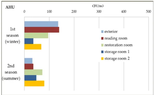
FIGURE 4.9–FUNGAL LOAD AND DIVERSITY ON AHU’S AIR SAMPLE,1ST SEASON, WINTER. ... 119
FIGURE 4.10–FUNGAL LOAD AND DIVERSITY ON AHU’S AIR SAMPLES,2ND SEASON, SUMMER. ... 120 FIGURES 4.11 AND 4.12–CEILING SHOWING THE FIRST SIGNS OF FUNGAL GROWTH AFTER A PIPE HAS
STARTED LEAKING. ... 121 FIGURES 4.13 AND 4.14–ONE YEAR AFTER, THE CEILING SHOWS PROFUSE FUNGAL GROWTH OF PENICILLIUM SP.
AND STACHYBOTRYS CHARTARUM... 121
FIGURE 4.15–FUNGAL LOAD AND DIVERSITY ON IHRU’S AIR SAMPLE,1ST SEASON,FEBRUARY ... 122
FIGURE 4.16–FUNGAL LOAD AND DIVERSITY ON IHRU AIR SAMPLE,2ND SEASON,APRIL.THE OFFICE ROOM RETURNED ZERO COLONIES. ... 123
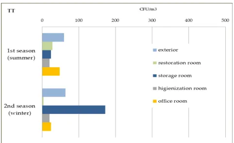
FIGURE 4.17–FUNGAL LOAD AND DIVERSITY IN TT’S AIR SAMPLE,1ST SEASON,APRIL. ... 124
FIGURE 4.18–TT AIR SAMPLE,2ND SEASON, WINTER.PLEASE MIND THE LOGARITHMIC SCALE. ... 125
FIGURE 4.19-AIR SAMPLES PERFORMED IN FOUR PORTUGUESE ARCHIVES AND ANALYSED IN TERMS OF FREQUENCY
(PRESENCE/TOTAL NUMBER OF SAMPLES) AND MAXIMUM NUMBER OF CFU/M3. ... 126
FIGURE 4.20-AIR SAMPLE FUNGAL FLORA CLASSIFIED ACCORDING TO THE CLASS DEFINITIONS PROPOSED BY THE
DECREE-LAW 78/2006 AND 79/2006 SUPPORTED BY THE NT-SCE-02. ... 129
FIGURE 4.21–AIR SAMPLE FUNGAL FLORA CLASSIFIED ACCORDING TO THE CLASS DEFINITIONS PROPOSED BY THE
ORDINANCE 353-A/2013,DECEMBER 4TH. ... 130
FIGURE 4.22–FUNGAL LOAD AND DIVERSITY ON SURFACE SAMPLES IN ADE1ST SEASON, WINTER.NO GROWTH: READING ROOM TABLE, RESTORATION ROOM TABLE.IN THE X-AXIS ARE THE FUNGI ASSOCIATED WITH PAPER BIODETERIORATION.OTHERS INCLUDE:BEAUVERIA SP.,CHRYSONILIA SP.,CHRYSOSPORIUM SP.,GRAPHIUM SP.,PHAEOACREMONIUM SP.,PHIALOPHORA SP.,PHOMA SP.,SCOPULARIOSIS SP.,SCEDOSPORIUM SP. AND
SCYTALIDIUM SP.PLEASE MIND THE LOGARITHMIC SCALE. ... 134 FIGURE 4.23–FUNGAL LOAD AND DIVERSITY ON THE ARCHIVAL CASES IN ADE,1ST SEASON, WINTER.NO GROWTH:
STORAGE ROOM 1 ARCHIVAL CASE.IN THE X-AXIS ARE THE FUNGI ASSOCIATED WITH PAPER
BIODETERIORATION.OTHERS INCLUDE:ARTHRINIUM SP.,CHRYSOSPORIUM SP.,PHOMA SP. AND SCYTALIDIUM SP.PLEASE MIND THE LOGARITHMIC SCALE. ... 135
FIGURE 4.24-FUNGAL LOAD AND DIVERSITY ON ADE SURFACE SAMPLES,2ND SEASON, SUMMER... 137 FIGURE 4.25–FUNGAL LOAD AND DIVERSITY OF ASPERGILLUS SPECIES IDENTIFIED IN THE SURFACES FROM ADE
(2ND SEASON, SUMMER). ... 138 FIGURE 4.26 -FUNGAL LOAD AND DIVERSITY ON ARCHIVAL CASES FROM ADE, SECOND SEASON, SUMMER. ... 139 FIGURE 4.27-DHPLC CHROMATOGRAM OF A MIXTURE OF TWO YEASTS.THE RUN WAS PERFORMED AT
61°C/55%B AT A 0.9ML/MIN RATE FLOW.THE FIRST PEAK, COLLECTED AT MINUTE 4 AT 63°C
CORRESPONDS TO CRYPTOCOCCUS SP. AND THE SECOND PEAK, COLLECTED AT MINUTE 4.2 CORRESPONDS TO
EXOPHIALA SP. ... 141
xxiii
FIGURE 4.29-FUNGAL LOAD AND DIVERSITY ON THE ASPERGILLUS SPECIES IDENTIFIED IN ADE’S SURFACE SAMPLES,
3RD SEASON, WINTER.SAMPLING WAS PERFORMED DURING CONSTRUCTION WORK.PLEASE MIND THE LOGARITHMIC SCALE. ... 143 FIGURE 4.30–FUNGAL LOAD AND DIVERSITY ON ADE’S ARCHIVAL CASES,3RD SEASON, WINTER. ... 144 FIGURE 4.31-FUNGAL LOAD AND DIVERSITY OF THE ASPERGILLUS SPECIES IDENTIFIED IN ADE’S ARCHIVAL CASES IN
THE 3RD SEASON, WINTER.IN THE X-AXIS ARE THE FUNGI ASSOCIATED WITH PAPER BIODETERIORATION. SAMPLING WAS PERFORMED DURING CONSTRUCTION WORK.PLEASE MIND THE LOGARITHMIC SCALE. ... 145
FIGURE 4.32-PERCENTAGE OF THE FUNGAL POPULATION ASSOCIATED WITH PAPER DEGRADATION IN THE ADE ARCHIVES FOR ALL THREE SEASONS... 146
FIGURE 4.33–FUNGAL LOAD AND DIVERSITY ON AHUSURFACES,1ST SEASON, WINTER. ... 148 FIGURE 4.34–FUNGAL LOAD AND DIVERSITY ON AHU’S SURFACE SAMPLES,2ND SEASON, SUMMER. ... 149
FIGURE 4.35–FUNGAL LOAD AND DIVERSITY ON AHU’S ARCHIVAL CASE FROM STORAGE ROOM 2.
PLEASE MIND THE LOGARITHMIC SCALE. ... 150 FIGURE 4.36–FUNGAL LOAD AND DIVERSITY ON THE ARCHIVAL CASES SAMPLED FROM STORAGE 1.BELOW IS A
DEPICTION OF THE LOCATION WHERE THEY WERE KEPT IN RELATION TO THE WATER AFFECTED AREA.PLEASE MIND THE LOGARITHMIC SCALE. ... 151
FIGURE 4.37-FUNGAL POPULATION DISTRIBUTION IN ALL SURFACES TESTED IN AHU FOR BOTH CONSIDERED SEASONS. ... 152 FIGURE 4.38–STORAGE ROOM 2 DISTRIBUTION OF FUNGAL TYPES ACCORDING TO THEIR BIODETERIORATION
POTENTIAL. ... 153 FIGURE 4.39 -FUNGAL LOAD AND DIVERSITY ON IHRU’S SURFACE SAMPLES,1ST SEASON, WINTER. ... 154
FIGURE 4.40-FUNGAL LOAD AND DIVERSITY ON IHRU’S DOCUMENT ARCHIVAL CASES,1ST SEASON, WINTER. ... 155
FIGURE 4.41-FUNGAL LOAD AND DIVERSITY ON IHRU’S SURFACE SAMPLES,2ND SEASON, SUMMER. ... 156
FIGURE 4.42-FUNGAL LOAD AND DIVERSITY ON IHRU’S SURFACE SAMPLES,2ND SEASON. ... 157
FIGURE 4.43–FUNGAL LOAD AND DIVERSITY ON IHRU’S ARCHIVAL CASE, STORAGE ROOM 2,2ND SEASON, SUMMER. NEGATIVE CULTURE: ARCHIVAL DOCUMENT CASE 1.PLEASE MIND THE LOGARITHMIC SCALE... 158
FIGURE 4.44-FUNGAL POPULATION DISTRIBUTION IN ALL SURFACES TESTED IN IHRU FOR BOTH CONSIDERED SEASONS. ... 158 FIGURE 4.45-FUNGAL LOAD AND DIVERSITY TT SURFACE SAMPLES,1ST SEASON, WINTER. ... 160 FIGURE 4.46-FUNGAL LOAD AND DIVERSITY ON TT’S SURFACE SAMPLES, ARCHIVAL CASES,1ST SEASON, SUMMER.
... 161 FIGURE 4.47-FUNGAL LOAD AND DIVERSITY ON TT SURFACE SAMPLES,2ND SEASON, WINTER.ALL CULTURES
POSITIVE. ... 162
FIGURE 4.48-FUNGAL LOAD AND DIVERSITY OF THE ASPERGILLUS SPECIES ON TT SURFACE SAMPLES,2ND SEASON, WINTER.PLEASE MIND THE LOGARITHMIC SCALE... 163 FIGURE 4.49-FUNGAL LOAD AND DIVERSITY ON TT’S ARCHIVAL CASES,2ND SEASON, WINTER. ... 164 FIGURE 4.50-FUNGAL POPULATION DISTRIBUTION IN ALL SURFACES TESTED IN TT ARCHIVE’S FOR BOTH
CONSIDERED SEASONS. ... 165 FIGURE 4.51 -DHPLC CHROMATOGRAM OF A MIXTURE OF TWO YEASTS. ... 167
FIGURE 4.52–FREQUENCY (%) THE FUNGAL FLORA ASSOCIATED WITH PAPER DAMAGE ACCORDING TO
FUNGAL/YEAST (GENERA AND/OR SPECIES) ON SURFACE SAMPLES. ... 174
xxiv
FIGURE 4.54-SURFACE SAMPLES AVERAGE AND STANDARD DEVIATION VALUES FOR FUNGAL CONTAMINATION. . 176
FIGURE 4.55–DOCUMENT ARCHIVAL SAMPLES AVERAGE FUNGAL CONTAMINATION.ONLY ONE SEASON WAS SAMPLED IN THE AHU ARCHIVE. ... 178 FIGURE 4.56–SAMPLED AREA FROM A DOCUMENT FROM THE ADE ARCHIVE.THESE PINKISH AREAS DID NOT
RETURNED A POSITIVE CULTURE BUT FUSARIUM OXSYPORUM’S PRESENCE WAS IDENTIFIED USING MOLECULAR BIOLOGY PROTOCOLS. ... 179
FIGURE 4.57-SAMPLED AREA FROM A DOCUMENT (XVIII CENTURY) FROM THE ADE ARCHIVE.THESE GREEN AREAS DID NOT RETURNED A POSITIVE CULTURE BUT TRICHODERMA VIRIDAE PRESENCE WAS IDENTIFIED USING MOLECULAR BIOLOGY PROTOCOLS. ... 180
FIGURE 4.58-DHPLC CHROMATOGRAM OF THE SAMPLE DEPICTED IN FIGURE 9, AFTER PEAK COL-LECTION
(MINUTE 13) AND NOW UNDER PARTIALLY DENATURING TEMPERATURE (59°C) AND 55%B,0.5 ML/MIN FLOW RATE.ONLY ONE PEAK APPEARED AND THE SAMPLE WAS COLLECTED AGAIN (MINUTE 12/13) AND SEQUENCED.THE RESULT WAS ASPERGILLUS VERSICOLOR. ... 183
FIGURE 4.59–DHPLC CHROMATOGRAM OF THE SAMPLE UNDER PARTIALLY DENATURING TEMPERATURE (61.5°C) AND 57.2%B,0.5 ML/MIN FLOW RATE.IT WAS POSSIBLE TO SEPARATE FUSARIUM SP. AT 3.7 MINUTES AND
ALTERNARIA SP. AT 3.9 MINUTES... 185
FIGURE 4.60FRAGMENT SIZE ANALYSIS OF A MIXTURE OF CANDIDA ALBICANS(283,73 BP),CANDIDA GLABRATA
(363,73 BP) AND CANDIDA KEFYR(376 BP)... 192
FIGURE 4.61–FRAGMENT LENGTH SIZES FROM THE YEASTS FOUND IN THE SAMPLED DAC’S ... 193 FIGURE 5.1-PARTICULATE MATTER DISTRIBUTION IN BOTH ARCHIVES AND SEASONS.ONLY THE LOCATIONS WHERE
STAFF OR VISITORS COULD BE FOUND ARE PRESENTED.PLEASE NOTE THE DIFFERENT SCALE ON PM10 SUMMER SEASON. ... 211 FIGURE 5.2-PARTICULATE MATTER DISTRIBUTION IN BOTH ARCHIVES AND SEASONS.ONLY THE LOCATIONS WHERE
DOCUMENTS ARE KEPT ARE PRESENTED. ... 212
FIGURE 5.3-OZONE LEVELS (PPM) FOR THE ANALYSED ROOMS IN BOTH STUDIED ARCHIVES.ALSO POINTED OUT IS THE LEGISLATED LIMIT (0.1 PPM) FOR THE OZONE LEVEL IN PORTUGAL.THE CONSERVATION GUIDELINE
(0.001 PPM) IS MUCH INFERIOR AND IS NOT VISIBLE IN THIS FIGURE. ... 213
FIGURE 5.4QUANTITATIVE AND QUALITATIVE FUNGAL ASSESSMENT IN EACH OF THE SPACES ANALYSED IN AHU, IN THE FIRST SEASON, WINTER. ... 216 FIGURE 5.5-QUANTITATIVE AND QUALITATIVE FUNGAL ASSESSMENT IN EACH OF THE SPACES ANALYSED IN AHU, IN THE SECOND SEASON, SUMMER. ... 217
FIGURE 5.6-QUANTITATIVE AND QUALITATIVE FUNGAL ASSESSMENT IN EACH OF THE SPACES ANALYSED IN IHRU, IN THE FIRST SEASON (WINTER). ... 218
FIGURE 5.7-QUANTITATIVE AND QUALITATIVE FUNGAL ASSESSMENT IN EACH OF THE SPACES ANALYSED IN IHRU, IN THE SECOND SEASON (SUMMER). ... 219
FIGURE 5.8-INFLUENCE OF PARTICULATE MATTER (PM10 AND PM2.5) ON FUNGAL CONTAMINATION. ... 220 FIGURE 5.9 -INFLUENCE OF PARTICULATE MATTER (PM10 AND PM2.5) ON FUNGAL CONTAMINATION (ADJUSTED). ... 221 FIGURE 6.1 -WATER ACTIVITY MEASUREMENT ON A DOCUMENT SAMPLE. ... 229 FIGURE 6.2–RELATIVE HUMIDITY LEVELS AND THE WATER ACTIVITY MEASURED IN THE DIFFERENT SURFACES IN
ALL THE ARCHIVES ANALYSED. ... 232
FIGURE 6.3-RELATIVE HUMIDITY LEVELS AND THE WATER ACTIVITY MEASURED CONSIDERING ONLY THE
xxv
FIGURE 6.4:PSYCHOMETRIC CHART. WATER ACTIVITY VALUES AND CORRESPONDING TEMPERATURES WERE SUPERIMPOSED ON THE CHART AND ARE DELIMITATED BY THE RED DOTS.(PSYCHROMETRIC CHART OBTAINED FROM HTTP://EN.WIKIPEDIA.ORG/WIKI/PSYCHROMETRICS#MEDIAVIEWER) ... 234 FIGURE 6.5-RELATIVE HUMIDITY LEVELS AND THE WATER ACTIVITY MEASURED IN THE ALTERED DOCUMENT
SAMPLES TESTED (CONTROL AREAS) ... 237
FIGURE 6.6-RELATIVE HUMIDITY LEVELS AND THE WATER ACTIVITY MEASURED IN CHROMATICALLY ALTERED DOCUMENT SAMPLES TESTED. ... 237
FIGURE 7.1-SAMPLE B SHOWS HIGHER COMPLEXITY THAN SAMPLES A AND D AS SMALL YET IMPERFECT PEAKS APPEAR WHEN TESTED AT 61°C.SAMPLE C(NOT SHOWN) BEHAVED SIMILARLY BUT WITH LOWER INTENSITY PEAKS AND, SO, THESE WERE CONSIDERED TO REPRESENT THE SAME FUNGAL COMMUNITY AND SAMPLE B WAS CHOSEN FOR FURTHER TESTING.THE FIRST PEAK IN ALL DHPLC CHROMATOGRAMS REFERS TO THE INJECTION PEAK AND DOES NOT CARRY ANY INFORMATION REGARDING THE SAMPLE BEING TESTED. ... 250 FIGURE 7.2 -PROTOCOL FLOWCHART FOR THE ITS2 FRAGMENT SIZE DATABASE CREATION AND ANALYSIS OF
CLINICAL SAMPLES. ... 263
FIGURE 7.3-SAMPLE 39_O SUBMITTED TO PARTIALLY DENATURING CONDITIONS AT 59°C/57.2%B AND A 0.9 FLOW RATE.THE DHPLC SYSTEM ALLOWED THE RECOVERY OF TWO PEAKS WHICH, AFTER RE-AMPLIFICATION AND SEQUENCING RESULTED IN CANDIDA KEFYR AND CANDIDA GLABRATA. ... 266
FIGURE 7.4-SAMPLE 79N SUBMITTED TO PARTIALLY DENATURING CONDITIONS AT 63°C/54%B AND A 0.9 FLOW RATE.THE DHPLC SYSTEM ALLOWED THE RECOVERY OF TWO PEAKS WHICH, AFTER RE-AMPLIFICATION AND SEQUENCING RESULTED IN CANDIDA GLABRATA AND CANDIDA KRUSEI(PINHEIRO ET AL.,2011C) ... 267 FIGURE 7.5-AFCE FRAGMENT LENGTHS DETERMINED FOR SAMPLE 82_O.TWO PEAKS WERE OBTAINED AT 365
AND 376 BASEPAIRS. ... 267 FIGURE 7.6-SAMPLE 82_O SUBMITTED TO PARTIALLY DENATURING CONDITIONS AT 59°C/57.2%B AND A 0.9
xxvii
Table List
TABLE 1.1 -LIMITS PROPOSED FOR AIR FUNGAL CONTAMINATION IN ARCHIVES AND LIBRARIES. ... 24
TABLE 1.2–PROPOSED MAXIMUM CONCENTRATIONS (CFU/M3) LIMITS AND CONSIDERATIONS FOR INDOOR AIR FUNGAL CONTAMINATION (INDOOR AIR QUALITY) ... 28 TABLE 2.2.1-FUNGAL GENERA FOUND IN THE AIR OF ARCHIVES GROUPED ACCORDING TO THE NUMBER OF STUDIES
IN WHICH THEY WERE IDENTIFIED. ... 39 TABLE 2.2-FUNGAL GENERA/SPECIES IDENTIFIED IN AIR SAMPLES IN ARCHIVES. ... 40 TABLE 2.3–AIR CONTAMINATION STUDIES PERFORMED IN ARCHIVES AND LIBRAIRIES. ... 47
TABLE 2.4 -FUNGAL GENUS/SPECIES FOUND ON DUST AND/OR SURFACE SAMPLES (BOOKS, BOOK BINDINGS OR SHELVES/FLOOR)... 49
TABLE 2.5-FUNGI ISOLATED FROM SETTLED DUST AND AIR... 51
TABLE 2.6–FUNGAL GENERA/SPECIES IDENTIFIED IN PAPER SAMPLES (CELLULOSE-BASED SAMPLES, DOCUMENTS AND BOOKS). ... 55 TABLE 2.7–ENZYMES AND METABOLITES PRODUCED BY SOME OF THE GENERA/SPECIES ASSOCIATED WITH PAPER
BIODETERIORATION. ... 64
TABLE 3.1SUMMARY OF SOME OF THE CHARACTERISTICS PRESENTED BY THE SELECTED ARCHIVES ... 82 TABLE 3.2-ARCHIVES SELECTED AND LOCATIONS WHERE SAMPLES WERE TAKEN. ... 84
TABLE 3.3-PCR PROTOCOLS USED FOR FUNGAL ANALYSIS... 93
TABLE 3.4–PARAMETERS ANALYSED AND EQUIPMENT USED TO PERFORM THE INDOOR AIR QUALITY ANALYSIS. . 103
TABLE 4.1–REQUIREMENTS FOR AIR QUALITY CERTIFICATION.THESE WERE BINDING UP UNTIL 2013 FOR ALL BUILDING TYPES AND WERE ESTABLISHED BY THE DECREE-LAW 78/2006,79/2006 AND THE NT-SCE-02. ... 108 TABLE 4.2-REQUIREMENTS FOR AIR QUALITY CERTIFICATION FOR OFFICE AND SERVICES BUILDINGS.THESE WERE
RECENTLY ESTABLISHED BY THE ORDINANCE 353-A/2013,DECEMBER 4TH. ... 109 TABLE 4.3-SUMMARY OF THE RESULTS OBTAINED IN THE SAMPLED AIR ACCORDING TO RELEVANT REQUIREMENTS
ESTABLISHED BY BOTH LEGISLATIONS (2006 AND 2013). ... 127 TABLE 4.4-FUNGAL GENERA/SPECIES FOUND ON SURFACE SAMPLES (ARCHIVAL CASES, BOOK BINDINGS, SHELVES,
FLOOR AND TRAYS) AFTER THE PRESENT STUDY AND COMPARED TO THE OTHER THREE EXISTING STUDIES ON THE SUBJECT. ... 168
TABLE 4.5–ANALYSIS PERFORMED ON THE ALTERED DOCUMENTAL SAMPLES IN THE ADE ARCHIVE. ... 181
TABLE 4.6-ANALYSIS PERFORMED ON THE ALTERED DOCUMENTAL SAMPLES IN THE AHU ARCHIVE. ... 182
xxviii
TABLE 4.8-ANALYSIS PERFORMED ON THE ALTERED DOCUMENTAL SAMPLES IN THE TT ARCHIVE. ... 185
TABLE 4.9-FUNGAL GENERA/SPECIES IDENTIFIED IN PAPER SAMPLES (CELLULOSE-BASED SAMPLES, DOCUMENTS AND BOOKS). ... 186 TABLE 5.1PARAMETERS USED TO DETERMINE INDOOR AIR QUALITY AND THEIR LEGISLATED LIMITS AS STIPULATED
IN THE DECREE LAW N.78/2006,APRIL 4TH AND THE TECHNICAL NOTE NT-SCE-02. ... 205 TABLE 5.2-PARAMETERS AND GUIDELINES USED FOR CONSERVATION IN MUSEUMS, ARCHIVES AND LIBRARIES ... 206
TABLE 5.3 -RELATIVE HUMIDITY AND TEMPERATURE DETERMINED AT EACH LOCATION IN BOTH SEASONS. ... 214
TABLE 6.1-MINIMAL WATER ACTIVITY FOR SOME OF THE FUNGAL SPECIES PRESENT INDOORS (FLORIAN 2002;
SAMSON ET AL,2010).ASP. FUMIGATUS PRESENTS TWO VALUES ACCORDING TO THE TWO CONSULTED SOURCES,IN BOLD ARE THE FUNGI (GENERA OR SPECIES) FOUND ON DOCUMENT ARCHIVAL CASES. ... 228 TABLE 6.2–WATER ACTIVITY LEVELS IN THE SURFACES SAMPLED.ALSO PRESENTED ARE THE IMMEDIATE RELATIVE HUMIDITY VALUES IN THE ROOM WHERE THE SURFACES ARE LOCATED. ... 230 TABLE 6.3-WATER ACTIVITY LEVELS AND CORRESPONDING TEMPERATURE FOR THE ANALYSED ... 235 TABLE 7.1-DHPLC PEAK RETENTION TIMES OF THE FOUR SAMPLES AMPLIFIED FOR THE D2LSU REGION.THIS
RUN WAS PERFORMED AT 50°C AND AT 40%B ELUENT AT A 0.9 ML/MIN FLOW RATE.THE PEAKS ARE SHARPER AND HIGHER IN SAMPLES A(FRAGMENT SUSPENSION) AND D(FRAGMENT) AND WIDER AND SHORTER IN SAMPLES B(PLATE CULTURE) AND C(PLATE CULTURE).THESE LAST TWO SAMPLES ALSO PEAK SLIGHTLY EARLIER THAN SAMPLES A AND D. ... 249
TABLE 7.2 SAMPLES A(FRAGMENT SUSPENSION) AND D(FRAGMENT) SHOWED THE SAME BEHAVIOUR AT BOTH SETS OF CONDITIONS AND BOTH PRESENTED ONE SINGLE PEAK.SAMPLE D WAS CHOSEN FOR FURTHER STUDIES. ... 249 TABLE 7.3-REFERENCE YEAST STRAINS SELECTED TO INITIATE THE ITS2 FRAGMENT SIZE DATABASE FOR THE
AFCE METHOD. ... 261
TABLE 7.4YEAST SAMPLES SELECTED TO CREATE THE ITS2 DATABASE FOR THE AFCE METHOD.THE YEAST SPECIES WAS ATTRIBUTED USING THE APIID32C, A BIOCHEMICAL ASSAY TEST (BIOMÉRIEUX,MARCI L’ETOILE,
FRANCE). ... 261
TABLE 7.5-CLINICAL YEAST SAMPLES IDENTIFIED TO THE SPECIES LEVEL USING THE BIOCHEMICAL METHOD (API
ID32C), THE ITS2 AMPLIFICATION USING PRIMER PAIR ITS83HEX/ITS4 AND SEQUENCING DATA OF THE
xxix
Abbreviations
ADE – Arquivo Distrital de Évora
AFCE – Automated Fluorescent Capillary Electrophoresis
AHU – Arquivo Histórico Ultramarino
CFU – Colony forming unit
DG18 – Dichloran-Glycerol media
DGGE – Denaturing Gradient Gel Electrophoresis
DHPLC – Denaturing High Performance Liquid Chromatography
HVAC – Heating, Ventilation and Air Conditioning
IAQ – Indoor Air Quality
IHRU – Instituto de Habitação e Reabilitação Urbana
MEA – Malt Extract Agar media
PCR – Polymerase Chain Reaction
PM – Particulate matter
rDNA – Ribossomal DNA
RH – Relative Humidity
TT – Torre do Tombo
VOCs – Volatile Organic Compounds
17
1.
Introduction
Fungal microorganisms are ubiquitous. Composed of thousands of genera and
species, the Eumycota Kingdom (true fungi) is particularly relevant by its ever chang-ing ability to adapt to harsh environments. Its presence can be noticed in a geyser’s hot temperature and in the coldness of our refrigerators and its persistence and natural
appetence for survival is remarkable.
Their structure is very diverse: they can be unicellular, multicellular, or
dimor-phic, which means the fungi is unicellular or multicellular depending on
environmen-tal conditions. Fungi’s morphological and sexual instability has had serious
conse-quences in fungal nomenclature which is now an extremely complex and evolving
dis-cipline. The advent of DNA analysis has proved invaluable in the classification of fungi
and their inter-relationships. Within the Eumycota, there are two divisions:
Zygomyco-ta (the common mildew or black bread mould caused by Mucor sp. or Rhizopus sp., for
instance) and Dykaryomycota, which comprehends both Ascomycotina (including the
Ascomycotina Class, where most of the fungi found in archives are represented;
ana-morphs and teleoana-morphs are presented for this class depending on the type of
repro-duction - asexual or sexual, respectively) and Basidiomycotina (mushrooms, not
rele-vant in fungal biodeterioration in archives) (Florian, 2002).
Fungal cell walls are rigid and contain complex polysaccharides called chitin and
18
Fungi in the morphological vegetative stage consist of a tangle of slender,
thread-like hyphae, whereas the reproductive stage is usually more obvious (Fig. 1.1). The
hy-phae acts like a true root for a better adherence to the substrate while the asexual
productive structures like spores allow wide distribution and increase fungal
re-sistance to adversity.
Figure 1.1 - Special areas were sampled for fungal presence analysis. A sterile cotton swab was used for the effect.
(Image retrieved from: http://www.virtualmuseum.ca/sgc-cms/expositions-exhibitions/champignons-mushrooms/Images/Fungus/Illustrations/Lg/penicillium_md.gif)
As saprophyte heterotrophs they use dead or decomposing organic matter as a
source of carbon and have the ability to deteriorate a wide variety of materials: from
wood and leather to synthetic and stone materials (Gallo et al., 2003). When they
inter-act with artefinter-acts in general they can be responsible for their biodeterioration which
brings structural, chemical and aesthetic changes to the materials (Zyska, 1997; Arai,
2000; Sterflinger, 2010). Biodeterioration or biological decay is defined as “any undesi
r-able change in the properties of a material caused by living organisms” (Hueck, 1965).
Metulae
Conidia
Phialide
Conidiophore
19
Fungi and the Biodeterioration of Paper
Although incredibly beneficial in some areas, the world is more accustomed to
see fungi as threats to our health and it is not wrong to think so. However, it is not just
human health that is in jeopardy when close contact is established with some species or
fungi or human presence is maintained in heavily contaminated environments
(Pinhei-ro et al., 2015a, in press).
Archives and libraries are the custodians of our written heritage and most of it is
recorded in the complex matrix we call paper. Invented in China around 100 AD, it is
of vegetal origin and mostly composed of cellulose. In the case of rag paper, used up
until the XIX century, the composition is almost entirely pure cellulose but modern
pa-per (made of wood pulp) contains not only cellulose but also lignin, hemicellulose,
pectin, waxes, tannins and proteins (Gallo et al., 2003).
Cellulose fibres are linear polymers of D-glucose monomers linked by glycoside
bonds. Hemicellulose is composed of several glucose monomers (glucose,
D-mannose, D-xylose, D-arabinose, etc...) and its fibbers are considered short. The
occur-rence of paper biodeterioration is dependent not only on the presence of organisms but
also on the materials that constitute the paper. The susceptibility of a given material to
colonization by organisms is based on its intrinsic properties and is defined by Guilitte
(1995) in a concept called bioreceptivity. This author subdivided this concept in three
types: primary bioreceptivity, as the intrinsic potential of a material to suffer biological
colonisation; secondary bioreceptivity, as the ability of an altered material, changed
over time by physical and chemical agents, to be colonised; and tertiary bioreceptivity,
as the potential of biological colonisation of material altered by human hand (e.g. after
a conservation treatment).
The differences in paper composition affect their bioreceptivity as
hemicellu-lose´s disorganized structure is more prone to attack than the structure assumed by the
aligned long microfibrils that constitute pure cellulose (Gallo et al., 2003; Klemm et al.,
20
Because modern wood pulp paper is of low quality, several substances are added
during production. For an opaque appearance, fillers such as clays and chalks are used
while for reducing the ink spread and even out the paper surface starch, gelatine or
rosin is used (Cappitelli and Sorlini, 2005). Due to the quality of its fibbers, paper made
of pure cellulose is less attacked but the substances added to the paper manufacturing
may increase its bioreceptivity, playing an important role in the biodeterioration by
fungi (Nitterus, 2000). If we add dust particles caught between the paper fibbers
dur-ing production and handldur-ing and the organic input brought by human manipulation, it
is safe to say microorganism have a wide variety of organic materials to sustain them.
Several studies have focused on effects microfungi (artificial, paraphyletic group,
distinguished from macrofungi only by the absence of a large, multicellular fruiting
body) can have on our written heritage which can include pigment’s and ink’s disco
l-oration (Florian and Manning, 2000; Pinzari et al., 2006; Mesquita et al., 2009), chemical
stress (Canhoto et al., 2004; Konkol, 2010), physical and mechanical stress
(Ponce-Jimenez et al., 2002a, 2002b; Caneva et al., 2003; Konkol, 2010).
The fungal hyphae penetrate and act mechanically on the paper fibbers leaving
them weakened and prone to further damage (Ponce-Jimenez et al., 2002a; Sterflinger,
2010). Fungal spores and early growth on paper are not immediately visible to the
na-ked eye, as damage can already have happened when the hyphae become visible
21
Figure 1.2 – All different life stages of fungi can be observed on paper: spores, germi-nated young hyphae or adult mycelium and spore forming structures. None of these were visible to the naked eye. Image obtained using an optical microscope Leica DMI 5000 M (50x
to 1000x), a courtesy of CENIMAT.
Some microfungi can, in certain conditions, produce enzymes known as
cellulo-lytic which can have a strong impact on paper-based heritage (Fabbri et al., 1997; da
Silva et al. 2006; Adamo et al., 2003). These enzymes promote the cellulose chain’s tear
and while this reaction takes place more acidic metabolites are being excreted and the
pH is lowered (Ponce-Jimenez et al., 2002b). Cellulose, however, is not the only means
of survival for fungi as glucanases, laccases, phenolases, keratinases and
mono-oxygenases can also be produced (Sterflinger, 2010) to degrade other molecular food
sources. The release of all these extracellular enzymes or pigments acts chemically on
the substrate.
Fungi can also be responsible for a deteriorative modification of paper with the
appearance of brownish and reddish stains known as foxing (Arai, 2000; Florian and
Manning, 2000; Rakotonirainy et al., 2007; Michaelsen, 2010a).
Actually, in the case of foxing spots, there is no active growth of the fungus and
these stained areas do not appear mouldy at all. It is now assumed that the foxed areas
are a result of a dyeing fungal structure and the chromatic changes induced in the
pa-per are a result of these structures’ ageing process (Rakotonirainy et al., 2007). These
22
and the number of stained areas increases with time and can ultimately deem the
doc-ument unreadable (Rakotonirainy et al., 2007). Foxing is not fully understood in its
na-ture as it could result either of the activity of fungi and/or the oxidation of heavy
met-als deposits (Cappitelli and Sorlini, 2005). Nevertheless, it is thought that the majority
of foxing stains are caused by fungi (Arai 2000; Corte et al., 2003; Rakotonirainy et al.,
2007).
As shown, mould growth on paper is defined as an active and destructive
pro-cess which can cause structural damage and also chromatic changes. Nutrients,
mois-ture, and temperature are the most important factors that influence fungal growth
(Khan et al., 2012). The requirement for moisture depends on the fungal genus or
spe-cies and is expressed in terms of water activity (aw) also known as equilibrated relative
humidity (see Chapter 6 for further details). This corresponds to the free non-bound
water available to the microorganisms and it ranges from 0 to 1. Higher aw materials
tend to support more microorganisms: bacteria require a substrate with at least 0.90
and fungi at least 0.65 (Rockland and Beuchat, 1987) or 0.64 (Samson et al, 2010).
Micro-climates with very high aw can be generated in a room with an otherwise
low relative humidity (RH). For this reason, a measurement of indoor RH alone can be
a poor predictor of mould problems (Nielsen et al., 2003) and merely decreasing might
just not be enough. The microclimates created in books or even between the paper
fib-bers can be distinct from the relative humidity values in the room where they stand
(Sterflinger, 2010), sometimes even after a very long time.
So, we know fungi can be dangerous…But how so? And how many are enough to cause damage?
To answer these questions we have to look at each fungus up close and examine
its particularities. Does it produce pigments? Does it have the means to deteriorate
cel-lulose, for instance? Several studies have focused on these issues and although we
23
Genera Cladosporium, Fusarium, Alternaria, Stachybotrys and Chaetomium display a
strong cellulolytic activity and produce a series of metabolites (protease, amylase, etc…) (Andersen et al., 2000, 2002; Borrego et al., 2010). Aspergillus sp.and Trichoderma sp. are also well known efficient producers of cellulose degrading enzymes (Jayant et
al., 2011). Most of the studies on fungal cellulase activity have been limited to species
of Aspergillus, Fusarium, Trichoderma, Myrothecium, and Penicillium (Chadeganipour et
al., 2013) so it is possible that the list of fungi able to produce this enzymatic complex is
actually much longer.
Aspergillus penicillioides and Eurotium sp. (herbariorum and others) have been
asso-ciated with foxing (Arai, 2000; Florian and Manning, 2000). Emericella nidulans is
known to produce a brownish pigment while the metabolite produced by A. versicolor
is of an orange tone (Michaelsen et al., 2009).
No globally accepted international standard for archives has yet been established
(Cappitelli et al., 2010) either regarding the species or amount of fungi that can be
con-sidered dangerous when present in an archival environment. However, some countries
have designed their own national guidelines (MIBAC, 2001) and some countries have
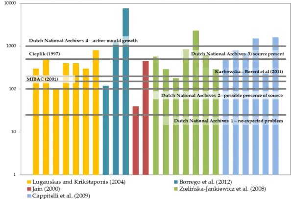
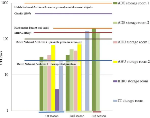
set them informally (Brokerhof et al., 2007; Harkawy et al., 2011) (see Table 1.1).
Several authors have proposed limits in Poland where the proposed indoor limit
value is the same as the limits stipulated for outside air. Karbowska-Berent et al. (2011)
based their values after studying the microbial quality on selected archives and
librar-ies and concluded that the higher concentrations of airborne fungi can signal the
24
Table 1.1 - Limits proposed for air fungal contamination in archives and libraries.
Each formed colony is called a colony forming unit (CFU).
Source Country Limit (CFU/m3)
Cieplik (1997) Poland 150 for a mixture of several species
50 for particular species
500 for common airborne fungal contaminants
Flieder and Capderou (1999)
France 100
MIBAC Italy 150
Dutch National archives (Brokerhoff et al., 2007)
Netherlands 0-25, no expected problems
25-100, possible presence of source, further testing needed
100-1000, source present, mould of-ten observed on objects
1000, active mould growth
Parchas (2008) France 120
Proposed guidelines (Harkawy et al, 2011)
Poland 5000
Karbowska-Berent et al. (2011)
Poland 200
The CFU (colony forming units) limits presented by the Dutch National archives
(Brokerhoff et al., 2007) refer to mixed flora and in case of just one species present, then
25-100 CFU/m3 already translates the presence of a source. As in other settings, there
are no proposed limits for surface contamination (CFU/m2). It could be very interesting
to think about this since it is the surface of books and their condition that we intend to
see preserved.
We have seen that in archives and libraries the presence of fungi can be
detri-mental to the documents and books safekept in these premises. But this principle is
du-25
ality makes it of the upmost importance to know the environment where books are
kept and people consult them or tend to their care.
Fungi and Human Health
Excerpts of this section are adapted from: Fungi in archives, a Double Concern,
In: Environmental Mycology in Public Health: An Overview on Fungi and Mycotoxins
for Risk Assessment and Management (Pinheiro 2015a, in press)
Fungal communities interact with paper based heritage. But what is the impact of
a fungal contaminated environment on the health of staff and visitors?
Fungi are considered as a serious threat to public health (Khan et al., 2012).
When in contact with the human skin, fungi can be responsible for the development of
dermatological problems such as tinea unguis, scalp disorders and dermal lesions with
varying severity degrees (Horner et al, 1995; Sousa et al, 2001). And fungi do not need
to establish close contact to induce health problems. Spores can be inhaled and, when
conditions are met, be responsible for lung and respiratory impairment. In cases of
re-duced immunity, this scenario can result in death (Horner et al., 1995). Both spores and
hyphae are potential allergens and their sole presence can trigger a histaminic response
(Horne et al., 1995; Khan et al., 2012). Sometimes difficult to diagnose, this affection can
last for weeks and bring about costs related to work absence and a general decrease in
life quality (Horner et al, 1995).
The mechanisms responsible for triggering mould induced illness include: type I
allergy, non-IgE mediated specific histamine release and inflammation, changes in
lymphocyte composition, generalized immunosuppression and toxic reactions. All of
these can be caused by fungal proteins, structural elements, microbial volatile organic
compounds (MVOCs) or mycotoxins and other secondary metabolites released from
fungal spores and colony fragments after inhalation (Nielsen et al., 2003). Mycotoxins
chem-26
ically heterogeneous assemblage that are grouped together because they can cause
dis-ease and death in human beings and other vertebrates (Bennett and Klich, 2003).
The subject of the health impact of fungal growth on the health of workers and
attendees of libraries and archives has been mentioned for almost a century but,
de-spite its relevance, there is still a lack of information concerning the fungal population
in libraries and archives (either quantitatively or qualitatively) and its relation to health
issues.
One of the first records on this concern dates back to the 1915 August 23 issue of
Every Week where Rose Murray, one of the six women referred to in the article "Wom-en Who Hold Down Unusual Jobs” is described as "the only woman in the world who holds the position of physician and surgeon to 'sick' books […]. She is the 'doctor' for all the volumes in the New York Free Public Library. There is a very lively element of danger in her
position, because books, like people, derive their sickness largely from germs and microbes. That
is why Miss Murray goes about her work dressed just like a surgeon at an operation. Her
equipment consists of a huge apron and a veil of cheesecloth." The image that illustrates the
text shows a woman nearly all enveloped in a white gown, with only her eyes, hands
and the bottom of her dress showing (Abbey Newsletter, 1994). Was she right in her
choice to heavily protect herself?
According to some authors, the most common malaises reported by staff
work-ing in libraries, archives or book containwork-ing premises are dermatitis, rhinitis, allergies
and asthma (Valentin, 2007). Allergic broncopulmonary aspergillosis and
hypersensi-tivity pneumonitis (Valentin, 2007) are probably the most serious. In a museum
envi-ronment, Wiszniewska et al. (2009) concluded that 30% of museum employees were
sensitized to at least one of the fungal allergens tested and that the prevalence of
aller-gic symptoms among the subjects was relatively high and frequently related to specific
sensitization. The most frequent symptoms reported by the examined subjects were:
conjunctivitis (68.5%), rhinitis (66%), skin symptoms (54%), chronic cough (26%) and
27
In 1997, Zyska performed an extensive review of the fungi encountered and
iden-tified in archives. Some of the fungi encountered are common air contaminants, such
as Penicillium sp. or Cladosporium sp., but others can be considered detrimental to
hu-man health – as Stachybotrys chartarum or Aspergillus fumigatus.
High level exposure to airborne viable fungi (106 CFU/m3) was determined as the
cause in a case of organic dust toxic syndrome in a museum staff handling mouldy
books (Kolstad et al., 2002). According to Zielińska-Jankiewicz et al. (2008), some of the
archive workers who participated in a survey conducted by Schata in 1995 reported
various skin, eye and respiratory symptoms which could have been associated with
occupational exposure to moulds. It was estimated that about one third of archive
workers might have developed allergy to moulds, which is about twice as high as in
the general population (Zielińska-Jankiewicz et al., 2008). The workers taking part in
the survey performed by Krake et al. in 1999, reported respiratory and sinus-related
symptoms which could have been associated with workplace exposure to moulds (Zielińska-Jankiewicz et al., 2008). In this study, mycological microflora belonging
mostly to the Penicillium, Cladosporium, Aspergillus, Alternaria, and Tritirachium species
was detected in levels that ranged from 200 to 450 CFU/m3, depending on the facility,
and for the Tritirachium species it approximated 800 CFU/m3.
Health-risk levels suggested in literature vary greatly from author to author and
through the years. Table 1.2 presents some of the available maximum fungal
concentra-tions (CFU/m3) proposals for indoor air (considering multiple building types).
The extreme variability in the mentioned values may be interpreted as a pointer
28
Table 1.2 – Proposed maximum concentrations (CFU/m3) limits and considerations for indoor air fungal contamination (Indoor Air Quality)
In Portugal, the ordinance 353-A/2013, December 4th, came to substitute
NT-SCE-02 for the regulation of the climatic certification of offices and service’s buildings. The former “legislation” (NT stands for technical note and complements the 2006 D e-cree-Law) was more rigorous than the present legislation since, for these types of
buildings, all the criteria had to be met at the time of inspection (see Chapter 5 for
fur-ther details). The Ordinance 353-A/2013 stipulates reference and conformity conditions
and now the indoor environment is considered safe when the indoor/outdoor (I/O)
fungal ratio is lower than one: higher than one ratios are not expected for fungi (Micalli
et al., 2003) since contamination comes normally from the atmosphere but study on
indoor fungal contamination is only taken further when this condition is not met.
When it is not, then there should be no visible fungal growth; fungal load should never
exceed the 500 CFU/m3; the presence of a mixture of relatively uncommon species
should not exceed 150 CFU/m3 and one uncommon species (Acremonium sp.,
Chrysonil-Proposed Limit Maximum fungal concentration
indoors
Holmberg (1984) (as cited by Flannigan, 2001) 2200 CFU/m3
Ohgke et al. (1987) Hurts et al. (1997) More than 100 CFU/m3 are a sign of
internal contamination
Reynolds, Streifel and McJilton (1990) 500 CFU/m3
Commission of the European Communities (1993) (as
cited by Zielińska-Jankiewicz et al., 2008).
2000 CFU/m3 (considered very high by
the Commission)
Yang, Hung and Lewis (1993); Etkin (1994) 200 CFU/m3.
Klánová (2000) 2000 CFU/m3 (higher should be
con-sidered a health threat)
World Health Organization (as cited by Goyer et al.,
2001)
29
ia sp., Tricothecium sp., Curvularia sp., Nigrospora sp.) should not exceed 50 CFU/m3. In
the former legislation uncommon species were defined as not being “Cladosporium sp.,
Alternaria sp. and Penicillium sp”.
As before, pathogenic (disease causing) species such as Cryptococcus neoformans,
Histoplasma capsulatum, Blastomyces dermatitidis and Coccidioides immitis are not
tolerat-ed. The presence of potentially toxinogenic (toxin emitting) fungi like Stachybotrys
chartarum (S. atra), Fusarium moniliforme, Fusarium culmorum, Trichoderma viride,
Asper-gillus versicolor, A.flavus, A. ochraceus, A. fumigatus and A. niger, in higher than 12
CFU/m3 is a sign of an environment of low(er) quality. By the former rules no
mini-mum was set for these species.
Some authors (Ren, Jankun e Leaderer, 1999) consider air sampling to be the best
method to determine fungal contamination and only air samples are accounted for in
these guidelines. Highlight in them is the need for fungal identification and not just a
mere quantification. The established relation between the minimum amount of spores
that can cause serious allergic reactions and the fungal species has been reported by
Valentin et al., 2007, as between 100/m3 for Alternaria alternata and 3000/m3 for
Cladosporium herbarum which reinforces the idea of identification rather than
quantifi-cation only (Micalli et al., 2003).
In most studies on indoor fungal contamination four main measures are applied:
total viable mould counts,
total mould counts (viable and nonviable),
specific mould species (qualitative or quantitative)
and β-(1,3)-d-glucan level (beta 1-3 glucans are structural elements of the fungus cell wall considered potent inflammatory agents (Valentin, 2003)
and responsible for hypersensitivity reactions (Salkinoja-Salonen et al.,
2003).
For the first three options, several methods can be chosen: gravitational
An-30
dersen sampler is the most popular), suction, filtration, electrostatic precipitation,
thermal precipitation and impingement (de Nuntiis et al., 2003) (see Chapter 3 for
fur-ther details).
The resulting samples can be analysed using the traditional culturing methods of
incubation and microscopic analysis or by molecular biology techniques, capable of
going further in terms of fungal identification and of sorting out the non-viable
frac-tion. Whenever possible, the best option is to perform both analyses.
For the determination of the atmospheric allergen load (β-(1, 3)-d-glucan level in-cluded) immunological techniques such as EIA (enzyme Immunoassay), ELISA
(En-zyme-linked Immunoassay) are frequently used (Favalli et al., 2003).
1.1 Research problems
Since the last review regarding fungi in archives was performed in 1997
and refers to fungi identified in museums and archives before the advent of modern
identification techniques, is it safe to say we know the fungal communities present in
our archives and libraries?
In fungal aerobiology studies what methodologies should be used?
Classic culturing methods, molecular biology protocols or both? Given the existing
choices in both fields, which particular methodologies should be pursued and
opti-mized?
Knowing the fungal flora present in archives and librairies is extremely
important to assess the risk and to devise mechanisms to eliminate/diminish it. What is
the present situation in Portuguese archives?
Are the fungal communities found in selected Portuguese archives in
agreement with international results?
What can we expect from the fungal flora identified in the selected
31
There are no existing guidelines on fungal contamination for Archives.
Can the collected data be a valuable tool in addressing this issue?
Fungal community studies are a pivotal part of any indoor air quality
study. Do archives and libraries comply with national legislation (for health purposes) and proposed limits (for conservation)?
1.2 Objectives
The main objective of this thesis is to assess the fungal communities present in
archives and study their impact on both paper conservation and human health. To
achieve these goals, it was necessary:
To review existing literature on fungal contamination in archives and
methodologies on fungal identification.
To design a study to assess fungal contamination in archives. The
se-lected methodology should include all steps of the experimental work: sampling,
sam-ple treatment and fungal identification.
To assess fungal contamination in selected Portuguese archives. This
evaluation should include air samples, surface samples and areas affected by fungal
growth and/or pigmentation.
To compare the resulting fungal assessment with the results obtained
abroad.
To evaluate the characteristics presented by the identified fungal flora
and estimate their influence on both the staff’s and attendees health and the doc
u-ment’s conservation.
To assess the indoor air quality in Portuguese archives, both from the
32
To investigate the influence of water activity on fungal development
and fungal load on documents. Also to study the relationship between the water
activi-ty on documents and relative humidiactivi-ty from the air.
To test the application of the molecular biology protocol developed
out-side the archives.
1.3 Thesis outline
It is essential to determine the fungal profile of a given setting to truly
un-derstand the impact fungi can have on both document’s conservation and human
health preservation. Indoor air quality studies do not omit this fact and include both
identification and quantification of fungal species/genera (Decree Law 78 /2006, April
4th and Ordinance 353-A/2013, December 4th). It was the purpose of this study to
ad-dress indoor air quality in archives and libraries with a special focus on the
characteri-zation of fungal flora communities and its relation to documents biodeterioration and
human health.
In 1997, almost 20 years ago, Zyska reviewed the fungi encountered and
identi-fied in archives and these results refer to analysis on books, documents, air and dust
samples in a time where fungal identification was only achieved by conventional
cul-turing methods. An updated review is imperative and presented in Chapter 2.
After having the knowledge about fungi identified in archives all over the world,
it is important to find out the Portuguese reality regarding this subject. Therefore, four
Portuguese archives were selected for study. Methodologies to perform this study are
presented in Chapter 3 and include classical culturing methods and modern molecular
biology protocols especially relevant to determine the non-viable fraction of the
biolog-ical contaminants. Within the latter, denaturing high performance liquid
chromatog-raphy (DHPLC), a very recent technology, first created to detect gene mutation has
al-ready been applied to the study of microbial community but only bacterial ones. study
of microbial communities. In this study, a new methodology was developed and firstly
33
Once presented the methodology, the results from a study on airborne and
set-tled fungi carried out in four Portuguese settings are presented. Considerations were taken from their presence, from both the document’s safekeeping and the human health perspective (Chapter 4).
Fungal communities are intrinsically related to water activity and other
pa-rameters such as particulate matter. This last one is a key component in any indoor air
quality evaluation - of which there are no records for Portuguese archives. Biological,
physical and chemical parameters must be kept within limits in order to obtain a
certi-fication for air quality. Certicerti-fication parameters were analysed in two Portuguese
ar-chives in order to assess both the indoor air quality for people attending or working in
these premises and for the valuable written heritage that must be kept for future
gen-erations (Chapter 5).
The study on fungal contamination showed a wide and varied fungal
communi-ty. Water activity is determinant to evaluate its potential for deteriogenic and
toxino-genic activity. An entire food industry relies on the principle of water balance between
substrate and the environment and this should also be considered when it is cultural heritage we’re discussing. This relevant issue is presented and discussed in Chapter 6.
Desirably, the breakthroughs achieved in a PhD project gain value when they can
also serve other scientific areas. As mentioned, this was a project meant to join together
the areas of health and written heritage conservation. Chapter 3 introduced the
DHPLC and its utility in the identification of fungi present in paper samples and the
developed methodology was applied successfully both in the field of clinical diagnosis
and fungal contamination in a gilded wood church structure. Both methodology
appli-cations are presented in Chapter 7.
The main results and conclusions of this study and the further research that
35
37
A review of the literature
The last formal review on the area of fungi in archives and libraries was
per-formed by Zyska in 1997 (almost 20 years ago) and recollects data from very diverse
origins. In fact, it is not just focused on books but on panoply of materials one can find
in museums, archives or libraries. It does have the great advantage (sometimes
forgot-ten by some of its recent followers) of presenting data from both air and surface
sam-ples which can be essential to truly know the fungal communities. Some spores, given
their characteristics, tend to aggregate and are not easily airborne which makes it
diffi-cult to find them in air samples although they can be present in the environment
(Duchaine and Mérieux, 2001). This could be deemed of lesser importance if this was
not the case for Stachybotrys chartarum, one the fungi most associated with Sick
Build-ing Syndrome (Bennett and Klich, 2003).
Because no other method was sufficiently evolved at the time, the identifications
gathered by Zyska include only classical culturing methods which leave out the trendy
and modern molecular biology protocols. And these do bring out in the open some
fungal species which were either unidentifiable (mycelia sterilia or sterile micelia) or
simply did not show any growth.
Between 1997 and 2013 several authors have embraced the task of characterizing
the fungal flora present in archives. A compilation of these studies is presented in the
next pages. Tables 2.1 to 2.6 refer to fungi identified:
in air samples (Tables 2.1, 2.2, 2.3 and 2.5)
surface samples (shelves, tables and floor) (Table 2.4 and 2.5)
archival document cases (Table 2.4 and 2.5)
38
2.1 Air studies
Due to the difficulty of achieving species identification - for reasons as simple as
the overwhelming variety of species within a fungal genus - many studies stop at this
taxonomic level. Penicillium species, for instance, are impossible to identify using just
simple culturing methods and/or relying on just one growth media.
Several countries have performed indoor air studies in other settings. While in
Scotland the most prevalent genus is Cladosporium (Stevens, 2004), in Great Britain the
most frequently isolated is Penicillium. In the Netherlands, North America, Canada,
Australia, Germany, Lithuania, Taiwan and Turkey, Penicillium and Aspergillus join
Cladosporium as the most common indoor fungi genera (Stevens, 2004).
With slightly different results and mentioning some species, the most common
indoor species for Singh (2001) were Cladosporium herbarum, Alternaria alternata,
Euroti-um herbariorEuroti-um, Penicillium sp., Aspergillus sp. (namely Aspergillus versicolor),
Aureo-basidium pullulans, Mucor sp., Phoma sp. and Wallemia sp.
In Portugal, indoor air quality studies have found Cladosporium, Aspergillus and
Alternaria as the most common genera (Viegas et al., 2012). The outdoor air is one of the main sources of fungi and in Lisbon’s outdoor environment the most predominant
genera are Cladosporium, Penicillium, Aspergillus and Alternaria (Viegas et al, 2012).
Zyska (1997) and Gallo et al. (2003, data recollected in 1998 for the first version of
the book) mention some of the most common fungi in archives and librairies. When to
these two reviews we add the identified fungal genera found in field studies (Gambale