Ciências da Saúde
Analysis of the expression of taste and olfactory
receptors in choroid plexus and orbitofrontal
cortex of Alzheimer’s disease patients
Victória da Cunha Alves
Dissertação para obtenção do Grau de Mestre em
Ciências Biomédicas
(2º ciclo de estudos)
Orientadora: Prof. Dra Isabel Gonçalves
Coorientadoras: Dra. Joana Figueiró Silva e Dra. Eva Carro, Instituto de
Investigación Hospital 12 de Octubre, Madrid
iii The content of this dissertation falls under the exclusive responsibility of the author.
v
Acknowledgments
Transformative achievements come from the concerted and at times unrecognized contributions of many. Any research is not an individual effort; it is a contributory effort of many hearts, hands and heads. I, therefore, attribute this successful endeavour to the vast array of significant players that I have had the grand fortune to have entered my life; this achievement is truly OURS. My deepest and sincere gratitude!
Specially note of thanks to Prof Dra Cecília Santos and to Prof Dra Isabel Gonçalves for placing this enormous opportunity in front of me and showing me the doors that might be useful to open.
I am as well forever grateful to Dra Eva Carro for welcoming me into her scientific group, for her endlessly guidance and encouragement, and for providing all necessary facilities for this research work.
Last but not least, I’d like to acknowledge and give my wholehearted gratitude to Joana Figueiró who has walked alongside me during this last months and whose guidance and continuous support could never be described in words. It has been a real pleasure and inspiration to work with such a brilliant mind. I thank you for challenging me intellectually, creatively and personally.
The bottom line is that without Joana’s encouragement and faith in my abilities, I’d have never become any kind of researcher; and it is to her that I gratefully dedicate this dissertation.
vii
Abstract
Alzheimer’s disease (AD) is a chronic neurodegenerative disease characterized by progressive memory loss and cognitive deterioration, attributed to neuropathological lesions within specific regions in the brain. However, other areas of the brain such as the choroid plexus and the orbitofrontal cortex, have gained much attention since the choroid plexus is a multifunctional tissue responsible for a wide range of functions crucial to the central nervous system and the orbitofrontal cortex is considered among the most polymodal regions of the brain. Previous studies have shown the expression of taste and olfactory transduction pathways in rat choroid plexus as well as taste and olfactory regulation and activation of orbitofrontal cortex. Therefore the present work aimed to study the expression of taste and olfactory receptors in the choroid plexus and orbitofrontal cortex of AD patients. Towards this aim, transcriptomic analysis of these chemoreceptors were performed by means of Real-time quantitative Polymerase Chain Reaction (RT-qPCR), and compared between AD patients (at different Braak stages of the disease) and age-matched controls. Transcriptomic analysis indicated that orbitofrontal cortical olfactory receptors (ORs) and taste receptors (TASRs) are expressed and regulated at different stages of AD in female patients, whereas in male this regulation was not observed. Moreover, olfactory receptors OR2K2, OR2H2, and OR1L8 and taste receptor TAS2R14 were downregulated at Braak stages I, V, VI, compared to age-matched controls in the orbitofrontal cortex of female AD patients. The strongest differences were found at Braak stage I of AD, suggesting that dysregulation of ORs and TASRs is an early event in the pathogenesis of AD in female patients. Also, that ORs and TASRs in the orbitofrontal cortex might be differently affected in male and female AD patients. These receptors we also found to be expressed in choroid plexus, however, due to their low expression we were not able to quantify the relative mRNA levels by RT-qPCR. Nevertheless further studies are needed in order to strengthen these findings and to elucidate their potential physiologic functions in this brain area.
Keywords
Alzheimer’s disease, olfactory receptors, taste receptors, choroid plexus, orbitofrontal cortex, Real Time quantitative PCR
ix
Resumo alargado
A doença de Alzheimer é uma doença neurodegenerativa crónica caracterizada por uma perda progressiva da memória e défice das capacidades cognitivas. Estes défices cognitivos são atribuídos a lesões neuropatológicas intrínsecas a regiões específicas do cérebro. No entanto, outras áreas do cérebro, como o plexo coroide e o córtex orbito-frontal, têm sido objeto de grande atenção uma vez que o plexo coroide é um tecido multifuncional responsável por uma vasta gama de funções cruciais ao sistema nervoso central e o córtex orbito-frontal é considerado uma das regiões mais polimodais do cérebro. Estudos anteriores demonstraram a expressão das vias de transdução do gosto e do olfacto no plexo coroide de ratos, bem como a ativação e regulação olfativa e gustativa do córtex orbito-frontal. Inclusivamente, num estudo anterior foi demonstrado que no plexo coroide de ratos a expressão de genes relacionados com o sabor e o olfato se encontram sob o controlo de hormonas esteróides sexuais.
Por esse motivo, o presente trabalho teve como objetivo estudar a expressão dos recetores olfativos e gustativos no plexo coroide e no córtex orbito-frontal de pacientes com doença de Alzheimer de ambos os sexos. A fim de atingir esse objetivo, foi necessário desenhar primers específicos para esses recetores e optimizar as várias condições para obter curvas de calibração eficientes para a análise transcritómica quantitativa. A análise do transcriptoma destes quimiorrecetores foi então efectuada através de reação em cadeia da polimerase quantitativa em tempo real (em inglês, Real-time quantitative Polymerase Chain Reaction), e os níveis de expressão foram comparados entre pacientes com doença de Alzheimer (em diferentes estágios da doença) e controlos pareados.
A análise do transcriptoma revelou que os recetores olfativos (ORs) e gustativos (TASRs) orbito-frontal corticais se expressam e se encontram regulados em alguns estágios da doença em pacientes do sexo feminino, enquanto que em pacientes do sexo oposto essa mesma regulação nao foi observada. Nomeadamente, a expressão dos recetores olfativos OR2K2, OR2H2 e OR1L8 e o recetor gustativo TAS2R14 encontra-se regulada negativamente nos estágios de Braak I, V e VI em comparação com os controlos pareados em córtex orbito-frontal de pacientes do sexo feminino com doença de Alzheimer. As diferenças mais evidentes foram encontradas no estágio de Braak I, sugerindo que a desregulação da expressão dos recetores olfactivos e gustativos é um evento que ocorre precocemente na patologia de Alzheimer em pacientes do sexo feminino. Apesar da sua tendência para aumentar em estágios mais avançados, possivelmente em resposta à progressão da doença (ao nível bioquímico e estrutural), o córtex orbito-frontal parece não ser capaz de restaurar os níveis observados em pacientes controlo. Além disso, estes resultados sugerem também que a expressão dos ORs e os TASRs no córtex orbito-frontal pode ser afetada de forma distinta entre pacientes de Alzheimer de um ou outro sexo.
Inclusivamente, verificou-se que estes recetores também se expressam em plexo coroide, no entanto, devido à sua baixa expressão não foi possível quantificar os seus níveis relativos de mRNA por PCR quantitativa em tempo real.
x
A utilização de tecidos post-mortem humanos para estudos de expressão génica é particularmente desafiador. Além do problema do RNA danificado, temos de enfrentar um elevadíssimo grau de variabilidade biológica dentro de um conjunto de amostras. As variações dos parâmetros individuais como a idade, a massa corporal, a saúde, mas também a causa e circunstâncias da morte e o intervalo post-mortem podem conduzir a um conjunto de amostras bastante inomogéneo.
Adicionalmente, mais estudos são necessários a fim de reforçar estas conclusões e elucidar as possíveis funções fisiológicas destes quimiorrecetores nestas áreas do cérebro.
Palavras-chave
Doença de Alzheimer, recetores olfativos, recetores gustativos, plexo coroide, córtex orbito-frontal, PCR quantitativa em tempo real
xi
Index
I. Introduction ... 1
1. Overview of Alzheimer’s Disease ... 3
1.1. Clinical phases and symptoms of AD ... 3
1.2. Sporadic and Familial AD ... 5
1.3. Pathological changes in AD ... 5
1.3.1 Amyloid β plaques and Tau neurofibrillary tangles ... 6
1.4. Risk factors for AD ... 7
1.4.1 Advancing age ... 7
1.4.2 Family history ... 8
1.4.3 Apolipoprotein E ε4 ... 8
1.4.4 Mild cognitive impairment ... 8
1.4.5 Head injury and traumatic brain injury ... 8
1.4.6 Cardiovascular disease risk factors ... 9
1.5. Gender differences in AD ... 10
1.6. Post-mortem diagnosis of AD ... 11
2. Choroid plexus ... 13
2.1 Location and structure ... 14
2.2 The choroid plexus as a 'bioreactor' to brain diseases - impact on AD ... 16
2.2.1 Morphological alterations ... 17
2.2.2 Aβ deposits ... 17
2.2.3 Impaired mitochondrial function ... 18
2.2.4 Oxidative stress and cell death ... 18
3. Cerebral cortex ... 19
3.1 The structure and functional localization of the cerebral cortex ... 19
3.2 Orbitofrontal cortex... 20
4. Olfactory and Taste Receptors ... 22
4.1 Olfactory Receptors ... 22
4.1.1 Olfactory receptor genes ... 22
xii
4.1.3 Olfactory signal transduction in olfactory neurons ... 24
4.2 Taste Receptors... 24
4.2.1 Taste receptor genes ... 25
4.2.2 Taste receptors expression ... 26
4.2.3 Taste signal transduction ... 27
II. Aim ... 29
Aim ... 31
III. Materials and Methods ... 33
1. Samples ... 35
2. Total RNA ... 36
2.1 Total RNA extraction ... 36
2.2 Quality assessment and quantification of total RNA ... 37
2.2.1 Agarose Gel Electrophoresis ... 37
2.2.2 Quantification ... 38
2.3 Removal of genomic DNA contamination ... 38
3. Reverse-Transcription ... 39
4. Polymerase chain reaction (PCR) ... 39
4.1 Primer design ... 40
4.2 Primer testing ... 42
4.3 Real Time quantitative PCR (RT-qPCR) ... 43
4.3.1 Conditions optimization ... 43 4.3.2 Standard curve ... 46 4.3.3 Data analysis ... 48 5. Statistical analysis... 48 IV. Results ... 49 1. Primer testing ... 51 2. RT-qPCR optimization ... 53
3. Housekeeping genes validation in orbitofrontal cortex ... 54
4. Olfactory and taste receptors expression in orbitofrontal cortex ... 56
4.1 Olfactory receptors ... 56
xiii
4.1.2 OR2H2 mRNA expression ... 57
4.1.3 OR1L8 mRNA expression ... 59
4.1.4 OR13A1 mRNA expression ... 60
4.1.5 OR7A17 mRNA expression ... 60
4.2 Taste receptors ... 61
4.2.1 TAS2R5 mRNA expression ... 61
4.2.2 TAS2R14 mRNA expression ... 62
5. Olfactory and taste receptors expression in choroid plexus ... 63
V. Discussion and Conclusions ... 65
VII. References ... 73
xv
Acronyms list
ABCA1 ATP binding cassette transporter 1
AC3 Adenylyl cyclase 3
AD Alzheimer’s disease
APOE Apolipoprotein E
APP Amyloid precursor protein
Aβ Amyloid β
BDNF Brain derived neurotrophic factor
cAMP Cyclic adenosine monophosphate
cDNA Complementary DNA
CNG Cyclic nucleotide-gated
CP Choroid plexus
CSF Cerebrospinal fluid
Ct Threshold cycle
dsDNA double-stranded DNA
EOAD Early-onset Alzheimer’s disease
FFA Free fatty acid
GAPDH Glyceraldehyde3-phosphate desydrogenase
GI Gastrointestinal
GPCR G protein-coupled receptor
HRT Hormone replacement therapy
IGF-1 Insulin-like growth factor-1
IP3 Inositol-1,4,5-triophosphate
IP3R3 IP3 receptor type 3
LOAD Late-onset Alzheimer’s disease
MCI Mild cognitive impairment
NO Nitric oxide
NPH Normal pressure hydrocephalus
OE Olfactory epithelium
OFC Orbitofrontal cortex
OR Olfactory receptor
PCR Polymerase chain reaction
PFC Prefrontal cortex
PGK1 Phosphoglycerate kinase 1
xvi
PSEN 1 Presenilin 1
PSEN 2 Presenilin 2
RT Room temperature
RT-qPCR Real Time quantitative polymerase chain reaction
SG Salivary gland
ssDNA single-stranded DNA
T1R Taste receptor type 1
T2R Taste receptor type 2
Ta Annealing temperature
TAE Tris-acetate-EDTA
TASR Human taste receptor
Tasr Rodent taste receptor
Tm Melting temperature
TR Taste receptor
TRPM5 Transient receptor potential ion channel, subfamily M, member 5 VGSC Voltage-gated sodium channels
1
I. Introduction
3
1. Overview of Alzheimer’s Disease
Alzheimer’s disease (AD) is a chronic neurodegenerative disease and the most common type of dementia (Wilson et al., 2012).
As a consequence of population aging worldwide, it has reached an incidence of a case every 7 seconds and a fourfold increase is expected to occur over the next decades (Cornutiu, 2015). Recent estimates foresee that more than 80 million individuals will be affected by the disease by 2040 (Wilson et al., 2012; Cornutiu, 2015).
Although AD was first identified more than 100 years ago, research into its symptoms, causes, risk factors, and treatment has gained momentum only in the past 30 years. Most experts agree that AD, like other common chronic diseases, develops as a result of multiple factors rather than a single cause. These factors include a variety of brain changes that begin as early as 20 years before symptoms appear, affecting one’s memory, behaviour, and ability to think clearly (Scheltens et al., 2016).
1.1. Clinical phases and symptoms of AD
According to recent propositions, three clinical phases of AD may be defined:
(i) Asymptomatic (or pre-clinical) AD, which may last for several years or decades until the overproduction and accumulation of Amyloid β in the brain reaches a critical level that triggers the amyloid cascade;
(ii) Mild cognitive impairment (MCI) (pre-dementia phase of AD) compatible with the definition of progressive, amnestic MCI, in which early-stage pathology is present, ranging from mild neuronal dystrophy to early-stage Braak pathology, and may last for several years according to individual resilience and brain reserve;
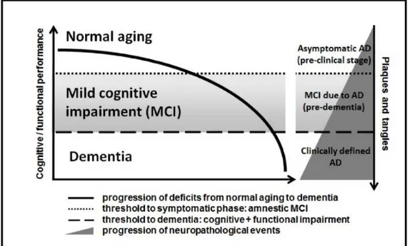
(iii) Clinically defined dementia phase of AD, in which cognitive and functional impairment is severe enough to surmount the dementia threshold; at this stage there is significant accumulation of neuritic plaques and neurofibrillary tangles in affected brain areas, bearing relationship with the magnitude of global impairment (Figure 1).
4
Figure 1 - Relationship between the progression of cognitive and functional symptoms and the neuropathological events in the transition from asymptomatic AD to mild cognitive impairment due to AD and clinically manifest dementia of the AD type. Adapted from Forlenza et al., 2010.
There is evidence of a long preclinical phase in AD, in which the aforementioned abnormalities gradually accumulate in affected brain areas prior to the presentation of significant cognitive decline and dementia (Tarawneh & Holtzman, 2012). Also, the neuropathological features of subjects with MCI are intermediate between those found in cognitively normal and demented individuals (Forlenza et al., 2010).
Recent models based on neuropathological, biochemical and neuroimaging methods have proposed that intracerebral amyloidosis precedes the onset of cognitive symptoms by several years, if not decades. Autopsy studies have shown that intracerebral amyloidosis may be observed in some subjects as early as in the third or fourth decades of life, with increasing magnitude in late middle age, and highest estimates in old age (Thal et al., 2000, Thal et al., 2002; Knopman et al., 2003). Therefore, the long pre-dementia phase in AD constitutes a unique time frame to search for clinical and neurobiological tools to reinforce the cross-sectional diagnosis and to predict the dementia outcome.
AD affects people in different ways, but the most common symptom pattern begins with gradually worsening ability to remember new information. This occurs because disruption of brain cell function usually begins in brain regions involved in forming new memories. As damage spreads, individuals experience other difficulties. The following are common symptoms of AD:
Memory loss that disrupts daily life. Challenges in planning or solving problems.
Difficulty completing familiar tasks at home, at work, or at leisure. Confusion with time or place.
5 Trouble understanding visual images and spatial relationships.
New problems with words in speaking or writing. Misplacing things and losing the ability to retrace steps. Decreased or poor judgment.
Withdrawal from work or social activities. Changes in mood and personality.
Individuals progress from mild AD to moderate and to severe at different rates. As the disease progresses the individual’s cognitive and functional abilities decline. Those in the final stages of the disease lose their ability to communicate, fail to recognize loved ones, and become bedbound and reliant on around-the-clock care. They even become more vulnerable to infections, including pneumonia. AD is ultimately fatal, and AD-related pneumonia is often a contributing factor (Castellani et al., 2010).
1.2. Sporadic and Familial AD
There are two types of AD: familial (also known as early onset) and sporadic (also known as late onset, LOAD). Although most cases of AD are sporadic, rare familial forms exists with an autosomal dominant pattern of inheritance. Familial AD accounts for less than 1% of all cases and is caused by any of three known genetic mutations (Bekris et al., 2010; Acosta-Baena et al., 2011). These mutations involve the gene for the amyloid precursor protein (APP) and the genes for the presenilin 1 (PSEN1) and presenilin 2 (PSEN2) proteins. Inheriting a mutation to the APP or PSEN1 gene guarantees that an individual will develop AD. Those inheriting a mutation in the PSEN2 gene have a 95% chance of developing the disease (Goldman et al., 2011). Individuals with mutations in any of these genes tend to develop AD before age 65 years, sometimes as young as 30 years (Acosta-Baena et al., 2011). In contrast to familial AD, LOAD is etiologically heterogeneous and results from a combination of many environmental conditions, lifestyle and genetic features of the individual (Huang et al., 2004; Liu et al., 2015). Although there is evidence of a strong genetic component to disease risk, until quite recently, the only established genetic factor for LOAD is apolipoprotein E (APOE). APOE-associated AD is due to
APOE ε4 allele, which is further explained in section 1.4.3.
1.3. Pathological changes in AD
AD is a disease with well-defined pathophysiological mechanisms, mostly affecting medial temporal lobe and associative neocortical structures (De-Paula et al., 2012). Neuritic plaques and neurofibrillary tangles represent the pathological hallmarks of AD, and are respectively related to the accumulation of the amyloid β peptide (Aβ) in brain tissues, and to cytoskeletal changes that arise from the hyperphosphorylation of microtubule-associated Tau protein in neurons (Takahashi et al., 2013). The accumulation of misfolded proteins in the aging brain
6
results in oxidative and inflammatory damage, which in turn leads to energy failure and synaptic dysfunction (Chen et al., 2012).
The accumulation of Aβ within structurally damaged mitochondria (Calkins et al., 2011) is consistent with other evidence of intraneuronal Aβ in AD (Gouras et al., 2012). Aβ is a potent mitochondrial toxic, especially affecting the synaptic pool (Querfurth & LaFerla, 2010). Dysfunctional mitochondria release oxidizing free radicals which cause considerable oxidative stress. Experimental models show that markers of oxidative damage precede pathological changes (Nunomura et al., 2001).
Vascular injury and parenchymal inflammation perpetuate the cycle of protein aggregation and oxidation in the brain in AD. Pervasive pathological changes include cerebral amyloid angiopathy (Greenberg et al., 2004), affecting more than 90% of AD patients, capillary abnormalities, disruption of the blood–brain barrier, and large-vessel atheroma (Roher et al., 2004).
1.3.1 Amyloid β plaques and Tau neurofibrillary tangles
According to the amyloid hypothesis of AD, the overproduction of Aβ is a consequence of the disruption of homeostatic processes that regulate the proteolytic cleavage of the amyloid precursor protein (APP). Genetic, age-related and environmental factors contribute to a metabolic shift favouring the amyloidogenic processing of APP in detriment of the physiological, secretory pathway. The neurotoxic potential of the Aβpeptide results from its biochemical properties that favour aggregation into insoluble oligomers and protofibrils. These further originate fibrillary Aβspecies that accumulate into senile and neuritic plaques. These processes, along with a reduction of Aβ clearance from the brain, leads to the extracellular accumulation of Aβ, and the subsequent activation of neurotoxic cascades that ultimately lead to cytoskeletal changes, neuronal dysfunction and cellular death (De-Paula et al., 2012). Neurofibrillary tangles, which are filamentous inclusions in pyramidal neurons, are also a pathologic marker of severity of AD. The major component of the tangles is an abnormally hyperphosphorylated and aggregated form of tau protein (Iqbal et al., 2010). Like Aβ oligomers, intermediate aggregates of abnormal tau molecules are cytotoxic (Khlistunova et al., 2006) and impair cognition (Querfurth & LaFerla, 2010). Neurofibrillary tangles appear first in allocortical structures, whereas amyloid plaques may first be found in the neocortex (Nelson et al., 2009; Forlenza et al., 2010). Interestingly, experimental evidence indicates that Aβ accumulation precedes and drives tau aggregation (Gotz et al., 2001; Lewis et al., 2001; Oddo et al., 2003). Increased oxidative stress, the impaired protein-folding function of the endoplasmic reticulum, and deficient proteasome-mediated and autophagic-mediated clearance of damaged proteins — all of which are also associated with aging — accelerate the accumulation of amyloid and tau proteins in AD (López et al., 2000; Hoozemans et al., 2005). In addition to amyloid accumulation
7 and neurofibrillary pathology, synaptic dysfunction leading to neuronal dystrophy are phenomena proxy to the structural changes of the brain, which ultimately triggers the clinical syndrome that characterizes incipient AD (Jack et al., 2010) – Figure 2. The cognitive manifestations associated with this process are compatible with subtle damage to hippocampal and related limbic and prefrontal structures, and may last for many years until the functional burden becomes severe enough to surmount the dementia threshold (Blass, 2002).
Figure 2 - Hypothetical model of the pathological processes in Alzheimer's disease (AD), focusing on the amyloid β peptide (Aβ) cascade. Adapted from Forlenza et al., 2010.
1.4. Risk factors for AD
With the exception of the rare cases of AD caused by genetic mutations, experts believe that AD develops as a result of multiple factors rather than a single cause. This section describes known risk factors for Alzheimer’s.
1.4.1 Advancing age
The risk of developing AD increases exponentially with age (Plassman et al., 2007). Although the greatest risk factor is advancing age, AD is not a normal part of growing older. Most people with AD are diagnosed at age ≥65 years. These individuals are said to have LOAD. However, people aged <65 years can also develop the disease. When AD develops in a person aged <65 years, it is referred to as “early-onset” AD (EOAD) (Apter et al., 2015).
8
1.4.2 Family history
Individuals who have a parent, brother, or sister with AD are more likely to develop the disease than those who do not have a first-degree relative with AD (Green et al., 2002, Loy et al., 2014). Those who have more than one first-degree relative with AD are at even higher risk of developing the disease (Goldman et al., 2012). When diseases run in families, heredity (genetics), shared environmental/lifestyle factors or both may play a role.
1.4.3 Apolipoprotein E ε4
Individuals with the ε4 allele of the gene apolipoprotein E (APOE ε4) are at increased risk of developing AD. APOE ε4 is one of three common variants (ε2, ε3, and ε4) of the APOE gene, which provides the blueprint for a protein that carries cholesterol in the bloodstream. The
APOE is an autosomal gene, and the presence of APOE ε4 variant increases the risk of developing
AD and of developing it at an earlier age (Spinney, 2014). Two APOE ε4 genes grant an even higher risk (Loy et al., 2014, Holtzman et al., 2012). Researchers estimate that between 40-65% of people diagnosed with AD have one or two copies of this form of the APOE gene (Olgiati et al., 2011). However, unlike inheriting a genetic mutation that causes AD, inheriting the ε4 form of the APOE gene does not guarantee that an individual will develop AD.
1.4.4 Mild cognitive impairment
MCI is a condition in which an individual has mild, but measurable, changes in thinking abilities that are noticeable to the person affected and to family members and friends, but do not affect the individual’s ability to carry out everyday activities (Summers et al., 2012). People with MCI, especially MCI involving memory problems, are more likely to develop AD and other dementias compared to non-impaired age-matched adults. Between 10% and 20% of adults above the age of 65 are diagnosed with MCI (Kirova et al., 2015), and approximately 10% of MCI adults progress to AD (Forlenza et al., 2010). In some cases, such as when MCI is caused by certain medications, MCI can be reversed. In other cases, MCI reverts to normal cognition on its own or remains stable. Revised criteria and guidelines for diagnosis of AD suggest that in some cases, MCI is actually an early stage of AD or another dementia (Mckhann et al., 2011, Albert et al., 2011, Sperling et al., 2011).
1.4.5 Head injury and traumatic brain injury
Head injury, head trauma, and traumatic brain injury are associated with an increased risk of AD and other dementias. Moderate head injuries are associated with twice the risk of developing AD compared with no head injuries, and severe head injuries are associated with
9 4.5 times the risk (LoBue et al., 2016). Moderate head injury is defined as a head injury resulting in loss of consciousness or posttraumatic amnesia lasting more than 30 minutes; if either of these lasts more than 24 hours, the injury is considered severe. Groups that experience repeated head injuries, such as boxers, football players, and combat veterans, may be at increased risk of dementia, late-life cognitive impairment, and evidence of tau tangles (a hallmark of AD) at autopsy (Guskiewicz, 2005; Vincent et al., 2014).
1.4.6 Cardiovascular disease risk factors
Growing evidence suggests that the health of the brain is closely linked to the overall health of the heart and blood vessels. The brain is nourished by one of the body’s richest networks of blood vessels. A healthy heart helps ensure that enough blood is pumped through these blood vessels to the brain, and healthy blood vessels help ensure that the brain is supplied with the oxygen- and nutrient-rich blood it needs to function normally.
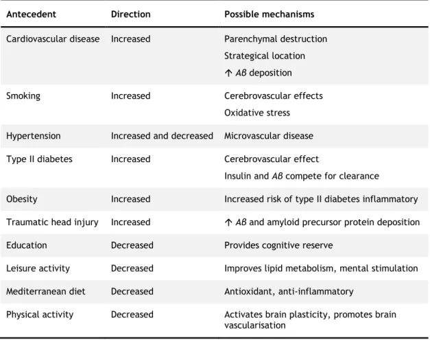
Some data indicate that cardiovascular disease risk factors are associated with a higher risk of developing AD and other dementias. These factors include physical inactivity (Reitz et al., 2011; Willis et al., 2012), high cholesterol (especially in midlife) (Solomon et al., 2009; Meng et al., 2014), diabetes (Pendlebury et al., 2009; Reitz et al., 2011; Sekita et al., 2010), smoking (Anstey et al., 2007; Pendlebury et al., 2009; Rusanen et al., 2011), and obesity (Anstey et al., 2011; Rönnema et al., 2011; Loef et al., 2013). Other factors such as a higher level of education, as well as Mediterranean diet were shown to decrease the risk of developing AD (Gu et al., 2010; Reitz et al., 2011; Lourida et al., 2013). Table 1 shows some identified factors that modify the risk of AD. Unlike genetic risk factors, many of these cardiovascular disease risk factors are modifiable, that is, they can be changed to decrease the likelihood of developing cardiovascular disease and, possibly, the cognitive decline associated with AD and other forms of dementia.
10
Table 1 - Factors that modify the risk of Alzheimer disease. Adapted from Mayeux et al., 2012.
Antecedent Direction Possible mechanisms
Cardiovascular disease Increased Parenchymal destruction Strategical location Aβ deposition
Smoking Increased Cerebrovascular effects
Oxidative stress Hypertension Increased and decreased Microvascular disease Type II diabetes Increased Cerebrovascular effect
Insulin and Aβ compete for clearance
Obesity Increased Increased risk of type II diabetes inflammatory Traumatic head injury Increased Aβ and amyloid precursor protein deposition
Education Decreased Provides cognitive reserve
Leisure activity Decreased Improves lipid metabolism, mental stimulation Mediterranean diet Decreased Antioxidant, anti-inflammatory
Physical activity Decreased Activates brain plasticity, promotes brain vascularisation
1.5. Gender differences in AD
The area of gender differences in AD and in neurodegenerative processes, although still largely unexplored, appears to offer great promise for the future development of better intervention strategies for patients. Prevalence studies on dementia generally show a higher risk in women than in men (Mielke et al., 2014). A possible gender difference in the risk of AD is further supported by recent evidence suggesting that the brain’s so called cognitive reserve is reduced in women. Also, in patients with MCI and AD, brain volumes have been found to decline faster in women than men, supporting the evidence of faster progression of women from MCI to AD (Skup et al., 2011). In addition, the majority of studies have reported that the effects of the
ε4 genotype - the strongest known genetic risk factor for LOAD (Liu et al., 2013) - are more
pronounced in women than in men (Ungar et al., 2013). It was also reported that women with one ε4 allele had about a four-fold risk of AD, whereas men showed little increased risk (Mielke et al., 2014; Ungar et al., 2013). The APOE ε4 allele also has a greater deleterious effect on hippocampal pathology, functional connectivity changes in the default mode network, cortical thickness, and memory performance in women compared with men at different stages of AD (Liu et al., 2010; Damoiseaux et al., 2012). Furthermore, a large autopsy study found that
11 amyloid plaque and neurofibrillary tangle pathology was greater among women who were ε4 carriers (Holland et al., 2013).
Other genes and polymorphisms have also been shown to increase risk and progression of AD in one sex, but not the other. A study reported that the Met66 allele of Brain derived neurotrophic factor (BDNF) gene, which reduces the transport of BDNF, is associated with an increased risk of AD in women, but not in men (Fukumoto et al., 2010). This finding is biologically plausible since oestrogen plays an important role in the expression of BDNF. Postmenopausal women with the Met66 allele would therefore have both reduced transport and expression of BDNF, thus causing an increased risk of AD. Also the 219K allele of the ATP Binding Cassette Transporter 1 (ABCA1) gene had a 1.75-fold increased risk of developing AD in women, but was found to be protective in men (Mielke et al., 2014). ABCA1-mediated pathways have been linked to some of the physiological benefits of oestrogen (Mielke et al., 2014).
In fact, most studies finding sex differences link the association to sex hormone levels. Oestrogens and other gonadal steroids act on target sites and groups of neurons in the brain that possess intranuclear oestrogens receptors (Jung et al., 2008). Most of these actions have the potential to contrast the neurodegenerative process that characterizes AD.
The beneficial effects of oestrogens on the brain might explain why AD in women is rarely seen before the menopause and why, in observational studies, hormone replacement therapy (HRT) is associated with a reduced incidence of AD (Maki & Henderson, 2012).
1.6. Post-mortem diagnosis of AD
The development of intraneuronal lesions at selectively vulnerable brain sites is central to the pathological process in AD. The lesions consist mostly of hyperphosphorylated tau protein and include pretangle material, neurofibrillary tangles in cell bodies, neuropil threads in neuronal processes, and material in dystrophic nerve cell processes of neuritic plaques (Braak et al., 2006).
AD-related neurofibrillary changes occur at predisposed cortical and subcortical sites. The distribution pattern and developmental sequence of the lesions are predictable and permit identification of six Braak stages, which can be subsumed under three more general units: I–II, III–IV, V–VI (Braak et al., 2006; Mufson et al., 2016).
In more detail, in stage I the involvement is slight and confined to the transentorhinal region, located on the medial surface of the rhinal sulcus (Figure 3a). In stage II, the lesions encroach upon the layer pre-α or layer II of the entorhinal region. The layer gradually sinks into a deeper position in the transentorhinal region. The lesions also make headway into the hippocampus (Figure 3b). In stage III, the lesions in the hippocampal formation worsen. Entorhinal layers pre-α and, additionally, pri-pre-α of the deep layers become strongly involved. Lesions extend through the transentorhinal region into the adjoining high order sensory association areas of the
12
temporal neocortex (Figure 3c). Additionally, it is revealed the severe involvement of the entorhinal cortex (anterior portions of the parahippocampal gyrus) at stage III and the tendency of the pathology to extend from there into the adjacent neocortex, i.e., occipito-temporal gyrus laterally and lingual gyrus posteriorly (Figure 3d). In stage IV, the third and fourth sectors of the Ammon’s horn and a large portion of the insular cortex become affected. The involvement of the neocortical high order sensory association cortex of the temporal lobe now extends up to the medial temporal gyrus and stops short of the superior temporal gyrus. The primary fields of the neocortex and, to a large extent, also the premotor and first order sensory association areas of the neocortex remain intact (Figure 3e). In stage V, in addition to the presence of AD-related lesions in all of the regions involved in stage IV, pathological changes appear in the superior temporal gyrus and even encroach to a mild degree upon the premotor and first order sensory association areas of the neocortex (Figure 3f). Also, in the occipital lobe, the peristriate region shows varying degrees of affection, and lesions occasionally can even be seen in the parastriate area (Figure 3g). Ultimately, in stage VI, the pathology reaches even the first order sensory association areas (e.g., the parastriate area) and the primary areas of the neocortex (e.g., the striate area) of the occipital neocortex (Figure 3h,i).
13
Figure 3 - Stages I–VI of cortical neurofibrillary pathology in 100 μm polyethylene glycol-embedded hemisphere sections immunostained for hyperphosphorylated tau (AT8, Innogenetics). a Stage I:
Lesions develop in the transentorhinal region. b Stage II: Lesions extend into the entorhinal region. c,d Stage III: Lesions extend into the neocortex of the fusiform and lingual gyri. e Stage IV: The disease process progresses more widely into neocortical association areas. f,g Stage V: The neocortical pathology extends fanlike in frontal, superolateral, and occipital directions, and reaches the peristriate region.
h,i Stage VI: The pathology reaches the secondary and primary neocortical areas and, in the occipital
lobe, extends into the striate area. Adapted from Braak et al., 2006.
2. Choroid plexus
For decades, the choroid plexuses have attracted much attention because of their contribution to cerebrospinal fluid (CSF) formation and because they are strikingly dissimilar to most other transporting epithelia. The choroid plexus (CP) epithelium is a secretory epithelium par
14
excellence with unique cellular transport mechanisms being the most efficient tissue in terms of secretory rate (Damkier et al., 2013).
2.1 Location and structure
The CP is a highly vascularised tissue that is located within each ventricle of the brain: one in each of the two lateral ventricles, one in the third, and one in the fourth ventricle (Figure 4). CSF flows from the lateral to the third ventricle via the interventricular foramina (also known as the foramen of Monro), and then through the cerebral aqueduct to the fourth ventricle (Figure 4). Subsequently, the CSF flows down the central canal of the spinal cord or circulates in the subarachnoid space, where it is reabsorbed by arachnoid villi and granulations (Lehtinen & Zappaterra, 2011; Damkier et al., 2013), either by classical lymphatics in sinonasal tissues that underlie the cribriform plate (Johnston et al., 2005; Koh et al., 2006) or by the recently described meningeal–dural sinus lymphatics (Louveau et al., 2015) back into the systemic circulation or to regional and cervical lymph nodes. Blood flow and CSF secretion are thought to be regulated in part by sympathetic and parasympathetic innervations (ter Laan et al., 2013; Damkier et al., 2013).
Figure 4 - Organization of the ventricular system of the brain. The brain parenchyma is shown in gray,
the ventricles and aquaducts are in yellow, and the choroid plexuses are marked in red. Cerebrospinal fluid is formed by the choroid plexus of the two lateral ventricles, the 3rd and 4th ventricles. Lateral ventricle fluid converges in the 3rd ventricle via the foramen of Monro and reaches the 4th ventricle via the aquaductus cerebri. The cerebrospinal fluid exits from the 4th ventricle through the foramina of Magendie and Luschka to the outer surface of the central nervous system. The majority of the fluid is reabsorbed in the arachnoidal granulations draining to the superior sagittal sinus. Adapted from Damkier et al., 2013.
15 The CP has a relatively simple structure, similar in the lateral, third and fourth ventricles. It consists of a single layer of cuboidal to low cylindrical epithelial cells that reside on a basement membrane and surround a core of capillaries and connective tissue (Figure 5) (Damkier et al., 2013; Mortazavi et al., 2014). The epithelial cells are joined by tight junctions, which form the blood–CSF barrier (Lun et al., 2015). Unlike the endothelium in the brain’s parenchyma, capillaries of the CP are fenestrated (Wolburg & Paulus, 2010). These endothelial fenestrae are connected by thin membranous diaphragms that are permeable to small molecules and water, thus enabling the rapid delivery of water via the blood to epithelial cells for CSF production. Solutes may cross from the blood into the stromal space by diffusion across endothelial fenestrae or by vesicular transport (Lun et al., 2015). As with capillaries in other tissues, pericytes are found in the CP and wrap around the endothelial cells. Finally, the CP harbours various immune cells and is considered to be a gateway for immune cell entry into the CNS (Ransohoff & Engelhardt, 2012; Lun et al., 2015).
Figure 5 - Choroid plexus ultrastructure. A: transmission electron micrograph of a rat CP epithelial
cell. The luminal surface contains numerous microvilli and is separated from the lateral intercellular space (LIS) by tight junctions (TJ). A labyrinth of plasma membrane infoldings is seen at the border between the relatively smooth lateral and basal membranes. The basement membrane (BM) separates the epithelium from the interstitium, which in this image lacks its connective tissue because of the preparation technique. B: differential interference contrast micrograph of the tip of a 4th ventricle (4th V) - CP villus with its capillaries (Cap) in the sparse interstitial connective tissue and the single layer of cuboidal epithelium. C: scanning electron micrograph of the ventricular surface of human CP. Examples of microvilli and motile cilia are indicated. D: cryo-fractured preparation of mouse CP. The capillaries of the sheetlike CP and the lateral ventricle lumen are indicated. Adapted from Damkier et al., 2013.
16
2.2 The choroid plexus as a 'bioreactor' to brain diseases -
impact on AD
Choroidal epithelial cells can be regarded as 'bioreactors' that respond to chemical changes in extracellular fluid. Their response includes the synthesis of peptides, growth factors and sundry molecules for homeostatically repairing injured neurons. CP epithelium together with other interfaces handle catabolites and peptide fragments (Johanson et al., 2004) by reabsorbing them into the systemic circulation for clearance or by sequestering toxic substances in lysosomes for metabolic conversion. Signalling molecules are also carried from diseased regions of the brain to the CP where they bind to specific receptors and consequently elicit a variety of bioreactive responses (Chodobski et al., 2001).
Accumulating evidence supports the idea that continuous decrease of CP function in advanced ageing exacerbates AD. Structural alterations and functional failures in CP as well as brain capillary transport systems adversely affect fluid dynamics and composition. Efficient CSF turnover is essential for a healthy brain; it depends upon an exquisite balance between CSF formation and reabsorption (Johanson et al., 2004). Compromised secretory phenomena at the CP predispose the brain to AD-type problems. On the other hand, defective clearance of CSF at the arachnoid membrane leads to normal pressure hydrocephalus (NPH) (Silverberg et al., 2003). The pivotal role of the CP in CSF homeostasis and brain viability becomes more evident when the system fails.
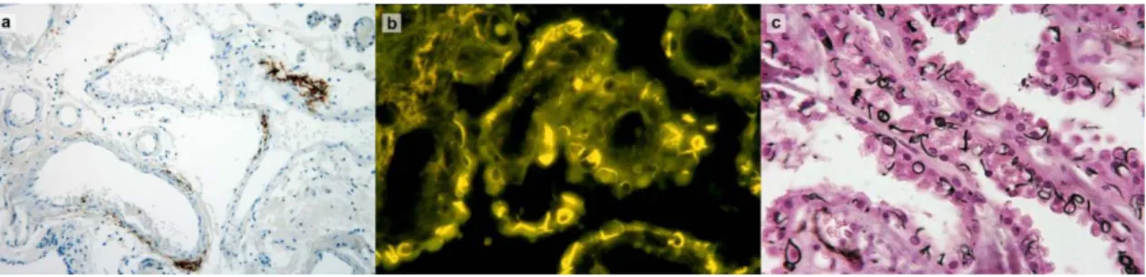
Involvement of the CP in AD is reflected by increased amyloid burden (Fig. 6a), impaired mitochondrial function and oxidative stress damage in CP tissue (Perez-Gracia et al., 2009; Krzyzanowska & Carro, 2012). These pathogenic processes along with morphological structural changes contribute to decreased efficacy of CP in clearing Aβ, thus resulting in increased Aβ accumulation in the brain. This in turn induces further pathological cascades of toxicity, inflammation and neurodegeneration, which may enhance the disease process.
Figure 6 - Involvement of the choroid plexus in Alzheimer’s disease. a: Deposition of Aβ in blood vessel
walls of choroid plexus stroma (immunohistochemistry for Aβ). b,c: Biondi ring tangles are annular, serpentine or curled inclusion in the cytoplasm of choroid plexus epithelial cells, as visualized using thioflavin S (b) and Jones’ methenamine silver stain (c). Modified from Wolburg et al., 2010.
17
2.2.1 Morphological alterations
The CP is subject to morphological and physiological changes that produce a wide range of effects. In AD the CP develops abnormalities similar to those observed with aging, although greatly enhanced. In LOAD, there are CP significant changes: epithelial atrophy, thickening of the basement membrane, and stroma fibrosis. Epithelial atrophy is significantly accentuated: a decrease in cell height is observed compared to age-matched controls (Krzyzanowska & Carro, 2012). Epithelial cells acquire Biondi bodies (ring-like structures) and numerous lipofuscin deposits (Alvira-Botero & Carro, 2010). Nuclei become irregular and flattened, and the basement membrane is thickened. The stroma thickens and contains collagen fibres, hyaline bodies, calcifications and psammomas (Serot et al., 2000). These modifications explain the decrease of functional capacities of CP, including synthesis, secretion, and transport of proteins and other molecules.
2.2.2 Aβ deposits
Besides accumulating in brain parenchyma, Aβ also accumulates in CP and in cerebrovascular walls, where it induces blood-brain barrier disruption (Spuch et al., 2012; Dietrich et al., 2008). Recent studies suggested the existence of a direct relationship between Aβ deposits at CP epithelium and the development of a functional and structural disruption of the organ (Dietrich et al., 2008; Vargas et al., 2010).
CP has been reported to produce several key enzymes involved in Aβ production, metabolism, and alternate processing (Crossgrove et al., 2007). Soluble Aβ, a product of the secretory pathway in amyloid precursor protein (APP) processing, is produced by the CP as observed in both rat and human post-mortem tissue (Krzyzanowska & Carro, 2012).
The source of Aβ deposited in the CP is unclear, but Aβ is probably taken up by CP epithelial cells from the CSF (Crossgrove et al., 2005). Deposition of Aβ in the CP may be mediated by interactions of Aβ and aquaporin-1, the latter being restricted to the CP in the normal brain but expressed by reactive astrocytes under pathological conditions (Misawa et al., 2008). Furthermore, there is evidence that circulating insulin-like growth factor-1 (IGF-1) participates in brain Aβ clearance by modulating CP function, because blockade of the IGF-1 receptor on CP exacerbates AD-like pathology in old transgenic mice (Carro et al., 2006). Substances produced by the CP and secreted into the CSF may bind Aβ and subsequently affect Alzheimer pathology. Furthermore, Biondi ring tangles are markedly increased in CP epithelial cells and ependymal cells during aging (with a sharp rise at about 45 years) and in AD. These annular, serpentine, or curled inclusions (Fig. 6b, c) with histochemical properties of amyloid are biochemically and ultrastructurally different from neurofibrillary tangles and from β amyloid, suggesting that they represent a distinct type of filaments accumulating in the aged and diseased CP. It has been suggested that Biondi ring tangles may be among the earliest manifestations of AD. The exact
18
molecular nature of these enigmatic structures and their functional consequences remain to be determined (Wolburg et al., 2010).
2.2.3 Impaired mitochondrial function
Mitochondrial dysfunction is one of the earliest deficits identified in AD brains (Wang et al., 2014; García-Escudero et al., 2013). It has been described that mitochondrial enzyme activity defects occur in hippocampal neurons and choroidal epithelial cells more frequently in AD patients (Cottrell et al., 2001). An increase in the number of COX-deficient choroidal epithelial cells provides strong evidence that a substantial decline of mitochondrial enzyme activity occurs more frequently in AD than in normal aging. This deficiency in mitochondrial enzyme activity is likely to result in decreased transport across the epithelial cells and thus have implications in choroidal Aβ clearance (Krzyzanowska & Carro, 2012). Aβ itself can also impair mitochondrial function (Readnower et al., 2011) and since Aβ deposits accumulate in the CP of AD patients (Dietrich et al., 2008), it is likely that Aβ interferes with their function. A recent study showed that mitochondrial dysfunction in CP from AD subjects occurs through a down-regulation of mitochondrial proteins and activity (Vargas et al., 2010).
2.2.4 Oxidative stress and cell death
Oxidative damage to proteins is a relative early phenomenon in the pathogenesis of AD. It has been reported in hippocampus and inferior parietal lobules of MCI and AD cases (Butterfield et al., 2006; Reed et al., 2008). In the CP it occurs at later stages of AD compared with other brain areas (Perez-Gracia et al., 2009). Such oxidation may result in impaired protein interaction, protein folding and protein kinase activity; abnormal function of endothelial and vascular smooth muscle cells, and impaired HDL-cholesterol metabolism, thus having important implications in the deterioration of CP functions (Perez-Gracia et al., 2009; Krzyzanowska & Carro, 2012).
Abnormal patterns of stress protein expression were found in cerebral cortex and hippocampus of AD subjects (Anthony et al., 2003). Moreover, heat shock proteins such as HSP90 and GRP94, presumably reflecting stress response, are upregulated in CP of AD patients (Johanson et al., 2004).
An increase in nitric oxide (NO) production by CP of AD patients was also reported and associated with Aβ deposits (Vargas et al., 2010). Excessive generation of NO has been implicated in the pathogenesis of AD (Aliyev et al., 2004), and plays an important role in Aβ-induced mitochondrial dysfunction (Keil et al., 2004). Aβ could interfere with oxidative phosphorylation, which results in oxidative stress.
Aβ is also involved in cell death pathway in CP, since increased expression of caspases 3 and 9
19 (Vargas et al., 2010; Krzyzanowska & Carro, 2012). Furthermore, inhibition of Aβ levels through viral-directed overexpression of gelsolin reduced cell death in CP of APP/PSEN1 transgenic mice (Antequera et al., 2009), suggesting that deposition of Aβ is neurotoxic for CP epithelial cells.
3. Cerebral cortex
The cerebral cortex plays a key role in the most complex neural functions, such as sensory perception, voluntary control of movement, language, personality traits, and other sophisticated mental events, such as thinking, memory, decision making, creativity, and self-consciousness. It is the highest, most complex, integrating area of the brain (Sherwood, 2013). The human cerebral cortex undergoes numerous structural changes over the lifespan. In AD, the loss of neurons and subsequent axonal degeneration leads to cerebral atrophy; the structural effects on the cerebral cortex include widening of sulci and thinning of the cortical ribbon (McGinnis et al., 2011).
3.1 The structure and functional localization of the cerebral
cortex
The cerebral cortex is a complex, highly organized, six-layered structure that contains hundreds of different neuronal cell types and a diverse range of glia (Gaspar & Vanderhaeghen, 2011). The six layers are numbered I-VI from superficial to deep (Figure 7). Layer I is the molecular layer, which contains very few neurons; layer II the external granular layer; layer III the external pyramidal layer; layer IV the internal granular layer; layer V the internal pyramidal layer; and layer VI the multiform, or fusiform layer. Each cortical layer contains different neuronal shapes, sizes and density as well as different organizations of nerve fibers (Barret & Simmons, 2015).
20
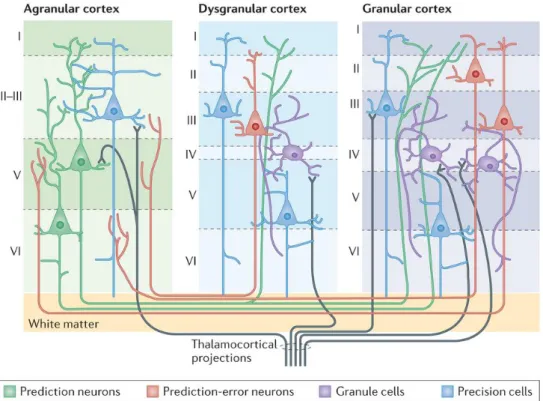
Figure 7 - Illustration of intracortical architecture and intercortical connectivity. Cortical columns
are defined by different numbers of layers, with each layer having characteristic cell types and patterns of intracortical and intercortical connectivity. Cortical areas that lack a layer IV are called agranular. Cortical areas that have only a rudimentary layer IV are called dysgranular. Adapted from Barret & Simmons, 2015.
3.2 Orbitofrontal cortex
The orbitofrontal cortex (OFC) is a region in the prefrontal cortex of the brain that is involved in the cognitive processing of decision-making. The OFC is so named because of its position on top of the eye orbits (Figure 8). In humans it consists of Brodmann area 10, 11 and 47 (Kringelbach, 2005). The prefrontal cortex (PFC), latest evolutionary addition to the mammalian brain, has long been known to endow qualities that differentiate human beings from all other animals. Yet the clinicians considered it "a silent" area in the absence of clinically discernible neurological deficits following its damage (Tandon, 2013).
21
Figure 8 - (a) A schematic of a mid-sagittal section through the head, depicting the OFC (red shaded region). (b) A post-mortem photograph of the ventral surface of the brain. The OFC is so named because of its position on top of the eye orbits. Adapted from Wallis, 2006.
The human OFC is among the least-understood regions of the human brain; but it is considered among the most polymodal regions of the brain. OFC receives multi-sensory inputs of taste, smell, auditory, visual and somatosensory as well as visceral signals, due to its wide and deep connections to functionally diverse cortical and subcortical regions, including the amygdala, cingulate cortex, insula, hypothalamus, hippocampus, striatum, as well as its neighbouring dorsolateral prefrontal cortex (Kringelbach, 2005; Nestor et al., 2013).
The OFC is located between the frontopolar gyri rostrally, the anterior perforated substance caudally, the inferior frontal gyrus laterally and the ventromedial margin of the cerebral hemisphere medially (Chiavaras & Petrides, 2000). Its anatomically heterogeneous sulcogyral morphology (Chiavaras & Petrides, 2000; Nakamura et al., 2007) is thought to be reflective of the rich molecular processes underlying neuronal migration, local neuronal connection, synaptic development, as well as lamination and formation of cytoarchitecture (Nestor et al., 2013).
Medial and lateral OFC regions can be dissociated, both functionally and anatomically. The medial OFC has its strongest connections to the hippocampus and associated areas of the cingulate, retrosplenial and entorhinal cortices and anterior thalamus, which would also be consistent with its involvement in higher order cognition, especially declarative episodic memory. On the other hand, while the lateral OFC also has strong connections to brain regions critical for higher order cognition, its links to the inferior parietal lobule and dorsolateral prefrontal cortex may suggest a special role in traits related to detecting and evaluating social threats to self-interest (Spitzer et al., 2007, Nestor et al., 2013).
When OFC connections are disrupted, a number of cognitive, behavioural, and emotional consequences may arise. In humans, damage to the orbitofrontal cortex causes major changes in emotion, personality, behaviour and social conduct. Patients often show lack of affect, social inappropriateness and irresponsibility (Hornak et al., 2003; Kringelbach et al., 2005).
22
Some dementias have been associated with OFC connectivity disruptions (Seeley et al., 2008). Some research suggested that later stages of AD are impacted by altered connectivity of OFC systems (Tekin & Cummings, 2002).Moreover, neurofibrillary tangles involving the OFC bilaterally have been associated with agitation and aggression in patients with MCI and AD (Trzepacz et al., 2013).
4. Olfactory and Taste Receptors
Olfactory receptors (ORs) and taste receptors (TRs) account for over half of the G protein-coupled receptors (GPCRs) repertoire (Kanageswaran et al., 2015). GPCRs are seven transmembrane-spanning proteins that represent the largest receptor superfamily in the human genome (Katritch et al., 2013). GPCRs recognize and bind an array of sensory inputs and ligands, including photons, ions, bioamines, lipids, carbohydrates, peptides and proteins, as well as a diverse range of volatile compounds. Ligand-induced activation of GPCRs converts extracellular stimuli into intracellular signals, mediating diverse cellular and physiological responses, including the senses of smell, taste, and vision. Not surprisingly, mutations and modifications of GPCRs, G proteins and their regulatory partners are linked to dysfunction and disease (Overington et al., 2006; Foster et al., 2013b), and the importance of these receptors is reflected in the fact that 40% of drugs on the market target GPCRs.
4.1 Olfactory Receptors
4.1.1 Olfactory receptor genes
With the sequencing of mammalian genomes, it is now clear that there are around 900 olfactory receptors (ORs) in humans (including pseudogenes) and ~1500 in rodents (representing 3–5% of all encoded genes) (Gaillard et al., 2004, Aloni et al., 2006, Henion et al., 2007, Niimura et al., 2007). In humans, there are 390 putatively functional (protein-coding) OR genes (Adipietro et al., 2012).
ORs are genes of around 1 kb in length and characteristically possess a hypervariable sequence
region in the transmembrane segments, which constitutes the putative odorant binding pocket (Lagerstrom & Schioth, 2008). They are distributed throughout the genome, residing in clusters of various sizes on nearly all chromosomes (except chromosomes 20 and Y) (Figure 9).
ORs are highly variable at a sequence level, and are further grouped into 18 families and
hundreds of subfamilies defined by their aminoacid identity (Olender et al., 2008). While forming the largest group of GPCRs, the OR repertoire is still small in comparison to the number of potential odorants. To some extent, this is explained by the capacity of ORs to detect odorants in a ‘combinatorial’ manner, whereby one OR recognizes multiple odorants and one odorant is detected by multiple ORs (Niimura, 2012).
23
Figure 9 - Chromosome locations of human OR genes. Six hundred thirty OR genes were localized to 51
different chromosomal loci distributed over 21 human chromosomes. OR loci containing one or more intact OR genes are indicated in red; loci containing only pseudogenes are indicated in green. The cytogenetic position of each locus is shown on the left, and its distance in megabases from the tip of the small arm of the chromosome is shown on the right (chromosome-Mb). The number of OR genes at each locus is indicated in parentheses, and the number of OR genes on each chromosome is indicated below. Adapted from Malnic et al., 2004.
4.1.2 ORs expression
The ORs were first identified in the olfactory system, being expressed in the cilia of olfactory sensory neurons in the olfactory epithelium (OE). However, soon after their initial discovery, reports surfaced of so-called ‘ectopic’ expression of “olfactory-like” receptors outside of the OE (Kang & Koo, 2012). Moreover, despite the label “olfactory receptor” only a subset (~75%) of the OR repertoire is actually expressed in the OE (Zhang et al., 2007).
While it is true that the expression level of ORs is highest within the OE, it is now clear that at least some ORs are expressed in a variety of additional tissues. With new techniques such as whole transcriptome sequencing, the list of “ectopic ORs” has greatly expanded in the last few years, and these sites now include, for example, the gastrointestinal (GI) tract, muscle, heart, pancreas, liver, lung, and skin (Spehr et al., 2003; Feldmesser et al., 2006; Griffin et al., 2009; Kang & Koo, 2012; Pluznick & Caplan, 2012; Kim et al., 2015; Wu et al., 2015; Shepard & Pluznick, 2016). An overview of identified human ORs expressed in non-chemosensory tissues is shown in more detail in Supplementary Table 1.
While the physiological functions that each of these ORs plays in non-chemosensory tissues remain an ongoing area of research, it is evident that ORs serve important roles in physiology. Notably, murine and human ORs have been found in sperm and play a key role in sperm chemotaxis (Spehr et al., 2003; Fukuda et al., 2004).
24
ORs have been also identified in several areas of the brain such as the cerebral cortex in mice
(Kang & Koo, 2012) and the murine choroid plexus (Gonçalves et al., 2016). The presence of
ORs and some obligate downstream components of OR signalling was also demonstrated in
neurons of the cerebral cortex and other regions in the human brain (Garcia-Esparcia et al., 2013). These studies speculated on the possibility that some predicted OR genes may not be olfactory receptors per se, but may subsume other functions.
4.1.3 Olfactory signal transduction in olfactory neurons
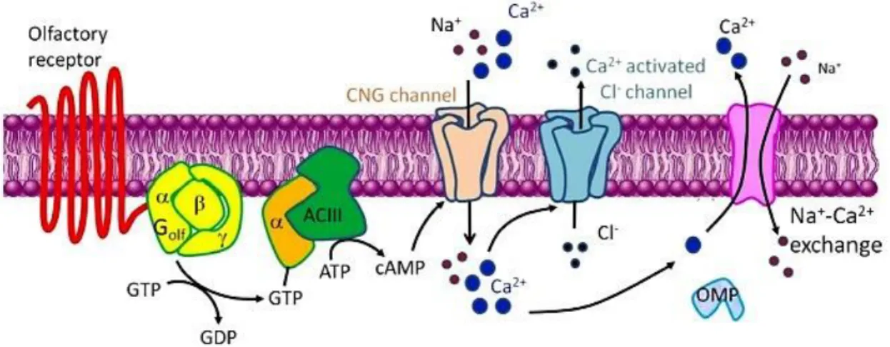
ORs display a highly specialized signal transduction pathway (Figure 10). This canonical pathway
involves the coupling of odorant-bound receptor to a G protein called Gαolf and activation of an olfactory isoform of adenylyl cyclase (AC3) to generate the second messenger cyclic adenosine monophosphate (cAMP) (Wong et al., 2000). Elevated levels of cellular cAMP in turn activate cation-selective cyclic nucleotide-gated (CNG) channels that lead to increased permeability to Ca2+ ions (Zheng & Zagotta, 2004), membrane depolarization and the generation of action
potentials in olfactory neurons.
Figure 10 - A schematic diagram of olfactory signal transduction. The olfactory signaling pathway
involves the coupling of ligand bound receptors to the olfactory isoform of the heterotrimeric G protein Gαolf and the activation of adenylyl cyclase (ACIII) to generate cAMP as a second messenger. This leads to influx of Ca2+ via cyclic-nucleotide gated channels (CNG), membrane depolarization and the generation of action potentials in neurons. Adapted from Kang & Koo, 2012.
4.2 Taste Receptors
The mammalian gustatory system recognizes five basic taste qualities: sweet, umami, bitter, salty and sour, which together enable the assessment of nutritional value of food constituents, while avoiding potentially harmful substances. In the past two decades, the molecular mediators of sweet, umami and bitter tastes have been identified as families of GPCRs, referred to collectively as taste receptor type 1 (T1R) and taste receptor type 2 (T2R). The T1R family has 3 members that form sweet and umami receptors, whereas the T2R family consists of 25 highly divergent GPCRs that mediate bitter taste. In addition, there is evidence that another
25 taste quality related to lipid sensing is mediated via the free fatty acid (FFA) GPCR family (Foster et al., 2014).
The five taste qualities are detected by specialized taste receptor cells located in the oral cavity. Here, taste receptor cells occur in morphological structures of 50–100 cells, called taste buds (Figure 11). The majority of taste buds are found on the tongue surface embedded in gustatory papillae, on the soft palate and on the throat (Chaudhari & Roper, 2010; Roper, 2013). Within each taste bud a variety of specialized cells are found, which can be classified by functional and morphological characteristics. The type II cells express taste receptors of the
GPCR type, type III cells form synapses with afferent nerve fibers, type I cells are believed to
have supporting functions and type IV cells, located at the basal parts of taste buds, are a pool of undifferentiated precursor cells devoted to replace the other cell types throughout life (Chaudhari & Roper, 2010).
Figure 11 - Schematic representation of a taste bud. The diagram shows an onion-like taste bud structure and the four subtypes of taste bud cells. Of the four subtypes, only type III taste bud cells form recognizable synapses with the afferent nerve fibres. Adapted from Calvo & Egan, 2015.
4.2.1 Taste receptor genes
Taste receptors are divided into two groups: GPCRs and channel type receptors (Chaudhari & Roper, 2010, Niki et al., 2010).
Sweet and umami taste are recognized by the taste receptor type 1 (T1R; genes are designated
TAS1 in humans and Tas1 in rodents) family - T1R1, T1R2 and T1R3 - that belongs to family C
of GPCRs (Vegezzi et al., 2014, Niki et al., 2010). T1Rs assemble into heterodimeric receptor complexes to function as sweet (T1R2+T1R3) or umami (T1R1+T1R3) taste receptors (Nelson et al., 2001, 2002; Niki et al., 2010). Thus the T1R2+T1R3 heterodimer is activated by various sweeteners (i.e., sugars, artificial sweeteners, sweet amino acids, and sweet proteins),
26
whereas the T1R1+T1R3 heterodimer is activated primarily by monosodium L-glutamate in humans and by amino acids in animals (i.e., mouse) (Nelson et al., 2002; Li et al., 2002; Niki et al., 2010).
Bitter taste is recognized by the taste receptor type 2 (T2R) family that belongs to family A of
GPCRs (Niki et al., 2010, Meyerhof et al., 2010). A list of human and mouse T1R e T2R genes,
including their names, synonyms, and orthologues, can be found in Supplementary Table 2, and can also be found in human and mouse genome databases. In humans, ~25 members of the T2R family may function as bitter taste receptors detecting and responding to an incredibly broad range of structurally diverse aversive and toxic compounds (Niki et al., 2010, Meyerhof et al., 2010; Clark et al., 2012; Vegezzi et al., 2014). These receptors are single exon genes that characteristically possess very short N-terminal extracellular domains. Although bitter taste is evoked by perhaps tens of thousands of synthetic and natural compounds (Clark et al., 2012), bitter ligands for some of the T2Rs (i.e., T2R41, T2R42, T2R45, T2R48, T2R60) are still unknown (Niki et al., 2010, Meyerhof et al., 2010). The number of compounds perceived by humans as bitter is much larger than the number of human TAS2R genes, implying that each human T2R responds to more than one bitter ligand (Behrens & Meyerhof, 2006). The same is likely to be true for other species. Some T2Rs interact with a wide range of bitter tasting ligands (e.g.,
TAS2R14 and TAS2R16; see Supplementary Table 3). However, some other T2Rs appear to have
narrow ligand specificities (e.g., TAS2R5).
4.2.2 Taste receptors expression
Taste GPCRs are often low abundance genes and appear below the limits of detection of arrays, compounded by the fact that arrays often are not optimized to detect taste GPCRs (Insel et al., 2012). In any case, small scale RT-PCR screens have provided ample transcript data to lay the foundations for investigation of taste receptors beyond the mouth. In addition to the oral cavity, TASRs expression has been detected in a variety of tissues including the respiratory and cardiovascular system, digestive tract, pancreas, liver, kidney, heart, testis, several mouse forebrain regions and choroid plexus (Ren et al., 2009; Behrens & Meyerhof, 2010; Meyer et al., 2012; Yamamoto & Ishimaru, 2013; Xu et al., 2013; Foster et al., 2013b; Tomás et al., 2016). Bitter TASRs together with downstream functional mediators were also found to be expressed in the brain of rats and in frontal cortex in humans (Singh et al., 2011; Dehkordi et al., 2012; Garcia-Esparcia et al., 2013) and in the choroid plexus of rats (Tomás et al., 2016). An overview of identified human TASRs expressed in non-chemosensory tissues is shown in more detail in