Universidade de Aveiro Departamento de Biologia 2007
Teresa Margarida
Pedrosa Cardoso
Estabilidade Conformacional da Proteinase
Aspártica – Arquétipo, Pepsina A
Conformational Stability of the Archetypal
Aspartic Proteinase, Pepsin A
Universidade de Aveiro 2007
Departamento de Biologia
Teresa Margarida
Pedrosa Cardoso
Estabilidade Conformacional da Proteinase
Aspártica – Arquétipo, Pepsina A
Conformational Stability of the Archetypal
Aspartic Proteinase, Pepsin A
Dissertação apresentada à Universidade de Aveiro para cumprimento dos requisitos necessários à obtenção do grau de Doutor em Bioquímica, realizada sob a orientação científica da Professora Doutora Marlene Maria Tourais de Barros, Professora Associada com Agregação do Centro Regional das Beiras da Universidade Católica Portuguesa, e do Professor Doutor Euclides Manuel Vieira Pires, Professor Associado do Departamento de Bioquímica da Faculdade de Ciências e Tecnologia da Universidade de Coimbra.
Apoio financeiro da FCT e do FSE no âmbito do III Quadro Comunitário de Apoio.
o júri
presidente Professor Doutor Vasile Staicu
Professor Catedrático do Departamento de Matemática da Universidade de Aveiro
Professora Doutora Maria Ana Dias Monteiro Santos
Professora Catedrática do Departamento de Biologia da Universidade de Aveiro
Professor Doutor Eduardo José Xavier Rodrigues de Pinho e Melo
Professor Associado com Agregação da Faculdade de Engenharia de Recursos Naturais da Universidade do Algarve
Professora Doutora Marlene Maria Tourais de Barros
Professora Associada com Agregação da Universidade Católica Portuguesa, Centro Regional das Beiras – Pólo de Viseu
Professor Doutor Euclides Manuel Vieira Pires
Professor Associado da Faculdade de Ciências e Tecnologia da Universidade de Coimbra
Professora Doutora Paula Cristina Veríssimo Pires
Professora Auxiliar da Faculdade de Ciências e Tecnologia da Universidade de Coimbra
Professora Doutora Ana Cristina de Fraga Esteves Sarmento
agradecimentos
À Universidade de Aveiro, por permitir o desenvolvimento deste projecto.À Fundação para a Ciência e a Tecnologia, pelo financiamento concedido. À Professora Doutora Marlene Barros, pelo apoio científico e logístico. Ao Professor Doutor Euclides Pires, pelas pertinentes sugestões.
Aos Professores Nicholas Price e Alan Cooper, pelos ensinamentos e proveitosas discussões. À Doutora Sharon Kelly, a Tommy Jess e a Mrs. Margaret Nutley, pelo indispensável auxilio no planeamento e realização de trabalho experimental. A todos, pelo caloroso acolhimento durante as minhas estadias em Glasgow.
À Cláudia Oliveira, Cristina Sarmento e Anabela Pereira, por apoios prestados durante a realização de trabalho experimental. À Sofia Duarte e Sofia Fraga pela disponibilidade.
À Isabel Velada, pela compreensão.
Ao Professor Doutor Carlos Pires, pelos conselhos.
Aos meus três anjos protectores, por tudo. Mas, sobretudo, pelo exemplo de coragem e força do meu Pai, pelo exemplo de optimismo e compreensão da minha Mãe, pelo exemplo de sinceridade e esforço da minha Irmã.
Aos meus Avós, por relevarem as minhas ausências. À restante Família, pela alegria em cada reencontro.
Errata da Dissertação
“Estabilidade Conformacional da Proteinase Aspártica - Arquétipo, Pepsina A”
Na primeira página, deveria ter-se incluído:“Apoio financeiro do POCTI no âmbito do III Quadro Comunitário de Apoio através do projecto POCTI/QUI/60791/2004.”
palavras-chave
Pepsina A; Acetonitrilo; Hidrocloreto de Guanidina; Ureia; Temperatura; Desnaturação de Proteínas; Estados Conformacionais Não-Nativos; Estabilidade Conformacional.resumo
A pepsina A tem sido considerada um protótipo do grupo das proteinasesaspárticas, cuja relevância ao nível da patofisiologia humana tem estimulado intensa pesquisa. As propriedades moleculares e catalíticas da pepsina A começaram a ser exploradas há cerca de um século. Contudo, a informação existente sobre a estabilidade conformacional desta proteína em condições nativas é insuficiente. Elucidação dos factores responsáveis pela manutenção do estado conformacional naturalmente enrolado e activo de proteínas é vital para estabelecer estratégias de modulação da estabilidade. Com o objectivo de obter novos conhecimentos acerca das bases da estabilidade conformacional da pepsina A, foi adoptada uma abordagem que consistiu na indução e monitorização da desnaturação da proteína no seu estado naturalmente enrolado. A severidade das condições ambientais foi gradualmente agravada por adição de desnaturantes e / ou modificação de propriedades físicas e químicas do sistema proteína-solvente. Após uma fase de incubação nas condições desnaturantes, com a duração típica de 1 h, mudanças nas propriedades espectrais, no comportamento hidrodinâmico, na estabilidade térmica e na actividade peptidolítica da pepsina A de suíno foram avaliadas. O solvente orgânico, acetonitrilo, induziu destruição não-cooperativa dos arranjos secundário e terciário desta proteína. Sugere-se que a drástica debilitação das estruturas de hidratação pelo acetonitrilo terá impelido o desenrolamento global. Adicionalmente, a pepsina A revelou-se particularmente resistente à acção desordenante de dois agentes caotrópicos clássicos, o hidrocloreto de guanidina (GdnHCl) e a ureia. Um estado intermediário estável foi identificado durante a reacção de desenrolamento da proteína nativa induzida pelo GdnHCl. A adição de acetonitrilo a uma molaridade intermédia permitiu a identificação de um diferente estado parcialmente desenrolado na presença de GdnHCl a concentrações pré-transicionais. Dados espectroscópicos indiciam promoção de ordem local a nível do esqueleto polipeptídico pela ureia a baixas e altas molaridades. Indução de extenso desenrolamento da proteína pela ureia requereu concentrações quase saturantes deste desnaturante e períodos de exposição de duração superior a 1 h. Foram detectados fenómenos de aparente estabilização, quando a fase de pré-incubação da proteína em ambos os agentes caotrópicos a baixas molaridades foi prolongada para 3 dias. A pepsina A no seu estado nativo revelou-se bastante termoestável. Contudo, 1 h de exposição da proteína, antes do seu aquecimento, a qualquer dos três desnaturantes a concentrações pré-transicionais foi suficiente para aumentar a termolabilidade da proteína. Contribuições de interacções hidrofóbicas e
keywords
Pepsin A; Acetonitrile; Guanidine Hydrochloride; Urea; Temperature; ProteinDenaturation; Non-Native Conformational States; Conformational Stability.
abstract
Pepsin A has been considered a prototype of the group of aspartic proteinases,whose significance in human pathophysiology has galvanized intensive research. Molecular and catalytic properties of pepsin A have been explored for about a century. However, there is only fragmentary data on the conformational stability of this protein under native conditions. Elucidation of the factors accountable for the maintenance of the natively ordered and active conformational state of a protein is vital for engendering strategies for tailoring stability. In order to gain new insights into the scaffold of the conformational stability of pepsin A, an approach based on induction and monitoring of denaturation of the natively folded protein was adopted throughout the investigation reported herein. Environmental stringency was gradually increased by adding denaturants and / or modifying chemical and physical properties of the protein-solvent system. After a period of incubation in the denaturing conditions, which lasted typically 1 h, changes in spectral properties, hydrodynamic behaviour, thermal stability and peptidolytic activity of porcine pepsin A were appraised. The organic solvent, acetonitrile, was found to induce non-cooperative destruction of secondary and tertiary arrangements of this protein. Drastic debilitation of hydration structures by acetonitrile is suggested to have triggered global unfolding. In addition, pepsin A revealed to be particularly resistant to the disordering action of two classic chaotropes, guanidine hydrochloride (GdnHCl) and urea. A stable, intermediary state could be identified in the pathway of GdnHCl-induced unfolding of native pepsin A. Upon addition of acetonitrile at an intermediary molarity, a different partially unfolded state could be identified in the presence of GdnHCl at pre-transitional concentrations. It is apparent from spectroscopic data that urea promoted local backbone order at low and high molarities. And, periods of exposure longer than 1 h to urea at near-saturating concentrations were required to onset gross unfolding by this denaturant. Phenomena of apparent stabilization were detected when submission of the protein to both chaotropes at low molarities was prolonged to 3 days. Furthermore, pepsin A in its native state was noted to be rather thermostable. However, 1 h - incubation at pre - transitional concentrations of any denaturant prior to heating caused a raise in the protein thermolability. Contributions from hydrophobic interactions and solvation structures to the conformational stability of pepsin A in its biologically active state are discussed in this dissertation.
Cover Illustration:
Transparent molecular surface of porcine pepsin A (PDB code 4pep) with the backbone rendered as a ribbon. This figure was generated by GRASS (Graphical Representation and Analysis of Structure Server available at http://trantor.bioc.columbia.edu/cgi-bin/GRASS/surfserv_enter.cgi) (Nayal, et al., 1999).
I
NDEX OF
C
ONTENTS
Symbols and Abbreviations
iNotation and Numbering
iiiChapter I
| I
NTRODUCTION 11. PROTEIN DENATURATION:INSIGHTS INTO FOLDING AND CONFORMATIONAL STABILITY 3
A. Theoretical Framework for Protein Denaturation 3
A.1. Phenomenological Aspects 4
A.1.1. Modes of Unfolding 4
A.1.2. Reversibility versus Irreversibility 5
A.2. Denatured States 5
A.2.1. Structural Diversity 5
A.2.1.1. Unfolded States 6
A.2.1.1.1. Random Coil 6
A.3. Pathways of Protein Unfolding 7
A.3.1. Two-State Unfolding and Cooperativity 7
A.3.2. Multi-State Unfolding and Intermediary States 8
A.3.2.1. Molten Globule States 8
B. Protein Denaturation in vivo 9
C. Drawing Inferences about Protein Folding 10
C.1. Insights into Protein Folding from Protein Unfolding in vitro 10
C.2. Simulation of Protein Folding by Protein Refolding in vitro 11
D. Protein Stability 12
D.1. Marginal Conformational Stability of Folded States 12
D.1.1. Physical and Molecular Foundations 13
D.1.2. Biological Advantages 14
2. PROTEIN HYDRATION 14
A. Structural and Dynamical Characteristics 14
A.1. Surface Hydration 15
A.2. Buried Water Molecules 16
B. Contributions to Protein Folding and Conformational Stability 17
3. PEPSIN A: AN ARCHETYPAL ASPARTIC PROTEINASE 17
A. Aspartic Proteinases 18
A.1. Classification and Distribution 18
A.2. General Insights into Molecular and Catalytic Properties 19
A.2.1. Zymogens and Mechanisms of their Activation 20
A.2.2. Three-Dimensional Structure 21
A.2.2.1. Catalytic Apparatus 21
A.2.2.2. Evolution 22
A.2.3. Catalysis 22
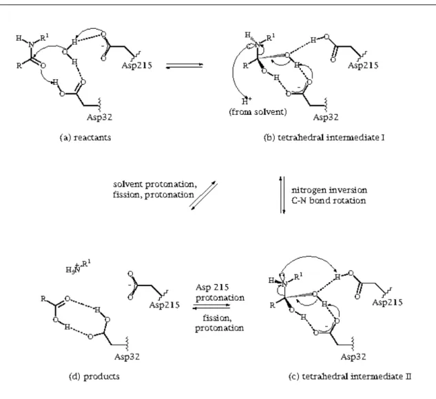
A.2.3.1. Mechanism for Peptide Hydrolysis 23
A.3. Physiological, Pathological and Biotechnological Significance 24
B. Pepsin A 25
B.1. Conversion of Pepsinogen A into Pepsin A 26
B.2. Structure: Specific Considerations 28
B.3. Catalytic Properties: Specific Considerations 30
Chapter II
| O
BJECTIVES&
O
UTLINE 31
1.MATERIALS 36
A. Proteins, Substrates and Amino Acids 36
B. Salts, Solvents and Other Chemicals 36
2.METHODS 37
A. Preparation of Solutions 37
B. Preparation of Stocks of Porcine Pepsin A 37
C. Quantification of Solutions of Porcine Pepsin A 37
C.1. Ultraviolet Absorption Spectroscopy 38
C.2. Micro BCATM Protein Assay Reagent Kit 38
D. Assessment of Purity and Autolysis of Porcine Pepsin A 39
D.1. Polyacrylamide Gel Electrophoresis 39
D.1.1. Polyacrylamide Gel Electrophoresis under Denaturing Conditions 39
D.1.2. Polyacrylamide Gel Electrophoresis under Near-Native Conditions 41
D.2. Protein Staining on Electrophoresis Gel 42
D.3. Analysis, Preservation and Storage of Electrophoresis Gels 43
E. Induction of Porcine Pepsin A Denaturation 43
F. Measurement of Spectroscopic Properties of Porcine Pepsin A 44
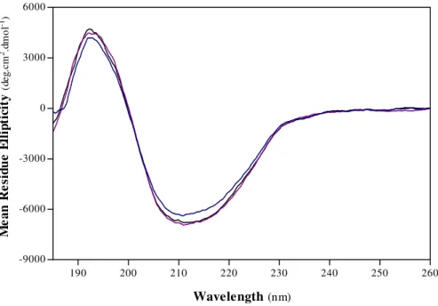
F.1. Circular Dichroism Spectroscopy 44
F.1.1. Measurements under Isothermal Conditions 44
F.1.2. Measurements in Thermal Gradients 45
F.1.3. Estimation of Secondary Structure from Far-UV Circular Dichroic Spectra 45
F.2. Intrinsic Tryptophanyl Fluorescence Spectroscopy 46
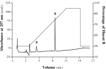
G. Size Exclusion Chromatographic Analyses of Porcine Pepsin A 46
H. Differential Scanning Calorimetric Assays of Porcine Pepsin A 48
I. Assessment of Peptidolytic Activity of Porcine Pepsin A 48
I.1. Aspects of Assay Design 49
I.1.1. Substrate 49
I.1.2. Temperature and pH 50
I.2. Standard Experimental Procedure 50
I.2.1. Preparation and Performance of Hydrolytic Reactions 50
I.2.2. Quantification of Hydrolytic Products 51
J. Solvent Accessible Surface Area Calculations for Porcine Pepsin A 52
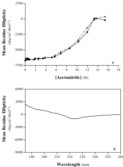
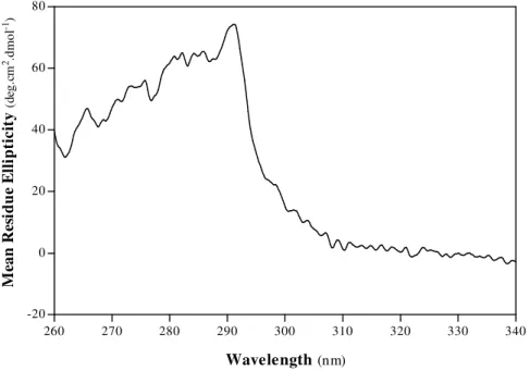
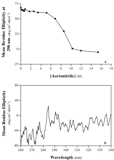
1.ACETONITRILE-INDUCED DENATURATION OF PORCINE PEPSIN A 57
A. Structural Properties 58
A.1. Secondary Structure 58
A.2. Tertiary Structure 63
A.3. Interpretative Remarks 71
B. Hydrodynamic Behaviour 74
B.1. Interpretative Remarks 78
C. Thermal Denaturation and Stability 79
C.1. Interpretative Remarks 86
D. Peptidolytic Activity 87
D.1. Interpretative Remarks 89
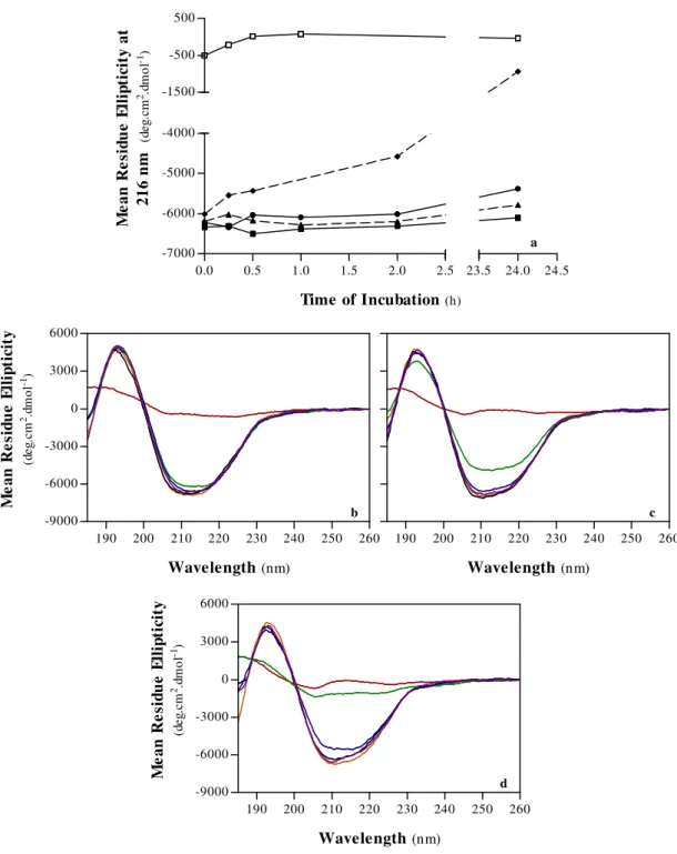
2.CHAOTROPE-INDUCED DENATURATION OF PORCINE PEPSIN A 92
A. Structural Properties 94
A.1. Secondary Structure 94
A.2. Tertiary Structure 96
A.3. Interpretative Remarks 103
A.3.1. Non-Conventional Effects of Urea: Improvement of Backbone Order 103
A.3.2. Conventional Effects of Chaotropes: Induction of Unfolding 104
A.3.2.1. Guanidine Hydrochloride 104
A.3.2.2. Urea 106 B. Hydrodynamic Behaviour 108 B.1. Interpretative Remarks 111 C. Thermal Stability 112 C.1. Interpretative Remarks 114 D. Peptidolytic Activity 115 D.1. Interpretative Remarks 116
E. Induction of Denaturation under Non-Standard Experimental Conditions 117
E.1. Structural Properties 118
E.1.1. Secondary Structure 118
E.1.2. Tertiary Structure 123
E.1.3. Interpretative Remarks 127
E.1.3.1. Prolonged Incubation in Aqueous Solutions of Chaotropes 127
E.1.3.1.1. Non-Conventional Effects of Chaotropes: Inhibition of Unfolding and Enhancement of Packing 128
E.1.3.1.2. Conventional Effects of Chaotropes: Induction of Unfolding 129
E.1.3.1.2.1. Guanidine Hydrochloride 129
E.1.3.2. Incubation in Non-Aqueous Solutions of Guanidine Hydrochloride
132
Chapter V
| G
ENERALD
ISCUSSION 1331. FROM DENATURATION TO CONFORMATIONAL STABILITY OF PORCINE PEPSIN A 136
A. Large-Scale Denaturation 136
A.1. Chemical Compounds 136
A.1.1. Resistance to Chemical Denaturation 138
A.2. Elevated Temperature 141
A.2.1. Thermal Stability 141
B. Denatured States 142
C. Small-Scale Destabilization Prior to Large-Scale Denaturation 144
C.1. Chemical Compounds 144
C.2. Transition from Mildly Acidic to Lowly Alkaline pH 145
2. ENHANCEMENT OF ORDER AND APPARENT STABILIZATION AT GLOBAL AND SUBGLOBAL LEVELS OF PORCINE PEPSIN A BY CHAOTROPES 146
Chapter VI
| C
ONCLUSIONS&
F
UTURED
IRECTIONS 148
i
S
YMBOLS AND
A
BBREVIATIONS
θ θ θ θ – Ellipticity ∆ ∆ ∆
∆λλλλmax – Difference between the Fluorescence Emission Maxima of Model Compound and Protein [θθθθ]MRW – Mean Residue Ellipticity
∆ ∆ ∆
∆Hcal – Calorimetric Enthalpy ∆
∆ ∆
∆HvH – van’t Hoff Enthalpy λ
λ λ
λmax – Wavelength of Maximum Fluorescence Emission
εεεε
y – Extinction CoefficientA – Absorbance
ANS – 8-Aniline-1-Naphthalenesulfonate ATP – Adenosine 5’ Triphosphate BCA – Bicinchoninic Acid BSA – Bovine Serum Albumin C – Concentration (M)
c – Concentration (mg/mL)
C-Terminus – Free α-Carboxyl Group (at one end of a protein) CE – Combinatorial Extension
DAPI – 4’, 6-Diamidino-2-Phenylindole DMSO – Dimethyl Sulphoxide
EC – Enzyme Commission F – Folded State
GdnHCl – Guanidine Hydrochloride
GRASS – Graphical Representation and Analysis of Structure Server HIV-1 – Human Immunodeficiency Virus Type 1
HPLC – High Performance Liquid Chromatography HT – High Voltage
I – Intermediary State
Ip – Alkaline-Denatured State of Pepsin Kav – Gel-Phase Distribution Coefficient l – Path Length
Me – Methyl
Mr– Relative Molecular Weight (or Mass) MRW – Mean Residue Weight (or Mass)
N-Terminus – Free α-Amino Group (at one end of a protein) NATA – N-Acetyl-L-Tryptophanamide
Nitro – NO2 P – Product
PAGE – Polyacrylamide Gel Electrophoresis PDB – Protein Data Bank
pKa – Acid Dissociation Constant PSI – Plant-Specific Insert rpm – rotations per minute S – Substrate
SASA – Solvent Accessible Surface Area SCOP – Structural Classification of Proteins SDS – Sodium Dodecyl Sulphate
Selcon3 – Self-Consistent Method for Protein Circular Dichroism Analysis,Version 3 TEMED – N, N, N’, N’-Tetramethylene-Ethylenediamine
TFA – Trifluoroacetic Acid
Tris – 2-Hydroxymethyl-2-Methyl-1, 3-Propanediol U – Unfolded State
UV – Ultraviolet
V0 – Column Void Volume Ve – Elution Volume Vt – Total Bed Volume
iii
N
OTATION AND
N
UMBERING
Schechter and Berger notation will be adopted to designate the subsites of a proteinase (….S3, S2, S1 and S1’,
S2’, S3’….), and the corresponding amino acid residues of a substrate (…P3, P2, P1 and P1’, P2’, P3’….), on
the N- and C-terminal sides of the scissile peptide bond (P1 – P1’) (Schechter & Berger, 1967).
Standard single- or three-letter codes will be used for amino acid residues whenever appropriate.
Unless otherwise stated, amino acid sequence numbering for porcine pepsin A (Sielecki, et al., 1990) will be assumed throughout the present dissertation. The suffix ‘p’ is used to denote residues in the prosegment of porcine pepsinogen A.
“In the end, this is all about finding out how and why nature contradicts universal entropy in order to create order.”
C
HAPTER
I
INTRODUCTION ---
The importance of proteins, primarily as macromolecules essential for the development and maintenance of life, is undeniable. Besides, their properties are so valuable that proteins have acquired a notable status as polymers with industrial and pharmaceutical applications for long. Indeed, nowadays proteins are used as catalysts, materials and therapeutic agents.
The development of protein science has been significantly fed by the challenge of devising strategies for modulation of folding, conformation, stability and activity of proteins. The successful attainment of these aims implies at least: (i) clarification of relationships between protein conformation and biological function (i. e. of the classic structural biology paradigm); (ii) decipherment of the folding code; and (iii) thorough establishment of the physical and chemical principles assuring the maintenance of the native and biologically active conformational state of a protein. Postulates regarding these issues have been pursued with great energy by molecular biologists and biochemists. The legacy of an increasing number of protein sequences available through the genome projects (Fulton, et al., 2005; Saghatelian & Cravatt, 2005) and growing evidence for the implication of protein misfolding and aggregation in many diseases (Ingelsson, et al., 2005; Kransnoslobodtsev, et al., 2005; Sambamurti, et al., 2006) are recent incentives.
In this vein, protein engineering is one of the strategies mostly adopted. A complementary approach is based on the influence of the chemical composition and physical characteristics of the surrounding environment on protein conformations. These methods have contributed to the inference of a number of requirements for specific functional, structural and biophysical properties of proteins. Increasing data in this direction have been reinforcing aspirations of theoreticians and experimentalists who are aiming to effectively enhance folding efficiency and fine-tune activity and stability of proteins in view of specific purposes.
In the end, the ultimate and ambitious goal of protein science, the rational design of de novo proteins, will hopefully be fulfilled, and many avenues leading towards useful technological applications of proteins will be improved or opened. From medicine and nutrition to industry, a wide range of fields are envisaged to be touched by the unpaired potential of the arising benefits.
INTRODUCTION
---
3
1.
P
ROTEIN
D
ENATURATION
:
I
NSIGHTS INTOF
OLDING ANDC
ONFORMATIONALS
TABILITYSupervision of induced order-to-disorder transitions in proteins, eventually followed by reconstitution of native-like conditions, and characterization of implicated conformational states may yield a wealth of information about the determinants (forces and molecular characteristics) of conformational stability, as well as shed some light on the prime factors driving folding, unfolding, misfolding and aggregation. Provided that care is taken when performing extrapolations, the presence of folding intermediates might be predicted and folding pathways might be decoded. Ultimately, a more detailed picture of the molecular events and energetics implied in any transition between conformational states might be sketched (vide, for example, the following references: Bhavesh, et al., 2003; Caflisch & Karplus, 1994; Finkelstein, 1997; Grant, et al., 1992; Pace, et al., 1990b; Stapelfeldt & Skibsted, 1999).
It is the goal of this subchapter to put current knowledge on protein denaturation into a comprehensive perspective, and to emphasize its potentials as an individual or complementary methodology for understanding folding and assessing conformational stability. Firstly, we should concentrate on the diversity of denatured states and unfolding pathways. Edifying information on protein folding obtainable from the employment of protein denaturation and renaturation techniques will also be outlined. Lastly, marginal conformational stability of protein native states will be explored in terms of its physical and molecular foundations, as well as biological rationales.
Before proceeding, it urges to clarify that the following discussion implies the classic axiom, by which a folded conformational state is demanded for adequate protein function. Furthermore, even though many principles lying beneath conformation, stability and unfolding / folding reactions are similar for different architectural categories of proteins, the prime targets of the subsequent description are globular proteins.
A. Theoretical Framework for Protein Denaturation
Modifications in pressure, pH, ionic strength or temperature of the surrounding environment (Arnold, et al., 1996; Bai, et al., 1998; Jayaraman, et al., 2006; Stapelfeldt & Skibsted, 1999; Stigter, et al., 1991), or addition of ligands, detergents, chaotropes, reducing agents or organic solvents to the protein-solvent system (Akhtar, et al., 2002; Bettati, et al., 2000; Li, et al., 1995; Mozhaev, et al., 1989; Otzen, 2002; Simon, et al., 2001) usually alter the delicate balance of forces accountable for preservation of the biologically active and native conformational state of a protein. The term ‘denaturation’ refers to any process by which a protein is perturbed. At most times, denaturation is viewed as an order-to-disorder conformational transition, whereby secondary, tertiary, and quaternary structures of a protein are disrupted; even though it
Protein Denaturation: Insights into Folding and Conformational Stability
---
does not necessarily lead to complete unfolding. Instead, structural disarray is sometimes restricted to definite regions within the biomacromolecule, insomuch that some interactions might remain unchanged or even be intensified, and new ones might be formed. And, thus, distinct non-native states with intermediary properties between those of the natively folded and the fully unfolded states might appear (Halle, et al., 2005; Uversky, 2002a). A reduction in or a complete loss of activity often accompanies protein disordering transitions (Chen, et al., 2003 ; Ternström, et al., 2005; Wei, et al., 2006).
By virtue of its primacy in elucidating principles governing protein folding, conformation and stability, protein denaturation has become a relevant research discipline (Fleming & Rose, 2005). Nowadays, conformational transitions can be followed step by step in great detail. A plethora of experimental techniques, including, for instance, circular dichroism or fluorescence spectroscopy (Rodger & Ismail, 2000; Szabo, 2000), have afforded delineation of folding and unfolding pathways, and physicochemical characterization of intermediary and final states implicated therein. The usefulness of theoretical methods in providing insights into these fields has also been recognized. Application of molecular dynamics simulations to protein denaturation using detailed atomic models for protein solvent is an evocative example (Brooks, 1998; Caflisch & Karplus, 1994). Merging of experimental and theoretical approaches is being implemented, in order to give atomic-level descriptions and predictions of protein conformational transitions (Fersht & Daggett, 2002).
A.1. Phenomenological Aspects
A.1.1. Modes of Unfolding
Global unfolding might be considered an extreme case of denaturation, for it results in a largely extended polypeptide chain characterized by the absence of any (or almost any) ordered structural elements. A second mode of unfolding has been distinguished, the so-called local unfolding, which corresponds to structural unravelling of confined regions within a protein (Neupert, et al., 2005).
Phenomena of local unfolding occur even in a protein under native conditions, where they are prompted by everlasting thermal fluctuations (Gaume, et al., 1998; Neupert, et al., 2005; Zhuang, et al., 2000). Conditions could occur in which the free energy barrier between folded and unfolded states is not surmounted, and refolding within a short period of time follows as a consequence (Dokholyan, et al., 2000). Otherwise, entrapment of local unfolded segments in a protein might occur due to, for instance, binding of denaturants (Zhuang, et al., 2000). Once these protein segments are trapped, unfolding will proceed, possibly leading to a completely unfolded state within milliseconds to minutes (Neupert, et al., 2005). Besides prompting global structural disarray, local unfolding events may as well facilitate aggregation (DeMarco & Daggett, 2005) and proteolysis processes (Fontana, et al., 1997; Vriend, et al., 1998).
INTRODUCTION
---
5 A.1.2. Reversibility versus Irreversibility
A reversible denaturation process entails total recovery of the starting state, in conformational and functional terms, through the reverse reaction (Ahern & Klibanov, 1988; Volkin & Klibanov, 1991). In the main, reversibility of protein denaturation depends on the environmental conditions and on the period of exposure to perturbants (Naik & Huang, 2004).
Several physical and chemical changes occurring upon an order-to-disorder transition might produce alterations in the folding properties of the polypeptide chain and hamper the back-reaction. Structural perturbations in the protein facilitate exposure of residues amenable to chemical modifications or peptide bonds liable to cleavage, hitherto more or less protected in the folded conformational state, thereby threatening the integrity of the polypeptide chain. A long list of covalent deteriorative mechanisms may be responsible, or co-responsible, for irreversibility of protein disordering processes, including, for example, hydrolysis of peptide bonds at the carboxyl terminus of aspartic residues at acidic pH, deamidation of amine residues, destruction of cystines, autolysis or degradation by exogenous proteinases, among others (Ahern & Klibanov, 1988; Mozhaev & Martinek, 1982; Tomazic & Klibanov, 1988; Vieille & Zeikus, 2001; Volkin & Klibanov, 1990; Volkin & Klibanov, 1991). Denaturation irreversibility is also often ascribed to intermolecular associations resulting in aggregated states (Ahern & Klibanov, 1988; Pace, 1986; Volkin & Klibanov, 1991). Unfolding leads to increased exposure of non-polar groups hitherto buried, which may cause the denatured protein to self-associate. These associations may either occur via hydrophobic interactions, so that the unfavourable contact between hydrophobic amino acid residues and water is minimized (Patro & Przybycien, 1994), or via chemical reactions, mainly through the formation of intermolecular disulphide bridges (Volkin & Klibanov, 1990).
A.2. Denatured States
Any incorrectly folded or ill-defined, disordered conformational state of a polypeptide chain might be simplistically referred to as a denatured state (Hamada & Goto, 2005; Uversky, 2002a). In general, each denatured state should be viewed as a collection of diverse interconverting conformations larger than the corresponding natively folded-state ensemble, due to higher structural heterogeneity (Fersht & Daggett, 2002; Shortle, 1996; Shortle, et al., 1998). In the past few years, substantial attention and effort have been devoted to the description of non-native states. Nevertheless, experimental endeavours are often threatened by technical difficulties arising from, for example, their insolubility or high flexibility (Dill & Shortle, 1991; Shortle, 1996). Therefore, fine structural details on denatured states are yet to be worked out.
A.2.1. Structural Diversity
Protein Denaturation: Insights into Folding and Conformational Stability
---
current literary review and for the remainder of this dissertation, shall be introduced. A semantic scheme was formalized mainly on the basis of reconciliation between different classification systems of conformational states assumed by several authors (Cooper, 1999; Uversky, 2002a). And, it relies on two central premises: (i) as aforementioned, any disordered or incorrectly folded conformation, different from the native conformation, is a denatured conformation; and (ii) an unfolded conformation is an unstructured conformation. In face of this, denatured states can be assorted into structurally diverse subsets, viz., molten globule states, misfolded states and unfolded states.
A description of molten globule states will be provided elsewhere. Misfolded states are produced in the course of protein folding, and result from entrapment of the folding molecule in aberrant conformations. These, however, might exhibit, to some extent, similarities to corresponding native folds (Cooper, 1999; Dobson, 2005; Privalov, 1996).
A.2.1.1. Unfolded States
Each unfolded state of a protein comprises a particularly large and heterogeneous ensemble of many open, irregular and fluctuating conformational substates (Bachmann & Kiefhaber, 2005; Cooper, 1999; Cooper, 2000a; Fleming & Rose, 2005; Ohnishi & Shortle, 2003). An unfolded form is fluid-like and the bonds of its amino acid side chains rotate quite freely (Haynie, 2001). Despite its high degree of unstructureness, an unfolded state may exhibit intramolecular hydrophobic interactions and specific hydrogen bonds. Non-covalent interactions between residues are, however, expected to be neither stable nor abundant (Creighton, 1990; Pace, et al., 1996). Owing to predominant exposure of protein groups to solvent, many intramolecular interactions within folded conformations are substituted by protein-solvent interactions in the unfolded state (Cooper, 2000a).
A more accurate definition of unfolded state should consider a set of conformational states characterized by any or almost any regular structure (Cooper, 1999; Uversky, 2002a), including premolten globule states (for further information, vide infra), aggregates and conformational states well-approximated by the random coil model.
A.2.1.1.1. Random Coil
Random coil is traditionally regarded as the ideal unfolded form for a protein. Apart from complete freedom of bond rotation (Fleming & Rose, 2005), the random coil model further assumes the absence of any local interactions along the polypeptide chain, and full exposure of all amino acid side chains (Haynie, 2001; Wirmer, et al., 2005). Yet, features attributed to the random coil model are not perfectly recognizable in real unfolded conformational states of proteins. In a real random coil (or random coil - like conformation) some atoms of the polypeptide chain may be in close proximity with each other, so that significant steric effects, and consequently, restrictions on the local flexibility may actually take place (Creighton, 1990; Tanford,
INTRODUCTION
---
7
1968; Uversky, 2002a). Besides some weak local interactions and few, if any, non-local interactions (Smith, et al., 1996; Wirmer, et al., 2005), random coil - like conformations have some tendency to display residual helicity (Creighton, et al., 1996).
A.3. Pathways of Protein Unfolding
This subsection is intended to overview unfolding pathways recognized for several proteins. From this point on, the phrase ‘unfolding transition’ will refer to a structured-to-unstructured transition, whose end-point is an unfolded state.
A.3.1. Two-State Unfolding and Cooperativity
If non-covalent stabilizing interactions in a protein are so dependent on each other, insomuch that disruption of a small number of interactions triggers concurrent disruption of all the remaining non-covalent interactions upon protein unfolding; then the folded-to-unfolded transition is said to be cooperative. This would be tantamount to saying that there is little or no partial unfolding preceding the achievement of the unfolded state. Any conformational strain that renders a partially folded state unstable might contribute to cooperativity. Disruption of an internal hydrogen bond without supplying comparable hydrogen-bonding partners to the acceptor or donor, or sufficient separation of two non-polar surfaces, which will engage comparable interactions with other surfaces or with the solvent, are evocative examples (Creighton, 1990; Doster & Friedrich, 2005).
Cooperativity of protein unfolding entails the occurrence of only two significantly populated conformational species at equilibrium in the transition region: the folded (F) and the unfolded (U) macrostates (Cooper, 1999; Maki, et al., 2005). Equilibrium two-state unfolding transitions are usually depicted by:
F U (1.1),
where kunfolding and kfolding stand for rate constants of the unfolding and folding reactions, respectively.
Apart from the folded and the unfolded macrostates, intermediary structures are always formed in single-step unfolding transitions (Genzor, et al., 1996; Neet & Timm, 1994; Vaintraub & Morari, 2003). Nonetheless, such partially unfolded transient conformations are only infinitesimally populated along equilibrium all-or-none transitions, for they are energetically unstable under all conditions. And, thus, these intermediary forms are undetectable by experimental measurements (Creighton, 1990; Milne, et al., 1999;
k
unfoldingProtein Denaturation: Insights into Folding and Conformational Stability
---
Vaintraub & Morari, 2003). The only species between the natively folded and the unfolded macrostates accessible to experimental study in a two-state unfolding reaction is the transition state. It is worth mentioning that the transition state is the species of the reaction pathway displaying the highest free energy. Such intermediate is unstable and corresponds to the folding core (Fersht & Daggett, 2002; Galzitskaya & Finkelstein, 1999). Transition states can be detected resorting to kinetic methods (Fersht & Daggett, 2002).
Unfolding of small, globular, monomeric proteins frequently conforms to the two-state model (Ganesh, et al., 1997; Naik & Huang, 2004; Zolkiewski, et al., 1996). Such model has also been applied to describe equilibrium, bimolecular unfolding processes of a group of dimers, in which the two stable macrostates consist of a folded dimer and an unfolded monomer (Mazzini, et al., 2002; Neet & Timm, 1994).
A.3.2. Multi-State Unfolding and Intermediary States
At each step of a structured-to-unstructured transition, reduction or even complete extinction of non-covalent interactions may be restricted to part of the whole molecule. Thereby, overwhelmingly populated, metastable, partially folded forms, with intermediary properties between those of folded and completely unfolded states, may sometimes appear along the course of an unfolding reaction (Halle, et al., 2005; Milne, et al., 1999; Uversky, 2002a). Intermediary states have been detected by both equilibrium and kinetic methods (Jennings & Wright, 1993; Maki, et al., 2005).
Unfolding transitions by which the initial folded state is converted into the final unfolded state via a series of discrete intermediary states, [I(i)], are designated equilibrium multi-state unfolding transitions, and are usually depicted by the following scheme (Makhatadze, 2005):
F I(1) I(2) … I(n) U (1.2).
Proteins composed of multiple individual cooperative elements are amenable to stepwise unfolding. Such behaviour is frequently observed for multidomain (Ahmad, et al., 2005; Akhtar & Bhakuni, 2003; Vlasov, et al., 1996) and multimeric proteins (Duan & Nilsson, 2005; Hsieh, et al., 2005; Mazzini, et al., 2002; Mei, et al., 2005).
A.3.2.1. Molten Globule States
Molten globule states have been observed in unfolding pathways of several globular proteins. These metastable intermediates have been found to accumulate under mild denaturing conditions, such as: (i) moderately low or high pH (Genzor, et al., 1996; Kuwajima, 1996; Marcos, et al., 2000; Morozova-Roche, et al., 1997; Poklar, et al., 1997); (ii) low concentrations of alcohols (Bychkova, et al., 1996; Uversky, et al., 1997); and (iii) mild concentrations of chaotropes (Kikuchi, et al., 1998; Kuwajima, 1996; Mizuguchi, et al., 1998; Uversky, 1993; van Dael, et al., 1993). Nonetheless, molten globule states have also been detected
INTRODUCTION
---
9
under high temperature (Maki, et al., 2005) as well as under high pressure (Lassalle, et al., 2003; Vidugiris & Royer, 1998).
General conformational and dynamical characteristics of a molten globule state are: (i) it is as nearly compact as the folded state (Bychkova & Ptitsyn, 1993; Liu, et al., 2006; Vassilenko & Uversky, 2002); (ii) its internal fluctuations are presumed to be much larger as compared to fluctuations in the folded state (Vassilenko & Uversky, 2002); (iii) it generally has a significant amount of secondary structural elements, but lacks rigid tertiary structure (Bychkova & Ptitsyn, 1993; Sheshadri, et al., 1999; Vassilenko & Uversky, 2002); (iv) it is stabilized by hydrophobic interactions, but its non-polar core is loosely packed (Lassalle, et al., 2003; Vassilenko & Uversky, 2002).
Almost certainly, molten globule states are in conformational equilibrium with natively folded states in living cells. Aside from protein folding, it has been deemed that molten globule states actively participate in diverse biological processes, namely, protein aggregation, degradation and transport (Bychkova, et al., 1988; Bychkova & Ptitsyn, 1993).
A.3.2.2. Premolten Globule States
A premolten globule state is a distinct thermodynamic state, whose overall conformation is surmised to be intermediary between molten globule and random coil - like states. Besides the absence of a rigid tertiary structure, the secondary structure contents of a protein in the premolten globule state are approximately 50% of that of the corresponding ordered state. Notwithstanding the compactness of the premolten globule state is considerably lower relative to the molten globule or folded states; a protein in this conformational state is still more compact than a random coil - like state. It has been further ascertained that in the premolten globule state the protein does not exhibit a globular shape, but it is less extended than a random coil - like state (Uversky, 2002a; Uversky, 2002b). Reports on the occurrence of premolten globule states in unfolding pathways are still rare (for an example, consult the following reference: Uversky & Ptitsyn, 1996).
B. Protein Denaturation in vivo
Besides the benefits of incisive research on disordering transitions and implicated conformational states pinpointed earlier in this subchapter, additional advantages may be envisaged. Suffice it to say that subtle interconversion between alternative forms is often required for efficacious functioning of a protein in vivo. Deep investigations have inclusively demonstrated relationships between functional properties of a protein, and conformational features of, not only the natively folded state of its free form, but also altered conformational states induced by binding of ligands or by action of effectors. Therefore, the elucidation of structural details of alternate functional states, along with the understanding of energetic and molecular
Protein Denaturation: Insights into Folding and Conformational Stability
---
transformations accompanying conformational conversions, grants edifying insights into in vivo processes promoted by proteins (Shortle, 1996).
Relevance of studies of protein denaturation in vitro is further reinforced by the pathophysiological significance of more severe order-to-disorder transitions in the cell. On one hand, ‘accidental’ denaturation events, whereby specific proteins or fragments change from non-toxic to toxic forms, occur and turn out to be key steps in the so-called conformational diseases. Prion disorders are illustrative examples (Ferreira & de Felice, 2001; Jackson, et al., 1999). On the other hand, regulated reactions of full and partial unfolding have proved to be crucial in several steps of the life cycle of many proteins, such as dismantling of protein complexes, protein degradation by ATP-dependent proteases and protein translocation across membranes (Fersht & Daggett, 2002; Gaume, et al., 1998; Huang, et al., 1999; Matouschek, 2003; Matouschek & Bustamante, 2003; Neupert, et al., 2005; Poklar, et al., 1997). A brief allusion to the role of partially (un)folded states in vivo had already been made under the heading ‘Molten Globule States’. Notwithstanding mechanisms of protein denaturation within the cell are not yet fully understood, it is well-established that unfoldases play a role in catalysis of in vivo unfolding. Such biocatalysts are able to increase the speed of conformational transitions by interfering with the unfolding pathways of their substrates (Huang, et al., 1999; Lee, et al., 2001). The local structure encountered by the unfoldase as it follows the polypeptide chain of its substrate from the targeting signal is believed to determine the protein resistance against unfolding (Lee, et al., 2001; Matouschek & Bustamante, 2003).
C. Drawing Inferences about Protein Folding
Thorough understanding of protein folding remains one of the major scientific contemporary challenges. Advances on this field have opened prospects of predicting protein folding reactions in the cell, and devising strategies to avoid and minimize protein misfolding, aggregation and instability, with implications for the combat of conformational diseases. Furthermore, if we are to one day solve the so-called protein folding problem and design new proteins with enhanced biophysical properties, one ought to be aware of the conformational changes involved in protein folding.
Application of structural techniques to monitor protein folding in vivo is hardly feasible. Nowadays, the most common experimental strategies involve protein denaturation in vitro, in conjunction or not with renaturation methodologies. These approaches and the limits of their applicability will be addressed in the following subsections.
C.1. Insights into Protein Folding from Protein Unfolding in vitro
At first sight, transfer of data from protein unfolding experiments and computer simulations to folding processes in vivo is a challenging and risky task. Nevertheless, it has been demonstrated that accurate
INTRODUCTION
---
11
information may derive from such indirect approach to protein folding. Firstly, the transition state is known to be the same for folding and unfolding reactions of a two-state folder protein. Thus, the transition state for folding can be defined by measuring or theoretically simulating unfolding reaction kinetics (Fersht & Daggett, 2002). In addition, integrated applications of protein engineering and chemically induced denaturation have proved to be very useful for interpreting the structure of, not only transition states (Best, et al., 2002; Fersht & Sato, 2004), but also other intermediary species (Best, et al., 2002; Campos, et al., 2004) along the protein folding pathway. Intermediary species with native-like features have been suggested to facilitate the search for the native conformational state (Creighton, et al., 1996; Roder & Colón, 1997).
Finally, an emerging view has been emphasizing the importance of a thorough characterization of residual structure in unfolded states for clarification of early events in protein folding, because it has been proposed to correspond to initiation points of the folding pathway (Wong, et al., 1996). Nuclear magnetic resonance has been considered the best method to map denatured states (Chatterjee, et al., 2005; Yi, et al., 2000).
C.2. Simulation of Protein Folding by Protein Refolding in vitro
In recent years, knowledge on the nature of protein folding processes has had important contributions from experimental data obtained through denaturation-renaturation strategies. In other words, induction of an order-to-disorder transition is followed by re-establishment of native (or quasi-native) conditions, and the ensuing conformational events undergone by the protein are monitored. On restoration of native conditions, a protein may or may not recover its original conformation. Possible causes for denaturation irreversibility had already been discussed in a foregoing section. In case the protein succeeds in reattaining its native conformational state and reacquiring its biological activity, the process is designated refolding (Lesk, 2004).
In general, the rate of refolding is dependent neither on conditions used to destabilize the natively folded state, nor on the final conformational state of the denaturation transition. Rather, it strongly depends on the protein covalent structure and on the final refolding conditions. On average, proteins have been found to refold on the second to minute time-scale (Creighton, 1988). Folding of some subpopulations within the starting denatured-state ensemble can be delayed by intrinsically slow events like, for example, cis-trans peptide bond isomerization (Creighton, 1988; Creighton, et al., 1996; Reimer, et al., 1998). And, many different factors may influence the final yield of in vitro refolding by driving the process to a kinetic partitioning into productive and non-productive events (Mendoza, et al., 1991; Sadana & Vo-Dinh, 2001). The latter could consist of misfolding and / or aggregation.
In vivo and in vitro folding mechanisms of small proteins up to 10,000 – 15,000 Da (which is approximately the size of a domain of a large protein) are similar. Thus, it has been taken for granted that knowledge obtained from studying mechanisms of folding of small, fast-folding proteins in vitro can be confidently applied to their folding in vivo, as well as to the folding of individual domains in larger proteins (Fersht & Daggett 2002). In vitro protein refolding has allowed reaching conclusions on folding mechanisms,
Protein Denaturation: Insights into Folding and Conformational Stability
---
as well as on formation of intermediates and on boundaries of the folding core. A telling example is that of a study performed by Campana and collaborators (2001) on refolding of frutalin, a tetrameric lectin. Their data have, not only hinted at a nucleation-condensation mechanism for frutalin folding, but also allowed the detection of an intermediary species and identification of a sugar-binding site as the folding core of the protein (Campana, et al., 2001).
D. Protein Stability
By disrupting a protein structure, one can study, not only its folding pathway(s) and architecture, but also its energetics. This impels the introduction of a new subject: protein stability. From an inclusive viewpoint, stability might be defined as the extent to which a protein is able to maintain its chemical and conformational integrity and native activity, when submitted to perturbation by physical and / or chemical factors. However, the term ‘stability’ has been adapted to a multitude of contexts in protein science, so that different expressions have appeared.
The concept lying beneath the phrase ‘conformational stability’ results from a constriction of the wide-ranging definition of stability to the conformational robustness of a protein folded state. The use of this expression is therefore restricted to situations in which no covalent changes occur (Pace, et al., 1990b; Pfeil, 1998). Whenever modifications, often irreversible, in the covalent structure of a protein take place, another aspect of protein stability is called into play, the chemical stability (Pace, et al., 1990b).
At most times, the concept of conformational stability claims a quantitative evaluation in energetic terms. However, throughout the current literary review and for the remainder of this dissertation, the expression ‘conformational stability’ will be employed in a qualitative sense. The difference between the Gibbs free energies of folded and unfolded states of a protein is more accurately coined thermodynamic conformational stability (Pfeil, 1998).
D.1. Marginal Conformational Stability of Folded States
Maintenance of the native state of a protein is generally restricted to specific environmental conditions, particularly, within narrow ranges of pH and temperature values, or concentrations of other compounds (Jaenicke, 1991). Subtly overstep these boundaries and proteins lose their ordered structure and functional efficiency. In fact, one of the most peculiar hallmarks of proteins is the marginal thermodynamic conformational stability of the folded state of proteins with respect to their unfolded state (under native conditions) (Doster & Friedrich, 2005). The free energy of stabilization of globular proteins in solution is tantamount to only a few weak intermolecular interactions (Jaenicke, 2000). Correspondingly, thermodynamic conformational stability values computed for globular and monomeric proteins under near- physiological conditions range between +6 and +14 kcal/mol. The positive sign reflects the energetic
INTRODUCTION
---
13
favourableness of the folded state vis-à-vis the unfolded state (Pace, et al., 2005; Pfeil, 1981); and the more positive the thermodynamic conformational stability value, the more stable the protein is (Betz, 1993).
D.1.1. Physical and Molecular Foundations
Inadequate stability and its consequences may restrain industrial, biotechnological and medical applications of proteins. Long-term stability and absence of aggregation under harsh conditions are often required. Sought of processes meant at positive regulation of protein stability, or stabilization, has become a challenging task. In search of clear-cut stabilization strategies, many principles, factors and mechanisms responsible for the folding and conformational stability of protein folded states have been unravelled. Even though they are still far from being completely understood, it is well-established that the natively active conformational state of a protein is a consequence of an intricate and fragile equilibrium between destabilizing and stabilizing forces and features (Vanhove, et al., 1995). And, different proteins appeal to different combinations of interactions and other intrinsic attributes in order to assure preference for the natively folded state over denatured states under physiological conditions, and to defy inappropriate environmental conditions (Jaenicke, 2000).
The numerous, albeit weak, non-covalent interactions that protein atoms engage, either with solvent or with other protein atoms, in any conformational state, contribute as an interdependent whole to the subtle energetic difference between folded and unfolded states (Genzor, et al., 1996; Neet & Timm, 1994). In this respect, optimization of the following types of forces is recognized to positively impact protein conformational stability: (i) van der Waals interactions in densely packed hydrophobic cores (Braxton, 1996; Chen, et al., 2000; Guerois, et al., 2005; Sneddon & Tobias, 1992); (ii) non-local charge-charge interactions (Vieille & Zeikus, 2001); (iii) cooperative networks of salt bridges and ion pairs in general at protein surface (Nakamura, 1996; Zhou, 2002); (iv) aromatic-aromatic interactions (Burley & Petsko, 1985; Serrano, et al., 1991; Vieille & Zeikus, 2001); (v) intramolecular cation-π interactions involving aromatic groups (Dougherty, 1996; Vieille & Zeikus, 2001); (vi) intramolecular charge-dipole interactions (Guerois, et al., 2005; Nakamura, 1996); (vii) intramolecular dipole-dipole contacts between polar groups not directly hydrogen-bonded to each other (Lockhart & Kim, 1993
);
and (viii) intramolecular hydrogen bonds, in particular, neutral-charged hydrogen bonds (Myers & Pace, 1996; Pace, 2001; Pace, et al., 1996; Tanner, et al., 1996; Vanhove, et al., 1995).Other stabilizing strategies consist of, for example: (i) oligomerization (Neet & Timm, 1994; Thoma, et al., 2000) (ii) loop shortening and tying down (Russell, et al., 1997; Schimmele & Plückthun, 2005); (iii) abundance of proline residues – the amino acid exhibiting the lowest conformational entropy (Jaenicke, 2000); and (iv) glycosylation (Solá & Griebenow, 2006). Aside from intrinsic structural determinants, protein conformational stability may also be incremented by water molecules (consult the following subchapter), salts, cofactors and substrates (Braxton, 1996; Takai, et al., 1997).
With regard to cellular life in particular, other specific mechanisms have been developed in order to keep proteins in their natively folded state and to enhance their intrinsic stability. Such in vivo stabilization
Protein Denaturation: Insights into Folding and Conformational Stability
---
mechanisms may involve molecular chaperones, protein repair enzymes and high concentrations of compatible solutes (Jaenicke, 2000; Sterner & Liebl, 2001).
D.1.2. Biological Advantages
From the biological standpoint, marginal conformational stability of proteins affords several gains, namely: (i) control of protein levels in the cell, for low conformational stability will facilitate degradation at specific rates (Makhatadze, 2005; Schimmele & Plückthun, 2005); (ii) transport of proteins across the membrane among diverse compartments in the cell, and other cellular processes involving folding-unfolding transitions (Matouschek, 2003); (iii) regulation of folding mechanisms, preventing accumulation of aberrant intermediary species such as aggregates (Doig & Williams, 1992); (iv) ready responses to environmental changes (Lazaridis & Karplus, 2003); and (v) balance between a well-defined and adequately rigid three-dimensional structure, required for functional specificity, and a tolerable flexibility, essential for conformational changes involved in ligand binding, catalysis, and allosteric regulation (Bettati, et al., 2000; Jaenicke 2000; Szeltner & Polgár, 1996; Závodszky, et al., 1998).
2.
P
ROTEIN
H
YDRATION
The primary, secondary, tertiary and quaternary structures cannot fully account for all observable properties of a protein. Surrounding environments unarguably influence protein folding, conformation, dynamics, stability and function. In vivo, proteins essentially exist either in aqueous environments such as the solvent within the cell, or in non-aqueous environments such as membranes (van Holde, et al., 1998). The intricate relationship between a protein and its surrounding environment is the subject to be covered here. Due to the vastness of this topic, the main goals of this subchapter are to succinctly review structural and dynamical aspects of hydration and to highlight its influence on protein conformational stability.
A. Structural and Dynamical Characteristics
The general solution process is referred to as solvation. Hydration is a term specifically used to designate solvation by water (Philips & Pettitt, 1995). The great majority of water molecules are bound by at least one hydrogen bond to proton acceptors like oxygen atoms, or donors like nitrogen centers, but with a preference for the former (Baker, 1995). Thus, water is mainly a proton donor. About half the hydrogen bonds are made with the α-carbonyl groups of the polypeptide backbone. The remaining hydrogen bonds are formed with α-amide groups and with polar atoms of side chains (Daune, 1999). Besides hydrogen bonds, it
INTRODUCTION
---
15
is of relevance to mention the occurrence of electrostatic (Koenig, et al., 1975) and van der Waals interactions between protein groups and water molecules (Pace, 2001).
A.1. Surface Hydration
A micellar model used to be considered when describing a folded protein molecule in an aqueous solution. According to this model, hydrophilic amino acid residues and polar heads of amphiphilic residues tend to seek the exterior, where they can be solvated through contact with the water (Daune, 1999). Conversely, exposure of hydrophobic moieties to solvent is minimized, as interactions between them and water are generally unfavourable. The tendency for burying and clustering hydrophobic side chains in order to sequester themselves in the interior of proteins, away from the contact with water, is designated hydrophobic effect (Spolar, et al., 1989). The hydrophobic effect is widely asserted as one of the major promoters of folding of globular proteins (Chan & Dill, 1990; Pace, et al., 1996). Nonetheless, it has been observed that hydrophobic residues are not strictly placed in the interior and the hydrophilic ones rigorously placed at the protein surface (Daune, 1999). In fact, protein surface is composed of hydrophobic and hydrophilic regions with comparable dimensions (Lins, et al., 2003).
In an aqueous solution, the surface of a soluble, globular protein is surrounded by several layers of water molecules non-covalently bound to the protein by hydrogen bonds – the hydration shell (or the water shell), with average residence times in the sub-nanosecond range
(
Denisov & Halle, 1996; Khmelnitsky, et al., 1991a; Khmelnitsky, et al., 1991b; Otting, et al., 1991). The set of ordered waters directly bound to electrically neutral or charged polar groups defines the first hydration shell (or the first water shell) (Eisenhaber, 1996). Such water layer is surmised to be an integral part of the protein (Eisenhaber, 1996; Pethig, 1995). Binding to the protein surface is stronger for the water molecules of the first hydration shell. The strength of binding will decrease in subsequent layers (Whitaker, 1994). Chalikian and collaborators (1996) proposed that hydration shells of polar-neutral groups in proteins are particularly enhanced, for these groups favour development of water networks which incorporate water molecules from the second and, possibly, even from the third coordination spheres. In these networks, it has been noted that a water molecule from the first hydration shell may simultaneously establish hydrogen bonds with two or more polar groups at the protein surface. On becoming highly immobilized, these water molecules behave similarly to protein polar groups, thereby facilitating formation of hydrogen bonds with water molecules from subsequent hydration layers (Chalikian, et al., 1996).The water shell around a protein molecule cannot be considered a rigid entity. Its constituent water molecules are in equilibrium with bulk water. Nonetheless, they are structurally, dynamically and thermodynamically altered by chemical and topographical properties of protein surface (Merzel & Smith, 2002). In reality, only at relatively large distances from the protein surface are the characteristics of bulk water conserved (Eisenhaber, 1996). For instance, the hydration shell may have locally higher density as