Sara Sofia Gonçalves Sousa Félix
Licenciada em Bioquímica
Formation of membraneless organelles
by liquid-liquid phase separation of
intrinsically disordered proteins
Dissertação para obtenção do Grau de Mestre em Bioquímica
Orientador: Prof. Doutor Eurico José da Silva Cabrita,
Professor Associado com Agregação, Faculdade de
Ciências e Tecnologia, Universidade Nova de Lisboa
Júri:
Presidente: Prof. Doutor Pedro António Brito Tavares Arguente: Prof. Doutor Douglas Vinson Laurents
iii
Formation of membraneless organelles
by liquid-liquid phase separation of
intrinsically disordered proteins
“Copyright”
Sara Sofia Gonçalves Sousa Félix
Faculdade de Ciências e Tecnologia
Universidade Nova de Lisboa
A Faculdade de Ciências e Tecnologia e a Universidade Nova de Lisboa têm o direito, perpétuo
e sem limites geográficos, de arquivar e publicar esta dissertação através de exemplares
impressos reproduzidos em papel ou de forma digital, ou por qualquer outro meio conhecido
ou que venha a ser inventado, e de a divulgar através de repositórios científicos e de admitir
a sua cópia e distribuição com objetivos educacionais ou de investigação, não comerciais,
v
Agradecimentos
O trabalho desenvolvido ao longo desta tese não teria sido possível sem a ajuda direta e indireta de várias pessoas, as quais quero deixar o meu enorme agradecimento.
Em primeiro lugar, gostaria de agradecer ao meu orientador, Professor Doutor Eurico Cabrita, por toda a confiança que depositou em mim e por acreditar que iria conseguir desenvolver este projeto desafiante. Quero agradecer pelo enorme apoio ao longo deste trabalho, pela constante disponibilidade e paciência, e por exigir sempre o melhor de mim. Gostaria também de agradecer por todo o conhecimento transmitido e por me ter proporcionado várias oportunidades de aprendizagem e crescimento profissional ao longo deste ano. A sua dedicação e vasto conhecimento científico foram uma enorme fonte de motivação.
Um agradecimento especial aos meus colegas de laboratório, Ana Diniz, Carmen Montoya, Helena Coelho, Micael Silva e Wagner Silva, por toda a ajuda e companheirismo, e pela extraordinária boa disposição diariamente. Obrigada por me terem aturado ao longo desse ano!
Gostaria de agradecer à Dra. Ana Sofia, Dra. Filipa Marcelo, Dr. Jorge Dias e Dr. Aldino Viegas, pelos indispensáveis esclarecimentos e por estarem sempre disponíveis a ajudar. Um obrigado especial ao Dr. Aldino Viegas pela ajuda essencial na parte do RMN. Obrigada ao Dr. Jorge Dias pelos esclarecimentos na parte da biologia molecular e por me fazer sempre rir quando estava “agarrada” ao AKTA!
Quero agradecer ao Professor Doutor Nicolas Fawzi, pela ajuda indispensável na parte de expressão e purificação da proteína, que se revelou bastante desafiante.
Um agradecimento ao Professor Doutor Mário Diniz pela incansável disponibilidade e auxílio na realização dos ensaios com microplacas.
Gostaria também de agradecer ao Professor Doutor Jaime Mota e à Doutora Irina Franco, pela disponibilidade e por toda a ajuda crucial na realização dos ensaios de microscopia.
Muito obrigada ao Professor Doutor Pedro Viana Baptista por me ter disponibilizado o RNA, que foi fundamental no desenvolvimento deste trabalho.
A toda a minha família, amigos e colegas que me acompanharam e apoiaram nesta jornada, um grande obrigado. Aos meus pais, que são uma fonte inesgotável de inspiração e motivação. Obrigada por todos os esforços e sacrifícios que fazem por mim, por acreditarem sempre no meu melhor, e por acreditarem que um dia vou ganhar um Prémio Nobel!
Ao meu pai, que me conhece melhor do que eu me conheço a mim própria, que sem ele nem sequer estava onde estou, muito obrigada. Obrigada pelo carinho, apoio inabalável e por todos os ensinamentos. Obrigada por lutares sempre por mim. Foste e és a minha maior motivação.
À minha mãe, por ter imenso orgulho em mim, e por estar sempre interessada em falar sobre organelos! Obrigada por todo o carinho e amor que me dás, e por, apesar de seres mãe galinha, saberes sempre o que é melhor para mim. A tua dedicação a tudo o que te é importante é uma enorme inspiração.
vi
playstation depois de um dia longo de trabalho, e por todas as viagens de carro a dançar com a música aos altos berros. O facto de adorares ciência por causa de mim e do meu trabalho, deixa-me extremamente orgulhosa. Fico à espera de um dia ler a tua tese de mestrado! Ao Lobo, por ser uma fonte constante de conforto, diversão e alegria.
Obrigada também aos meus avós, por serem as pessoas maravilhosas que são e por serem um exemplo para mim. Obrigada pela compreensão ao longo deste ano, por me desculparem por todas as vezes que faltei a almoços devido ao trabalho!
vii
Resumo
A compartimentalização sub-celular permite a organização de reações bioquímicas complexas no espaço e no tempo, subjacentes a processos vitais como homeostasia, divisão e desenvolvimento. No entanto, vários compartimentos não contêm uma barreira física. Estes organelos não membranares são normalmente constituídos por mRNA imobilizado e por proteínas que são sujeitas a separação de fase líquido-líquido (LLPS), sendo denominados de grânulos de ribonucleoproteína (RNP). Estes líquidos proteicos estão geralmente envolvidos na regulação da expressão genética e no processamento de ácidos nucleícos.
Proteínas que guiam o processo de LLPS contêm normalmente uma composição não estruturada, sendo referidas como proteínas intrinsecamente desordenadas (IDPs). Fused in sarcoma (FUS) é uma IDP ubiquamente expressa, composta por vários domínios desordenados, como o domínio de baixa complexidade (LC), três domínios arginina-glicina-glicina (RGG) e um sinal de localização nuclear de prolina-tirosina (PY-NLS). FUS contém ainda dois domínios globulares envolvidos em funções relacionadas com RNA, o domínio de reconhecimento de RNA (RRM) e o domínio zinc finger (ZnF). Em certas condições de stress, a FUS sofre LLPS no citoplasma levando à formação de grânulos de stress. A formação destes grânulos de RNP aumenta o risco de formação de fibrilas proteícas detetadas em doenças neurodegenerativas. Embora se saiba que o ambiente celular desempenha um papel crucial na mediação da formação de grânulos de RNP, os mecanismos e determinantes moleculares que levam a LLPS ainda não são claros. Neste contexto, o objetivo principal deste trabalho é elucidar a influência do ambiente na formação de grânulos de FUS e compreender os mecanismos por detrás do processo de LLPS. Para tal, explorou-se a influência da temperatura, pH e metabolitos celulares abundantes, no processo de LLPS.
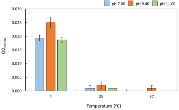
Usando ensaios de turbidez em microplacas, foi possível avaliar o grau de LLPS da FUS sob diferentes condições. A FUS apresentou uma temperatura de solução crítica superior (UCST), submetendo-se a separação reversível de fases a baixa temperatura. Observou-se que a separação de fases é significativamente intensificada quando a FUS apresenta uma carga global neutra (pH 9.40), e na presença de concentrações ótimas de metabolitos carregados, indicando que o processo de LLPS é mediado por interações eletrostáticas. Além disso, os metabolitos que induzem compactação de proteínas e que destabilizam interações hidrofóbicas, inibiram a LLPS da FUS, sugerindo que o processo possui também um caráter hidrofóbico e que a desordem estrutural da FUS é crucial para a formação de grânulos. Através de espetroscopia de RMN, verificou-se que os domínios globulares sofrem desnaturação reversível a frio, a uma temperatura em que o processo de LLPS é promovido. Juntamente com os ensaios de separação de fase, que demonstraram que o RNA e o Zn2+ promovem a LLPS, propõe-se que tanto o domínio RRM e o ZnF
estão envolvidos no processo de separação de fases, mediando interações intermolecular eletrostáticas ao sofrerem desnaturação. Notavelmente, foi também demonstrado que a FUS mantém a sua estrutura geral após a separação de fases. Através de ensaios de microscopia, foi possível observar os grânulos de FUS e identificar características de líquidos, como capacidade de molhar e fusão. No geral, foi demonstrado que o processo de LLPS é extremamente sensível e controlado pelas condições circundantes.
ix
Abstract
Subcellular compartmentalization allows the organization of complex biochemical reactions in space and time, underlying vital cell processes such as homeostasis, division and development. Several compartments, however, lack a physical barrier. These membraneless organelles are usually an assembly of stalled mRNA and proteins that undergo liquid-liquid phase separation (LLPS), being termed as ribonucleoprotein (RNP) granules. These proteinaceous liquids are usually involved in regulation of gene expression and nucleic acid processing.
Proteins that drive LLPS process normally exhibit an overall unstructured composition, being referred as intrinsically disordered proteins (IDPs). Fused in sarcoma (FUS) is a ubiquitously expressed IDP, composed of several disordered domains such as the low complexity (LC) domain, three arginine-glycine-glycine (RGG) boxes and a proline-tyrosine nuclear localization signal (PY-NLS). In addition, FUS contains two globular domains involved in RNA-related functions, the RNA recognition motif (RRM) and the zinc finger (ZnF) domain. In certain stress conditions, FUS can undergo LLPS in the cytoplasm leading to formation of stress granules. Formation of these RNP granules increases the risk of self-templating protein fibrils that underpin fatal neurodegenerative diseases.
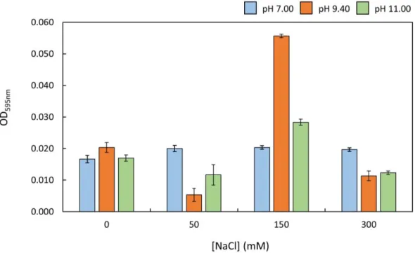
Although the cell environment is known to play a crucial role in mediating RNP granule formation, the mechanisms and molecular determinants that drive LLPS are still unclear. In this context, the main objective of this work is to elucidate the influence of the environment on the formation of FUS granules and to comprehend the mechanisms behind LLPS process. For that purpose, it was explored the influence of the temperature, pH and abundant cellular metabolites, on the LLPS process. Using turbidity microplate assays it was possible to assess the degree of FUS LLPS under different conditions. FUS presented an upper critical temperature solution (UCST) phase separation, undergoing reversible phase separation at low temperature. It was observed that phase separation is significantly enhanced when FUS presents an overall neutral charge (pH 9.40) and in the presence of optimal concentration of charged metabolites, indicating that LLPS is mediated through electrostatic interactions. Moreover, stabilizing metabolites that induce protein compaction and the destabilization of hydrophobic interactions inhibited FUS LLPS process, suggesting that this process has also a hydrophobic character and that FUS structural disorder is crucial for FUS granule formation. Through NMR spectroscopy it was found that FUS globular domains undergo reversible cold denaturation at a temperature in which LLPS is enhanced. Together with the phase separation assays that demonstrated that RNA and Zn2+ enhance LLPS, it is proposed that both RRM
and ZnF domain are involved in the phase separation process, undergoing electrostatic intermolecular interactions upon unfolding at low temperature. Remarkably, it was also showed that FUS maintains its overall structure upon phase separation. Using microscopic imaging, it was possible to observe FUS granules and to identify liquid-like characteristic such as wetting and fusion. Overall it was demonstrated that FUS LLPS process is extremely sensitive and controlled by the environmental conditions.
xi
Table of contents
Agradecimentos ... v
Resumo ... vii
Abstract ... ix
Table of contents ... xi
List of figures ... xiii
List of tables ... xv
List of abbreviation and symbols ...xvii
1. Introduction ... 3
1.1 Stress granules ... 4
1.1.1 Morphology and function of stress granules ... 4
1.1.2 Stress granules in disease ... 5
1.2 Formation of membraneless organelles ... 6
1.2.1 Liquid-liquid phase separation ... 6
1.3 Fused in Sarcoma protein ... 9
1.3.1 FUS structural architecture and function ... 9
1.3.2 FUS localization and nuclear-cytoplasmatic shuttling ...12
1.3.4 FUS in neurodegenerative disease ...14
1.4 Outline and aim of the study ...17
2. Experimental procedures ... 21
2.1 General methodologies and materials ...21
2.2 Protein expression and purification ...22
2.2.1 General expression ...22
2.2.1.1 Non-labeled FUS expression ...22
2.2.1.2 Uniformly 15N-labeled FUS expression ...23
2.2.2 Purification of non-labeled and uniformly 15N-labeled FUS ...23
2.3 FUS phase separation assays ...24
2.3.1 Temperature influence on FUS liquid-liquid phase separation ...24
2.3.2 Metabolite influence on FUS liquid-liquid phase separation ...25
2.4 Imaging of FUS stress granules ...25
2.5 NMR experiments ...26
2.5.1 Preparation of NMR samples ...26
xii
2.5.1.2 Phase-separated FUS ...26
2.5.2 NMR experiments on dispersed FUS ...26
2.5.3 NMR experiments on phase-separated FUS ...27
3. Results and discussion... 31
3.2 Temperature dependence of FUS liquid-liquid phase separation ...33
3.3 Metabolite influence on FUS liquid-liquid phase separation ...35
3.4 Imaging of FUS granules ...53
3.5 NMR experiments ...55
3.5.1 Temperature influence on FUS structure ...56
3.5.2 Phase separation impact on FUS structure ...61
4. Conclusions and future perspectives ... 67
5. Bibliographic references ... 71
6. Appendix ... 87
6.1 Supplementary materials and methods ...87
6.2 Supplementary production of FUS protein ...88
6.2.1 General Expression ...89
xiii
List of figures
Figure 1 – Liquid-like properties of P granules ... 3
Figure 2 – Representative illustration of a stress granule composition ... 5
Figure 3 – Thermodynamics of liquid-liquid phase separation ... 7
Figure 4 – Schematic representation of human FUS structure ...10
Figure 5 – Structural architecture of FET proteins across different species ...10
Figure 6 – RRM domain of human FUS ...11
Figure 7 – Overview of FUS biological functions ...12
Figure 8 –Structure of Kapβ2 in a complex with FUS PY-NLS ...13
Figure 9 – Pathological mechanisms of ALS-FUS and FTLD-FUS ...16
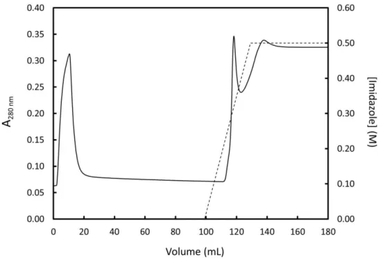
Figure 10 – First purification step of 15N-labeled FUS by Ni-NTA IMAC ...31
Figure 11 – Second purification step of 15N-labeled FUS by Ni-NTA IMAC ...32
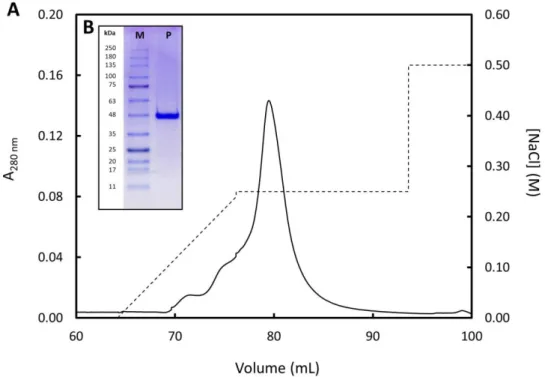
Figure 12 – Final purification step of 15N-labeled FUS by IEX...33
Figure 13 – Influence of temperature on FUS phase separation ...34
Figure 14 – Relative content of amino acids in FUS protein ...35
Figure 15 – Influence of NaCl on FUS phase separation ...37
Figure 16 – Schematic representation of DLVO interaction potential energy ...38
Figure 17 – Plot of FUS net charge as a function of pH ...39
Figure 18 – Cationic and anionic Hofmeister series ...40
Figure 19 – Influence of glucose on FUS phase separation...41
Figure 20 – Influence of ZnCl2 on FUS phase separation...43
Figure 21 – Influence of CaCl2 on FUS phase separation ...45
Figure 22 – Divalent Irving-Williams series ...45
Figure 23 – Influence of NaGlu on FUS phase separation ...47
Figure 24 – Plot of free glutamate net charge as a function of pH ...48
Figure 25 – Influence of LysHCl on FUS phase separation ...49
Figure 26 – Plot of free lysine net charge as a function of pH ...50
Figure 27 – Influence of total RNA on FUS phase separation ...51
Figure 28 – Observation of in vitro FUS phase separation by confocal microscopy ...54
Figure 29 – Edge detection of the 200 µM FUS microscope image ...55
Figure 30 – 1H-15N HSQC spectra of dispersed FUS in the presence of decreasing temperature (25oC → 15oC → 5oC) ...57
Figure 31 – Superposition of the 1H-15N HSQC spectra of dispersed FUS in the presence of decreasing temperature (25oC → 15oC → 5oC) ...58
Figure 32 – 1H-15N HSQC spectra of dispersed FUS in the presence of increasing temperature (5oC → 15oC → 25oC) ...60
Figure 33 – Reversible cold denaturation of dispersed FUS ...61
Figure 34 – Superposition of the 1H-15N HSQC spectra of dispersed FUS and phase-separated FUS ...62
xiv
xv
List of tables
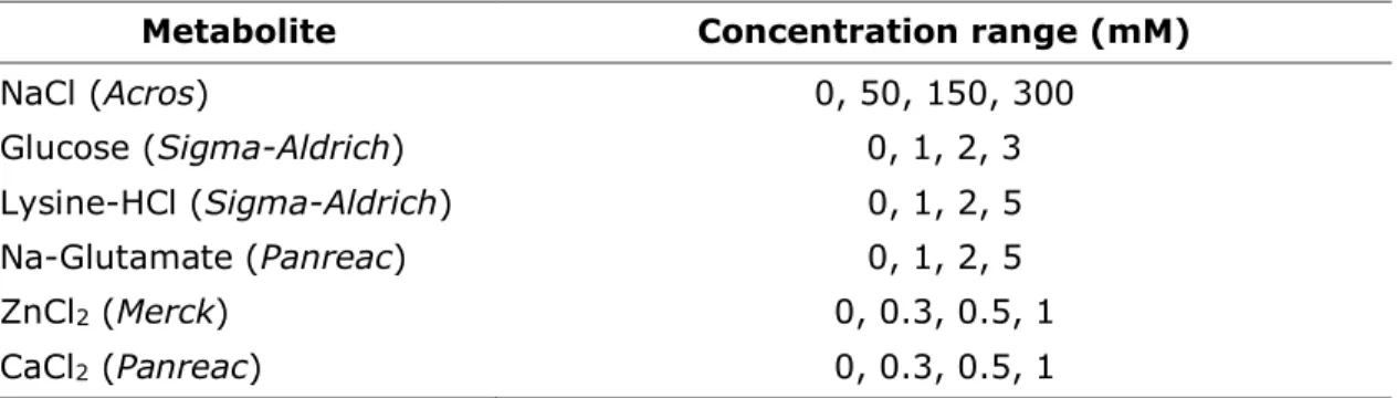
Table I – Metabolite concentration range applied in FUS microplate turbidity assays
...25
Table A. I – Composition of a discontinuous 10% Tris-SDS-PAGE gel (quantity for one gel) ...87
Table A. II – Composition of the Tris-tricine buffer for Tris-SDS-PAGE (per liter) .87 Table A. III – Composition of the sample buffer for Tris-SDS-PAGE (per 50 mL) .87 Table A. IV – Staining and destaining solutions for visualization of protein bands in SDS-PAGE gels ...88
Table A. V – Molecular weight and extinction coefficient values of non-labeled and 16N-labeled MBP-FUS and FUS ...88
Table A. VI – Composition of LB medium (per liter) ...89
Table A. VII – Composition of LB-agar ...89
Table A. VIII – Composition of M9 salts (per liter) ...90
xvii
List of abbreviation and symbols
1H Hydrogen proton
13C Carbon-13 isotope
15N Nitrogen-15 isotope
A280nm Absorbance at 280 nm
AD Alzheimer’s disease
ALS Amyotrophic lateral sclerosis Atm Standard atmosphere unit
βME β-mercaptoethanol
CAPS N-cyclohexyl-3-aminopropanesulfonic acid CTD C-terminal domain
DLVO Derjaguin-Landau-Verwey-Overbeek DNA Deoxyribonucleic acid
DSS 4,4-dimethyl-4-silapentane-1-sulfonic acid
E. coli Escherichia coli
EDTA Ethylenediaminetetraacetic acid FET FUS/EWS/TLS
FL Full-length
FTLD Frontotemporal lobal degeneration FUS Fused in sarcoma
G Gibbs free energy His6 Polyhistidine tag
HSQC Heteronuclear multiple quantum coherence IDP Intrinsically disordered protein
IEX Ion exchange chromatography
IMAC Immobilized metal affinity chromatography Kapβ Karyopherin β protein
kDa Kilodalton LB Luria-Bertani LC Low complexity
LCST Lower critical solution temperature LLPS Liquid-liquid phase separation LysHCl Lysine hydrochloride
mRNA Messenger ribonucleic acid MBP Maltose binding protein MW Molecular weight
xviii
NMR Nuclear Magnetic Resonance OD595nm Optical density at 595 nm
OD600nm Optical density at 600 nm
PDB Protein Data Bank pI Isoelectric point
pKa Logarithmic acid dissociation constant
Ppm Parts per million
RGG Arginine-glycine-glycine RNA Ribonucleic acid
RNP Ribonucleoprotein RRM RNA recognition motif SDS Sodium dodecyl sulfate
SDS-PAGE Sodium dodecyl sulfate polyacrylamide gel electrophoresis TEV Tobacco etch virus protease
Tris Tris(hydroxymethyl)aminomethane UCST Upper critical solution temperature UV-Vis Ultraviolet-visible
WT Wild-type ZnF Zinc finger
µ Chemical potential
Amino acids abbreviations
Alanine Ala A
Arginine Arg R
Asparagine Asn N
Aspartate Asp D
Cysteine Cys C
Glutamate Glu E
Glutamine Gln Q
Glycine Gly G
Histidine His H
Isoleucine Ile I
Leucine Leu L
Lysine Lys K
Methionine Met M
Phenylalanine Phe F
Proline Pro P
Serine Ser S
Threonine Thr T
Tryptophan Trp W
Tyrosine Tyr Y
3
1. Introduction
Inside the human body, millions of chemical reactions occur per second in each cell. In order to organize these reactions, eukaryotic cells contain a series of membrane-limited organelles which provide an independent and distinct chemical environment.1
This compartmentalization allows the organization of complex biochemical reactions in space and time.2,3 Subcellular organization of macromolecules is essential for vital
cellular processes such as homeostasis, division and development.4-6
In the cell, a compartment must have a boundary that permits separation from the cytosolic environment. Moreover, the components must be able to diffuse freely across it, so that chemical reactions can take place.2,7 Many organelles are delimited
by a lipid bilayer membrane including the mitochondria, the lysosome and the nucleus.8 Such membranes allow passive and active transport of large and small
molecules.9
Surprisingly, several cellular compartments lack a physical barrier. These non-membrane-limited organelles, can compartmentalize and concentrate specific set of molecules without the presence of a physical boundary.10
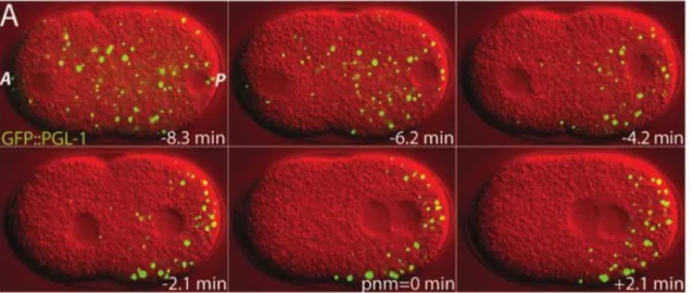
Membraneless organelles behave as fluid droplets, having liquid-like properties, such as shear flow deformation, fusion, high viscosity and rapid exchange of components with the cytoplasm (Figure 1).11,12 The first example of a liquid
compartment was the P granules from Caenorhabditis elegans embryos.13 P granules
are assembly of RNA and RNA-binding proteins that play a role in germ-cell specification.11 P granules can target mRNA for decapping and degradation under
regular and stress conditions.14
Figure 1 – Liquid-like properties of P granules – P granules (in green; GFP tagged) exhibit liquid-like properties such as diffusion and fusion overtime in C. elegans embryos. Time relative to pronuclear meeting (pnm). A – anterior, P- posterior. Modified from Brangwynne et al.11
4
chromatin20, histone locus bodies (HBLs)21, nuclear stress bodies (nSBs)22,
Oct1/PTF/transcription (OPT) domains23, polycomb bodies (PcG bodies)24,
perinucleolar compartment (PNC)25, promyelocytic leukaemia nuclear bodies (PML
nuclear bodies)26 and the Sam68 nuclear bodies (SNBs)25. In the cytoplasm, RNP
granules are less numerous and diversified. These comprise centrosomes27, neuronal
RNA bodies28, P-bodies29 and stress granules30. Moreover, in the mitochondria is
observed a unique subdomain structure, the mitochondrial RNA granule.31 Hence, a
variety of RNP bodies are present in the cytoplasm, mitochondria and nucleus of eukaryotic cells. Despite their diversity in structure, morphology, function and distribution, all of these membraneless subcellular structures are ubiquitous in cells, and contribute to numerous vital biological functions, including processing and storage of RNA and other biomolecules.4,32
1.1 Stress granules
Stress granules are RNP cytosolic bodies that assemble from pools of untranslated mRNA and RNPs.30,33 In the cytoplasm, stress granules regulate RNA stability and
protein translation in response to stress stimuli.34
Arising from mRNA stalled in translation initiation, stress granules contain several translation initiation factors, RNA-binding proteins (comprising ~ 50%) and non-RNA-binding proteins.35 Non-RNA-binding proteins are vital for RNA processes, including
post-translation modification enzymes, metabolic enzymes, and protein or RNA remodeling complexes.35-37
1.1.1 Morphology and function of stress granules
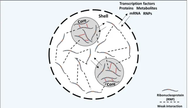
Stress granules are not uniform structures, normally containing two distinct layers: a core protein-rich structure and a dynamic shell.33,37 The shell is thought to be a
scaffold for dynamic exchange with the outside, while the core maintains the stress granule structure and provides further confinement for biomolecule sequestration (Figure 2).38
Stress granule formation is a dynamic and reversible process. Assembly of stress granules begins with the formation of the core followed by the shell. The reverse process is also a stepwise process that begins with the less-stable shell dissipation followed by core clearance.38
5 Figure 2 – Representative illustration of a stress granule composition –Stress granules exhibit a biphasic composition, structured by a protein-rich core surrounded by a dynamic shell.38
Stress granule assembly is known to be highly redundant since several and distinct post-translational modifications such as protein methylation, phosphorylation and glycosylation, influence differently the stress granule assembly.40-42 In fact, several
stress-granule-promoting proteins contain specific motifs for such modifications, such as RGG motifs, which are sites for arginine methylation.43
Stress granules have a broad effect on the physiology of the cells, not accounting for mRNA processing. Due to the high local concentration of components, stress granules can recruit numerous proteins in certain stress responses. For example, during viral infections, stress granules recruit several antiviral proteins such as RIG-1 and PKR, enhancing the immune response and viral resistance.44 Moreover, stress
granules can sequester numerous components, inhibiting several signaling pathways such as the apoptosis stress-responsive MAPKS pathway, or the TNR-α-mediated
NG-KB proinflammatory signaling pathway, as a protective mechanism against the
apoptotic or inflammatory response under stress conditions.45,46 In fact, having this
into account, there were developed chemotherapeutic agents that promote controlled stress granule formation.47,48
1.1.2 Stress granules in disease
Mutations that lead to functional impairment of stress granules have been implicated with many neurodegenerative diseases such as amyotrophic lateral sclerosis (ALS), frontotemporal lobar degeneration (FTLD) and inclusion body myopathy (IBM).49 In
most cases, the mutations are directly on the RNA-binding proteins (e.g. FUS, TDP-43, TIA1), increasing the stress granules self-assembly.49-51 Ultimately, this leads to
6
in misregulation of RNA processing and signaling pathways, or defects in nuclear-cytoplasmic transport of RNPs.52,53
Stress granules present an evident mechanism of assembly through regulated protein aggregation. However this process can soon evolve into pathological protein aggregates.54
The classical model for pathological protein aggregation, proposed by Dobson and Lansbury, describes the conversion mechanism of soluble protein into insoluble protein deposits.55,56 In this model, monomers of aggregation prone proteins can
undergo random misfolding, under certain conditions.10,11 The misfolded species
oligomerize and aggregate to form fibrils, through nucleated growth mechanism, leading to non-functional deposits.11
The rate of aggregation is dictated by mass action and energy minimization.54
Stress granules are comprised of proteins containing high number of charged and aromatic residues. These residues increase the propensity of oligomerization through electrostatic and aromatic interactions. Paired with increased cytoplasmatic concentration of these proteins, favored through mutation or persistent stress, the assembly of pathological aggregates is promoted.54,57
Hence, although stress granule formation is crucial for cell survival, their assembly pathway through regulated protein aggregation is vulnerable to disruption, which can lead to severe neurotoxicity.
1.2 Formation of membraneless organelles
As stated earlier a compartment must have a boundary and the components must diffuse freely across it. For that purpose, and in order to understand the compartmentalization through membraneless organelles, we need to understand the mechanisms behind their assembly and how molecules can diffuse in and out. Moreover, it is necessary to comprehend how such compartments can stay stable under aqueous cytosol and what are the properties that drive their assembly.
1.2.1 Liquid-liquid phase separation
The liquid nature of nuclear and cytosolic membraneless organelles seems counterintuitive. Thermodynamically, two miscible liquids mix in order to evolve towards the state of higher entropy, according to the second law of thermodynamics.58 However, stress granules can coexist in the cytosol without fusing
with the surroundings.3
Interestingly, liquids can undergo demixing process. In these cases, the phase separation is driven by interactions between the molecules that constitute each liquid. Thermodynamically, the system progresses towards lower energy if molecules of the same species interact. Here, the gain in energy reduction overcomes entropy increase tendency, driving the system towards demixing.2
The current accepted process that drives the cytosolic demixing and the consequent formation of stress granules is liquid-liquid phase separation (LLPS).10,59,60 The LLPS
7
LLPS is a process mediated both by thermodynamics and kinetics.61,62 The
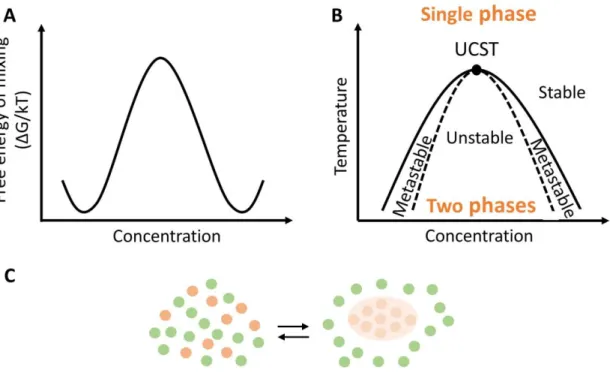
composition of the two phases at equilibrium is dependent on temperature. For this purpose, the LLPS mechanism is normally explained by temperature-composition diagram. This phase diagram is characterized by a coexistence curve, in which the boundaries show the composition of the phases that are in equilibrium in various temperatures (at a constant pressure, normally 1 atm). The coexistence curve has a critical point defined by a critical temperature (Tc) and a critical concentration (Cc),
temperature and concentration in which phase separation occurs.60,61
Proteins can exhibit two distinct types of phase separation behavior: upper critical solution temperature (UCST) type of behavior, or a lower critical solution temperature (LCST) type of behavior. The UCST is the highest temperature at which phase separation occurs. Above this temperature the system is homogenous, and hence, phase separation occurs at temperatures lower that the UCST. At LCST regime, which is less common, phase separation occurs when temperature is higher than LCST, and protein solubility is higher at lower temperatures.60,61
Phase diagrams often represent also the spinodal curve. This curve separates the metastable from the unstable region. Inside the unstable region occurs spontaneous phase separation (i.e. the mixture is unstable and the phase separation occurs spontaneously), whereas in the metastable region demixing only occurs if seeds are present. Depending on the type of phase separation behavior, the LCST or UCST point is where the coexistence and spinodal curves intersect (Figure 3).63
8
At Tcthe two phases are in equilibrium and the Gibbs free energy of the system is
minimized. Considering a simple mixture, the molar Gibbs energy (Gm) is equal to
the chemical potential (µ). In brief, µ is the measure of the potential that a substance has for undergoing change in a system, in this case, phase separation. LLPS is achieved when the system is at equilibrium, i.e. the chemical potential of the substance is the same throughout the sample, regardless of how many phases are presented. Hence, even when protein concentration is differently distributed, with a protein-rich phase and a protein-power phase, µ at those different locations is equal.61 As stated earlier, in a cellular compartment, components must diffuse freely
through it. Considering a membraneless organelle formed by a protein in a cell, the chemical potential of this protein is µ1 inside the organelle, and µ2 in the cytoplasm. In order to keep the stability of this membraneless organelle, µ1 must be equal to
µ2. Yet, since the protein can diffuse in and out the organelle, Gibbs energy of the system changes, and thus µ1 and µ2 should be different.2,61
For example, when an infinitesimal amount dp of the protein is transferred from the organelle to the cytoplasm, the Gibbs energy of the system changes by -µ1dp, considering the removal of the protein from the organelle. On the other hand, the Gibbs free energy of the system also change by +µ2dp considering the protein that is added to the cytoplasm. Hence, the overall change in Gibbs energy of the whole system is dG=(µ2- µ1)dp. In these cases, the phase stability (when G=0) is maintained since molecules move stochastically from one phase to another. Although there is diffusion of protein in and out of the phase, Gibbs energy is zero since equal number of molecules are going in and out of the phase. So, when the chemical potential in one location is raised, the components diffuse into the other location, maintaining the chemical potentials equal across the cell.2,61
The tendency to maintain G=0, and thus, maintain the phase separation, is dictated by the competition of interactions between the protein and the solvent (i.e. the cytoplasm).64
The polypeptide backbone of protein is an array of dipoles. Since aqueous-based solvent are also dipolar, there are three possible dipolar interactions: polypeptide-solvent, polypeptide-polypeptide and solvent-solvent. The energetic balance between the three types of interactions is quantified by the Flory-Huggins interaction
parameter, χ.65,66 The poor solubility and preference of polypeptide backbones for
globules is due to the poor solvent feature of water (χ > 0). Hence, when χ > 0, the
net attraction between protein dipoles overcomes the protein-solvent interaction. Consequently, the energetic polypeptide-polypeptide term surpasses the mixing entropy, favoring the bulk phase separation at higher concentrations.64
Therefore, even when there is a protein concentration gradient across the cell, and when there is diffusion in and out of the phase, the net attraction between the protein dipoles drives the system towards equilibrium, reaching phase separation.2
As a result, it is easy to comprehend that not all proteins benefit from interacting with other proteins instead of the solvent, and hence do not undergo LLPS.
Protein associated with membraneless organelles normally exhibit an overall unstructured composition, being referred as intrinsically disordered proteins (IDPs).67,68
9
regions, termed low complexity (LC) domains , are remarkably overrepresented in proteins that drive LLPS.12,40,70
In the LC domains, the sequences are often repetitive and enriched in glycine (G). The side-chain of this amino acid does not participate in hydrogen bonding when glycine is in the context of a protein and thus it cannot be regard as a polar residue. However, it cannot also be estimated as hydrophobic, since the methane side-chain is perhaps too small to have hydrophobicity contribution. Hence, since there is not an unequivocal agreement on glycine polarity across several hydropathy scales, in this study glycine will be regarded as having neutral polarity.71-74
LC domains also contain additional polar side-chains such as glutamine (Q), serine (S) and asparagine (N), positively charged side-chains such as arginine (R) and lysine (K), negatively charged side-chains such as aspartic acid (D) and glutamic acid (E), or aromatic side-chains such as phenylalanine (F) and tyrosine (Y).64
The LC domain of the IDPs has been reported to play an imperative in the mediation of RNP granules formation.57 In fact, specific sequences play an imperative role in
driving the LLPS process, such as YG/S-, Q/N-, FG-, RG-, GY-, KSPEA- and SY- rich motifs, as well as sections of alternating charges. The repetitive patterns promote multivalent inter-molecular interactions such as charge-charge, π-charge and π-π stacking.64,67
1.3 Fused in Sarcoma protein
Fused in sarcoma (FUS), also known as Translocated in liposarcoma (TLS), is a nuclear nucleic acid binding protein involved in regulation of gene expression, DNA/RNA processing and maintenance of genomic integrity.75-78 In certain stress
conditions, FUS undergoes liquid-liquid phase separation leading to stress granule formation in the cytoplasm.79,80
FUS has been implicated with several neurodegenerative diseases. Remarkably, FUS is a major component of pathological inclusion in over 5% of all types of ALS and 9% of FTLD cases.53,81
1.3.1 FUS structural architecture and function
Initially identified in 1993 as a fusion oncogene in human myxoid liposarcomas, FUS is a ubiquitously expressed 526 amino acid (53.42 kDa) nuclear RNP.82,83
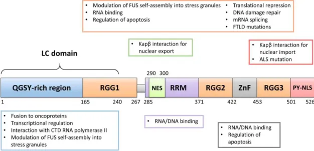
This multidomain protein contains a low complexity (LC) domain that is enriched with glutamine, glycine, serine and tyrosine (QGSY) residues, a nuclear export signal (NES) inserted in a RNA recognition motif (RRM), a cysteine2-cysteine2 zinc finger
(ZnF) domain and a proline-tyrosine nuclear localization signal (PY-NLS).84,85 FUS has
10 Figure 4– Schematic representation of human FUS structure – Representation of the organization of FUS several domains. QGSY = glutamine, glycine, serine and tyrosine; RGG: = arginine-glycine-glycine box; NES = nuclear export signal; RRM = RNA recognition motif; ZnF = zinc finger; PY-NLS = proline-tyrosine nuclear localization signal.
FUS is a member of the FUS/Ewing’s sarcoma (EWS)/TATA-binding protein-associated factor 15 (TAF15) FET family, a group of abundant and ubiquitously expressed RNPs.77,87 FET proteins are found in multicellular organisms including
vertebrates, plants, nematodes and insects, and all share an homologous domain architecture (Figure 5).77
Figure 5 – Structural architecture of FET proteins across different species – The structure of FET proteins is conserved among several multicellular organisms. Domain organization, excluding Arabidopsis thaliana, is consistent. The number of LC domains repeats can vary, which is accountable for different lengths of this domain within species. The number and length of RGG domains can also diverge among organisms.77
The RRM domain of FET proteins are distinguished from other RNP RRMs due to the
extended “KK-loop” between the α1 and β2 of the β1α1β2β3α2β4 fold, and also the
lack of aromatic amino acids on β3 (Figure 6).88,89
Lysine residues in the KK-loop of FUS are known to be crucial for RNA/DNA binding, by providing an interacting positively-charged surface. Remarkably, substitution of these residues impairs nucleic acid binding and, interestingly, FUS subcellular localization.17
11
nucleic acid binding in most RRM domains, suggests binding that diverges from the canonical RNA/DNA-RRM interaction.88,90,91
Figure 6 – RRM domain of human FUS – The RRM domain of FUS displays a β1α1β2β3α2β4 type of secondary structure, with a unique KK-loop between α1 and β2. Consecutive lysine residues in this loop (K315 and K316) are key for nucleic acid binding (PDB ID: 2LCW).88
The RGG motif and the ZnF domain also play a crucial role in FUS nucleic acid binding.86,92
RGG are a very abundant and evolutionary conserved intrinsically disordered regions (IDRs), being present in more than 1000 human proteins.43 The RGG motifs
are the second most common RNA-binding domain in the human genome, mediating interaction with distinct types of RNA, such as single- and double-stranded RNA and G-quartets.93,94 The high flexibility of the RGG domain provides a larger interacting
surface that can target a variety of RNAs.95 RGG motif-containing proteins are often
associated with a vast range of nucleic acid processing, such as regulation of apoptosis, translational repression, regulation of transcription, DNA damage signaling, and precursor mRNA splicing.43
The ZnF is among the most abundant domains in eukaryotic proteins.96 This domain
mediates a variety of processes such as DNA recognition and RNA packing, and regulates apoptosis, RNA transcription and protein folding.96,97 All FET members share
a C4-type ZnF characterized by four cysteine residues coordinating a zinc ion.77,92 In
FUS, the ZnF binds to GGUG-containing RNA and, interestingly, plays a more dominant role in RNA recognition that the RRM domain.92
A characteristic domain found in FUS, and all FET members, is the N-terminal LC domain. This unstructured region usually contains repeats of consensus motifs (Figure 4).77 In FUS, the LC domain (1-239 residues) is enriched with glycine
residues (28%), and contains several polar amino acids, such as serine (22.6%), glutamine (18%) and tyrosine (12.1%).98 The FUS LC usually contains 20 repeats of
(S/G)Y(S/G) motif, spread across two tandem repeats (RAC1 and RAC2) and 16 single motifs.99
The LC domain of FET proteins have an important role on protein self-association through LLPS.100,101 Also, the LC can function as a transcriptional activation domain
12
Recently, Burke et. al demonstrated that RNA polymerase II CTD interacts not only with fibrillar forms of FUS LC but also with the phase-separated state. Moreover, RNA polymerase II nucleates the self-assembly of FUS LC.102
The FUS LC domain has also a role in oncogenesis. Aberrant chromosomal translocations can lead to fusion of the FUS LC domain to DNA-binding domains of transcriptional regulators such as CHOP. In this case, the LC domain also functions as a transcriptional activator, leading to abnormal tumor development, such as in human myxoid liposarcoma.82,83,103
Finally, the LC domains have an important role on protein self-association through LLPS. By promoting stress granule formation, these domains have large impact in the modulation of the biological and pathological function of FET proteins.77
The multidomain character of FUS provides an exceptional array of broad functions that mediate vital cellular processes, that go from transcriptional regulation to stress response (Figure 7). Therefore, it is intuitive to comprehend that missense mutations upon FUS gene can disrupt cellular homeostasis, triggering severe pathogenesis.104
Figure 7 – Overview of FUS biological functions– FUS contains several functional domains involved in an array of cellular processes. Functions of each color-coded domain are depicted in boxes with the same representative color.
1.3.2 FUS localization and nuclear-cytoplasmatic shuttling
FUS is primarily localized in the nucleus where it regulates several nuclear functions such as transcription, pre-mRNA splicing and DNA repair.103 Still, FUS can shuttle
between the nucleus and the cytoplasm, participating in the biogenesis, transport and processing of cytoplasmatic mRNA.103,105
The nuclear-cytoplasmatic shuttling of FUS across the nuclear membrane is mediated by nuclear transport proteins of the Karyopherin β family. This family is responsible for most nuclear-cytoplasmatic translocation of proteins through the nuclear pore complex.106Karyopherin βs interaction with FUS are made upon the
13
import of FUS is achieved through interaction of its non-classical C-terminal PY-NLS (500-526 residues) with the nuclear transporter Karyopherin β2 (Kapβ2), also known as Transportin 1 (Figure 8).108,109
Figure 8 – Structure of Kapβ2 in a complex with FUS PY-NLS – FUS PY-NLS (yellow) occupies the C-terminal arch of Kapβ2 (red-grey-blue), interacting through the PY-NLS N-terminal PGKM hydrophobic motif, a central arginie-rich α-helix and the C-terminal PY-motif (PDB ID: 5VYG).110
Unlike the classical Importin α/β system which recognizes the compact and conserved
NLS, Kapβ2 interacts with PY-NLS, a more complex domain that lacks a consensus sequence.107 Therefore, PY-NLS domains are normally described as a collection of
weak consensus motifs composed of a structural disordered hydrophobic or basic N-terminal and a C-N-terminal RX2-5PY motif.111 Kapβ2 can recognize and bind diverse
basic and hydrophobic N-terminal motifs, due to its acidic/hydrophobic surface.109
Impairment of FUS nuclear import due to mutations that affect the PY-NLS, can lead to accumulation of abnormal protein inclusions in the cytosol, which is found in ALS and FTDL patients.112 Moreover, in 2012 Dormann D. et al. demonstrated that
Kapβ2 can also recognize the RGG motif that precedes the PY-NLS, and that this recognition is dependent on post-transcriptional arginine methylation of the RGG
motif. If the RGG motif is methylated, Kapβ2 is unable to recognize FUS, which halts
its nuclear import, leading to its accumulation in the cytoplasm.113
1.3.3 FUS stress granules
In order to reestablish homeostasis in stress conditions, cells activate several pathways that normally involve gene expression regulation or, ultimately, apoptosis.104,114 As stated earlier (see section 1.1 Stress granules) stress granules are
transient membraneless cytosolic structures that mediate the fate of gene expression, degradation, or suppression, in response to cellular stress.10
Mediating several stages of gene expression, is not surprising that FUS plays an imperative role in cellular stress response.104 Cells exposed to environmental stress,
such as oxidative stress, hypoxia, viral infection or osmotic stress, actively recruit cytoplasmatic FUS into stress granules.115-117 However, the stress environment does
14
must be in the cytoplasm and already poised to enter stress granules during the stress condition. Therefore, mutations that affect the PY-NLS domain of FUS, and consequently, the reimport of FUS into the nucleus, contribute to FUS stress granule accumulation.110,117
FUS undergoes LLPS in vivo and in vitro, self-assembling into liquid-like stress structures, at incredibly low concentration (1 µM).119,120 Cytoplasmatic demixing of
FUS allows rapid and local concentration of FUS and, presumably, a myriad of additional factors required for stress response.121 However, high local concentration
of FUS might trigger liquid-to-solid transition, from droplets granules into aggregated inclusions. Hence, stress granules are presumably precursors to pathological inclusions found in neurodegenerative diseases.120
The LC domain of FUS is crucial for self-assembly of FUS.102 The aromatic residues
of FUS LC mediate short range interactions that promote the LLPS. On the other hand, the abundant hydrophilic character of this domain dictates the solubility of FUS, consequently opposing LLPS once the aromatic interactions are degenerated. Furthermore, phosphorylation of tyrosine and serine residues in FUS LC disassembles liquid droplets.100,102 Thus, the LC domain of FUS seems to modulate the protein
phase separation.
The RGG domains are also crucial for FUS LLPS.101,122 In fact deletion of the RGG
domains, specially RRG1, reduces significantly FUS phase separation.123
FUS stress granules are highly dynamic, displaying constant exchange of components with the cytoplasm and rapid internal rearrangement. Therefore, if the cell uses a dynamic liquid compartment to perform physiological relevant functions, it will have to constantly contradict thermodynamic driven aggregation. Hence, FUS-related diseases are often associated with late onset pathology, when the quality control machinery starts to degenerate.10,124
1.3.4 FUS in neurodegenerative disease
FUS cytoplasmatic inclusions is a common hallmark of both amyotrophic lateral sclerosis (ALS) and frontotemporal lobar degeneration (FTLD).103,104
ALS is the most frequent neurodegenerative disease in the human motor system.125
Common manifestations encountered in ALS patients include muscle weakness and atrophy, and fasciculation in the motor neurons and brain stem.125,126 Progressive
weakening of the respiratory muscles leads to dyspnea and consequently respiratory failure, leading to low survival rate.125 About 50% of ALS patients die within 30
months of symptom onset and about 20% of patients survive between 5-10 years after the symptoms appear.127
FTLD is the second most common cause of dementia in presenile age group
(<65%), behind Alzheimer’s disease (AD). This neurodegenerative disease is
characterized by progressive degeneration of the frontal and anterior temporal lobes. This leads to progressive alterations and disturbances in personality, and/or language problems.128 Although the memory preservation in the initial phase of FTLD is higher
15
ALS and FTDL form a clinical disease continuum, since up to 15% of ALS patients meet the clinical criteria of FTLD and 15% of FTLD patients meet the clinical criteria of ALS.129
ALS can be divided in two major groups: sporadic ALS and familial ALS. Sporadic ALS accounts for about 90% of all ALS diseases, but its aetology is unknown. The rest 10% of ALS cases are familial, which is a form of hereditary ALS.130
In the familial ALS, several defects on genes have been identified in patients. These include mutation in SOD1 that encodes for copper/zinc superoxide dismutase (20% of familial ALS) and TARDBP that encodes for TDP-43 (5-10% of familial ALS).125,131
In 2009, the FUS gene was identified as the primary disease-associated gene for familial ALS6, which accounts for 5% of familial ALS.132FUS-linked ALS and FTLD is
designated as ALS-FUS and FTLD-FUS, respectively.
Cytoplasmatic depositions of FUS stress granules containing is a common hallmark for both FTLD-FUS and ALS-FUS subset.103,133 However, there are several distinctions
between the two diseases (Figure 9). In contrast to ALS-FUS, FTLD-FUS is rarely associated with FUS gene mutations.134
In FTLD-FUS there is a neuronal cytoplasmatic co-deposition of FUS and all other
FET family members (EWS and TAF15), along with Kapβ2 Moreover, this
neurodegenerative disease also displays low nuclear concentration of all three FET proteins.135-137 Interestingly, protein inclusions in FTLD-FUS patients contain
unmethylated FUS (see section 1.3.2 FUS localization and nuclear-cytoplasmatic shuttling).113 In this case, unmethylated RGG3 domain can cause overly tight binding
of the FET proteins to Kapβ2, inhibiting the FET-Kapβ2 dissociation in the nucleus. Hence, FET proteins are continuously re-exported to the cytoplasm which leads to cytoplasmatic FET deposits.103
Opposed to FTLD-FUS, ALS-FUS show an exclusive deposition of FUS, excluding
EWS, TAF15 and Kapβ2.136 In this case, point mutations in the FUS gene that truncate
or modify the FUS PY-NLS domain, can lead to impairment of nuclear import. Consequently, FUS accumulates in the cytoplasm and clusters into cytoplasmatic aggregates.112,116
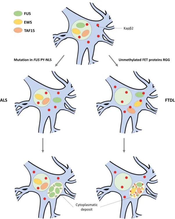
16 Figure 9 – Pathological mechanisms of ALS-FUS and FTLD-FUS – In homeostatic conditions, FET proteins (FUS, EWS and TAF15) are properly imported into the nucleus by
Kapβ2. In ALS-FUS, missense mutations in the FUS gene that affect the FUS PY-NLS domain, cause impairment of the FUS-Kapβ2 interaction. This hinders the re-import of FUS into the nucleus, which ultimately leads to cytoplasmatic deposits of aggregated FUS; In FTLD-FUS, unmethylated RGG domain of FET proteins leads to irreversible binding to Kapβ2 which inhibits the release of FET proteins in the nucleus, leading to cytoplasmatic deposits of aggregated FET proteins.103
17
The first model proposes that dysregulated assembly of FUS leads to fibrillar aggregates that perturb the physiological stress responses. Thus, there is a gain of toxic properties of FUS protein aggregates.123,138
The loss-of-function model focus of the loss of FUS physiological role. As this protein is recruited in stress granules, there is an impairment in the FUS cytoplasmatic function.139,140
Lastly, nuclear loss-of-function is consistent with the fact that FUS has a crucial physiological role in the nucleus. In both ALS-FUS and FTLD-FUS there is a depletion of nuclear FUS, which impairs several nuclear processes such as transcriptional regulation.53,75
Despite the cause of neurotoxicity, the inclusion of stable fibrillar FUS assemblies is a requisite in motor neuron cells in ALS-FUS and FTLD-FUS diseases.79 Several
efforts are being carried in order to understand the biochemical mechanisms and pathways that leads to FUS cytoplasmatic inclusions, with the hope of develop solutions to mitigate these neurodegenerative diseases.123,141-143
1.4 Outline and aim of the study
Given the importance of FUS stress granules in both physiological and pathological conditions, the aim of this study is to elucidate the mechanisms and determinants that drive their assembly.
The concept is based on the fact that cells have a complex milieu that impact and tune several processes. The conviction is that different environments, and consequently different interactions, can uniquely influence the FUS LLPS mechanism. Therefore, the main objective is to assess the influence of diverse environmental conditions in the formation of FUS stress granules and attempt to unravel the role of the different FUS domains in their assembly. For this purpose, it is explored the transition between dispersed and phase-separated WT-FUS using different biochemical techniques such as NMR, microplate and microscopic assays.
Insights on the environment role can prove valuable in understanding the physiological and pathological conditions that can trigger WT-FUS stress granule assembly. This comprehension can help us link different cellular conditions to the appearance of cytoplasmatic FUS stress granules, that are crucial templates for pathological inclusions found in neurodegenerative diseases.
For that purpose, the main tasks set for this work are:
• Expression and purification of non-labeled and 15N-labeled WT-FUS
• Study of the environment influence on FUS self-assembly, including pH, temperature and abundant cellular metabolites, through microplate turbidity assays
• Elucidation of the mechanisms involved in FUS LLPS
• Observation of FUS stress granules through microscopic imaging
• Comprehension of the impact of temperature on FUS overall structure by NMR spectroscopy
21
2. Experimental procedures
2.1 General methodologies and materials
In this section the general methodologies and commonly used materials will be described and any alteration to the general procedures will be referred in the appropriate section.
Buffers were prepared using distilled water or Milli-Q water, both obtained from laboratory facility instruments. The pH measurements were made with Docu-pH Meter (Sartorius).
Centrifugation steps were executed in different centrifuges according to the sample
volume. Microcentrifuge tube centrifugations (≤ 2 mL) were carried out by a MIKRO 120 centrifuge(Hettich). Centrifuge canonical tubes (≤ 30 mL) were centrifuged in a centrifuge 5804R (Eppendorf) with a F-34-6-38 fixed-angle rotor (Eppendorf). For
higher volumes (≤ 500 mL), centrifugations were performed in an Avanti J-26 XPI centrifuge (Beckman Coulter) with JA fixed-angle rotors (Beckman Coulter). Besides from small volume samples (≤ 2 mL), centrifugations were carried out in Nalgene PPCO canonical tubes (Thermo Fisher Scientific).
Small volume incubations were carried out in an Orbital Shaker-incubator ES-20 (Grant-Bio). For bacterial growth, incubations were executed in an Orbital Shaker-incubator (Optic-Ivymen System). Optical density was measured in a UltroSpec 2100 Pro UV-Vis spectrophotometer (GE Healthcare) in polystyrene cuvettes ( Sigma-Aldrich).
Sodium dodecyl sulfate polyacrylamide gel electrophoresis (SDS-PAGE) was prepared in a discontinuous manner, employing a Tris-tricine buffer system.144 In
brief, 10% separating gel and 4% stacking gel are independently prepared and respectively casted at the bottom and at the top of the electrophoretic glass apparatus. The separating gel allows effective separation of proteins with molecular weights ranging from 1 to 100 kDa. Detailed composition of the Tris-SDS-PAGE gel and Tris-tricine buffer system are described in the appendix (Appendix - Table A. I and Table A. II). For the Tris-SDS-PAGE procedure, samples were prepared by mixing 15 µL of protein sample and 5 µL of SDS-PAGE sample buffer (Appendix - Table A. III). The samples were subsequently heated at 100oC for 5 min and
centrifuged at 14000 rpm for 1 minute. From the prepared sample, 18 µL were applied to the gel along with 2 µL of protein marker (NZYTech Colour Protein Marker II, NZYTech) with reference bands covering a range of molecular weights from 11 to 245 kDa. The Tris-SDS-PAGE electrophoresis were executed in an electrophoresis apparatus (Bio-Rad). The power supply settings employed were 200 mA, 200 V and 50 W. Each electrophoretic run proceeded for about 40 min. The gels were stained and destained by Coomassie Blue Staining Method for 1 and 2 hours, respectively, at 37oC and 50 rpm (Appendix - Table A. IV).
Protein purifications were carried out in ÄKTA start chromatographic system (GE Healthcare) following A280nm using UNICORN Start software (GE Healthcare). Both
22
All dialysis steps were achieved using 7K MWCO SnakeSkin Dialysis Tubing (Thermo Fisher Scientific). Concentration and diafiltration of samples were performed using Vivapsin Turbo 15 10 MWCO (Sartorius) for sample volumes up to 15 mL, and Vivapsin Turbo 4 10 MWCO (Sartorius) for sample volumes between 2 and 4 mL.
Protein concentrations were obtained from A280nm measured on a NanoDrop
ND-1000 UV-Vis spectrophotometer (Thermo Fisher Scientific), using the molar extinction coefficients estimated by ProtParam ExPASy software (Appendix - Table A. V).145
2.2 Protein expression and purification
Recombinant MBP-FUS-FL-WT construct was kindly gifted by Nicolas Fawzi (Addgene plasmid #98651). The plasmid was constructed using a PETM-41 vector, to express the human WT-FUS full-length protein which included a His6-MBP moiety and a TEV
cleavage site (MBP-FUS) (Appendix - Figure A. 1).
2.2.1 General expression
Escherichia coli strain BL21 (DE3) competent cells were transformed with the MBP-FUS plasmid through the heat shock method. For the transformation 1 µL (50 ng/µL) of plasmid DNA encoding WT-MBP-FUS was mixed with 50 µL pf E. coli BL21(DE3) competent cells and kept on ice for 30 min. Then, cells were incubated at 42oC
(AccuBlock Digital Dry Bath, Labnet International)for 40 s and transferred to ice for 5 min. Finally, 950 µL of Luria-Bertani medium (LB) (Appendix - Table A. VI) was added to the samples, and subsequently incubated at 37oC for 60 min.
The transformed E. coli cells were plated overnight at 37oC in agar LB (Appendix
- Table A. VII) supplemented with 50 µg/mL kanamycin (NZYTech). A single isolated colony from the plated E. coli cells was chosen and incubated overnight at 37oC and
200 rpm, in 150 mL LB supplemented with 50 µg/mL kanamycin.
2.2.1.1 Non-labeled FUS expression
Non-labeled recombinant FUS was expressed in 1 L LB (x6) until mid-exponential growth (OD600nm~0.8), and subsequently induced with 0.5 mM isopropyl-β
-d-1-thiogalactoside (IPTG) (NZYTech) for 16 h at 18oC and 180 rpm. Cells were recovered
by centrifugation at 6700 rpm (JA-10 rotor) for 15 min, at 4oC. The cell pellet for
each liter of culture was resuspended in 30 mL of lysis buffer containing 20 mM sodium phosphate buffer (Na2HPO4–Honeywell, NaH2PO4–Sigma-Aldrich), 300 mM
NaCl (Acros), 40 mM imidazole (Affymetrix), 10% glycerol (Scharlau), 5 mM β
-mercaptoethanol (βME) (Sigma-Aldrich), 2 mM benzamidine (Sigma-Aldrich) and 1 protease inhibitor tablet (cOmplete Mini EDTA-free Protease Inhibitor Cocktail, Roche), at pH 7.50.
23
centrifugation at 20000 rpm (JA-25.50 rotor) for 60 min, at 4oC. The supernatant
was recovered for the subjacent purification steps.
2.2.1.2 Uniformly
15N-labeled FUS expression
Uniformly 15N-labeled recombinant FUS was expressed in M9 minimal medium
(Appendix - Table A. VIII and Table A. IX), using 15NH
4Cl as the only nitrogen
source. The expression followed the previously described process (see section 2.2.1.1 Non-labeled FUS expression), being retrieved the supernatant for purification.
To maximize the yield of labelled protein, FUS was also recovered from inclusion bodies. To accomplish this, after the sonication step a mild solubilization of the inclusion bodies was performed, following the procedure reported by Singh et al.146
In summary, the resulting pellet from 1 L of culture was resuspended in 1 mL H2O
Milli-Q and solubilized in 9 mL of solubilization buffer containing 100 mM CAPS (Alfa Aesar), 2 M urea (Sigma-Aldrich), 100 mM NaCl, 20% glycerol, 5 mM βME and 0.1% (w/v) SDS (Panreac), at pH 12.00. The solubilized proteins were incubated at room temperature for 30 min and later centrifuged at 11000 rpm (F-34-6-38 rotor) for 30 min, at 4oC. Then, for the refolding step, 10 mL of solubilized protein was added to
90 mL of refolding buffer containing 50 mM Tris-HCl (Fluka), 2 M urea, 100 mM NaCl,
10% glycerol, 5 mM βME and 1 protease inhibitor tablet, at pH 8.00. The refolding was carried out at 4oC in a pulsatile manner, adding the solubilized protein at 1
mL/min to the refolding buffer, with constant stirring. The refolded protein was cleared by centrifugation at 11000 rpm (F-34-6-38 rotor) for 45 min, at 4oC. Cleared
samples were diafiltrated against 20 mM sodium phosphate buffer, 40 mM imidazole, 300 mM NaCl, 10% glycerol, 5 mM βME, 2 mM benzamidine and 1 protease inhibitor tablet at pH 7.50. The refolded protein was then added to the labeled protein retrieved from the supernatant, for subsequent joint purification.
2.2.2 Purification of non-labeled and uniformly
15N-labeled FUS
The purification of non-labeled and uniformly 15N-labeled FUS followed the same
protocol. Recombinant MBP-FUS was primarily purified by ion metal affinity chromatography (IMAC) using two 5 mL HisTrap FF crude Ni-NTA columns (GE Healthcare), connected in series. The protein was applied to the equilibrated columns and washed with the binding buffer containing 20 mM sodium phosphate buffer, 40 mM imidazole, 300 mM NaCl, 10% glycerol, 5 mM βME, 2 mM benzamidine and 2 protease inhibitor tablets, at pH 7.50. Protein elution was achieved with an imidazole gradient (0-500 mM) settled by elution buffer containing 20 mM sodium phosphate buffer, 500 mM imidazole, 300 mM NaCl, 10% glycerol, 5 mM βME, 2 mM benzamidine and 1 protease inhibitor tablet, at pH 7.50. Fractions containing MBP-FUS protein were pooled and further concentrated.
Protein desalting was performed to remove the excess imidazole and the protease inhibitors, that interfered with the subsequent step. The protein was applied to a HiTrap Desalting column (GE Healthcare) and eluted with a buffer containing 20 mM sodium phosphate buffer, 300 mM NaCl, 10% glycerol and 5 mM βME at pH 7.50.
The removal of the MBP moiety was accomplished by TEV cleavage. The TEV protease was added to the protein in a ratio of 1 A280nm of TEV protease per 100 A280nm