Soraia Alexandra
Araújo Martins
Proteínas interactoras da CD81
CD81 interacting proteins
2015
Soraia Alexandra
Araújo Martins
Proteínas interactoras da CD81
CD81 interacting proteins
Tese apresentada à Universidade de Aveiro para cumprimento dos requisitos necessários à obtenção do grau de Mestre em Biomedicina Molecular, realizada sob a orientação científica da Professora Doutora Sandra Vieira, Professora Auxiliar convidada do Departamento de Ciências Médicas da Universidade de Aveiro
Este trabalho contou com o apoio do grupo de Neurociências e Sinalização - iBiMED, Departamento de Ciências da Sáude da Universidade de Aveiro, e foi financiando pelo fundo PTCD/BEX-BCM/0493/2012.
o júri
presidente Professora Doutora Ana Gabriela da Silva Cavaleiro Henriques
Professora Auxiliar Convidada, Universidade de Aveiro
Professora Doutora Sandra Isabel Moreira Pinto Vieira
Professora Auxiliar Convidada, Universidade de Aveiro
Professora Doutora Ana Cristina de Fraga Esteves
agradecimentos À minha orientadora, Professora Doutora Sandra Vieira, agradeço por toda a disponibilidade, amizade, apoio, dedicação e ensinamentos transmitidos. O meu mais sincero e profundo agradecimento por todo o tempo perdido e pela paciência prestada durante o decorrer deste trabalho.
À Professora Doutora Odete da Cruz e Silva pela oportunidade de participar neste projecto, assim como pelo encorajamento e motivação transmitida. Ao Professor Doutor João Ramalho-Santos pela colaboração neste projecto. A todos os meus colegas do Laboratório de Neurociências, por todos os conselhos, pela amizade, pelo apoio e por todos os sábios ensimentos que partilharam comigo ao longo desta minha etapa. Um agradecimento especial ao Roberto, à Pipa, à Rocha e à Oli sobretudo pela paciência que tiveram, por nunca terem rejeitado um pedido de ajuda, por todas as gargalhadas partilhadas e por me mostrarem o verdadeiro significado de “ciência”. À Renata, por toda a ajuda incansável e conhecimento que me me transmitiu sobre o mundo dos espermatozóides.
A todos os meus colegas de mestrado, em especial ao Hélder, Catarina, Hugo e Daniela, pelo companheirismo e apoio demostrado ao londo desta minha estadia em Aveiro.
Um especial agradecimento à Joana, por ter partilhado esta aventura comigo, pela ajuda sempre disponibilizada, por todos os conselhos, apoio e espiríto critico demostrado. Por todas as gargalhadas que animaram os nossos serões, pelas narrativas ouvidas, mas sobretudo pela paciência e boa disposição que sempre me transmitiu.
À Guida e à Rafa, com quem tive o prazer de partilhar esta nova estapa em Aveiro. Por terem sido o meu apoio, pela preocupação mostrada e por todos os bons momentos vividos.
À Cátia, por me ter mostrado o verdadeiro significado de amizade e por ter estado sempre presente, apesar da distância.
Aos meus pais e à minha irmã, por toda a força, ternura, coragem e confiança que me transmitiram. Obrigada por todo o apoio e por estarem sempre presentes em todos os momentos da minha vida, por me ensirarem a contornar os obstáculos, e acima se tudo, a ser feliz. Porque tudo o que tenho é a vocês que o devo. Muito obrigada.
palavras-chave Proteína CD81; interacção proteína-proteína; Proteína Percursora de Amilóide (PPA); espermatozóides; células SH-SY5Y; fecundação; remodelação do citoesqueleto
resumo A fecundação é um processo complexo e faseado que culmina na fusão celular das membranas dos gametas, do citoplasma e do genoma. A CD81 é uma proteína tetraspanina que participa na interacção espermatozóide-oócito, estando presente na superfície do oócito. A CD81 também tem sido associada a outros processos biológicos, contudo a sua função específica e os seus mecanismos de acção não estão elucidados. A ligação entre a CD81 e as suas proteínas interactoras fundamenta o envolvimento da CD81 numa variedade de processos celulares e funções específicas. O desenvolvimento de um sistema de Dois Híbrido em Leveduras, anteriormente realizado no nosso laboratório, mostrou que a CD81 potencialmente interage com a Proteína Percursora de Amilóide (PPA).
No presente trabalho, foi realizada a análise bioinformática das proteínas interactoras da CD81. A informação obtida permitiu a construção de uma rede de interações proteína-proteína, bem como a análise de enrequecimento de Ontologia de Genes. Adicionalmente, a expressão da CD81 foi avaliada nas linhas celulares CHO, GC-1 e SH-SY5Y e em espermatozóides humanos. A sua localização subcelular foi também analisada em espermatozóides humanos e na linha de neuroblastoma SH-SY5Y. Foram ainda realizados ensaios de co-imunoprecipitacão nas linhas celulares CHO e SH-SY5Y, com a tentativa de provar a intercação física entre a CD81 e a PPA. A interação funcional entre estas duas proteínas foi estudada através da análise do efeito da sobreexpressão da CD81 nos níveis de PPA. Foram também realizados estudos de colocalização entre a CD81 e algumas proteínas interactoras, nos espermatozóides humanos e na linha celular SH-SY5Y. Os interatores analisados, PPA, AKT1 e proteínas relacionadas com o citoesqueleto, foram obtidos da análise bioinformática previamente realizada. O efeito da CD81 na remodelação do citoesqueleto foi avaliado através da monitorização dos efeitos da sobre-expressão da CD81 nos níveis de actina e tubulina, bem como através da análise da colocalização entre a CD81 sobre-expressa e a F-actina.
Os nossos resultados mostram que a CD81 está expressa em todas as linhas celulares testadas, providenciando a primeira evidência da presença da CD81 em espermatozóides humanos. A marcação da CD81 foi predominantemente detectada na cabeça do espermatozóde e na peça intermédia, onde colocaliza com a PPA, bem como na região pós-acrossómica. Em adição, a CD81 colocaliza com a PPA na membrana plasmática e nas projecções celulares das células SH-SY5Y, onde a sobre-expressão da CD81 influencia os níveis de PPA, efeito também observado nas células CHO. A análise de proteínas interactoras da CD81, como a AKT1 e proteínas relacionadas com o citoesqueleto, evidenciou que a CD81 está envolvida na remodelação do citoesqueleto, nomeadamente na motilidade dos espermatozóides, na migração celular e na neuritogénese. Estes resultados permitiram aprofundar o conhecimento das
keywords CD81 protein; protein-protein interaction; Amyloid precursor protein (APP); sperm cells; SH-SY5Y cells; fertilization; cytoskeleton remodeling
abstract Fertilization is a multistep and complex process culminating in the merge of gamete membranes, cytoplasmic unity and fusion of genome. CD81 is a tetraspanin protein that participates in sperm-oocyte interaction, being present at the oocyte surface. CD81 has also been implicated in other biological processes, however its specific function and molecular mechanisms of action remain to be elucidated. The interaction between CD81 and its binding partner proteins may underlie the CD81 involvement in a variety of cellular processes and modulate CD81/interactors specific functions. Interestingly, in a Yeast two Hybrid system previously performed in our lab, CD81 has emerged as a putative interactor of the Amyloid Precursor Protein (APP).
In the work here described, bioinformatics analyses of CD81 interacting proteins were performed and the retrieved information used to construct a protein-protein interaction network, as well as to perform Gene Ontology enrichment analyses. CD81 expression was further evaluated in CHO, GC-1 and SH-SY5Y cell lines, and in human sperm cells. Additionally, its subcellular localization was analyzed in sperm cells and in the neuronal-like SH-SY5Y cell line. Subsequently, co-immunoprecipitation assays were performed in CHO and SH-SY5Y cells to attempt to prove the physical interaction between CD81 and APP. A functional interaction between these two proteins was accessed thought the analyses of the effects of CD81 overexpression on APP levels. A co-localization analysis of CD81 and some interactors proteins retrieved from the bioinformatics analyses, such as APP, AKT1 and cytoskeleton-related proteins, was also performed in sperm cells and in SH-SY5Y cells. The effects of CD81 in cytoskeleton remodeling was evaluated in SH-SY5Y cells through monitoring the effects of CD81 overexpression in actin and tubulin levels, and analyzing the co-localization between overexpressed CD81 and F-actin.
Our results showed that CD81 is expressed in all cell lines tested, and also provided the first evidence of the presence of CD81 in human sperm cells. CD81 immunoreactivity was predominantly detected in the sperm head, including the acrosome membrane, and in the midpiece, where it co-localized with APP, as well as in the post-acrosomal region. Furthermore, CD81 co-localizes with APP in the plasma membrane and in cellular projections in SH-SY5Y cells, where CD81 overexpression has an influence on APP levels, also visible in CHO cells. The analysis of CD81 interacting proteins such as AKT1 and cytoskeleton-related proteins showed that CD81 is involved in a variety of pathways that may underlie cytoskeleton remodeling events, related to processes such as sperm motility, cell migration and neuritogenesis. These results deepen our understanding on the functions of CD81 and some of its interactors in sperm and neuronal cells.
List of contents
Abbreviations ... v
Introduction ... 1
1.1 Sperm cells and fertilization ... 1
1.1.1 Sperm Capacitation ... 3
1.1.2 Acrosome reaction ... 4
1.1.3 Proteins involved in sperm-oocyte interaction ... 5
1.2 CD81: a member of the protein tetraspanin superfamily ... 10
1.2.1 CD81 structural features ... 10
1.2.2 The tetraspanin web: a dynamic network of molecular interactions ... 12
1.2.3 CD81 physiological role ... 13
1.3 Amyloid Precursor Protein (APP) ... 16
1.3.1 APP superfamily and isoforms ... 16
1.3.2 APP Proteolytic Processing ... 18
1.3.3 APP functions ... 20
1.3.4 Putative APP-CD81 interactaction in human testis ... 22
Aims of the Thesis ... 23
Materials and Methods ... 25
3.1 Bioinformatics analyses ... 25
3.1.1 Collection of literature-curated CD81 interacting proteins and constructing a protein-protein network ... 25
3.1.2 Gene ontology and pathways... 25
3.2 Preparation of CD81 cDNAs ... 25
3.3 Mammalian cell assays ... 26
3.3.1 Culture and maintenance of the cell lines ... 27
3.3.3 CHO cells growth curve ... 27
3.3.4 Cells transient transfection using the TurboFect™ reagent ... 28
3.3. Antibodies ... 29
3.4 Proteomic assays ... 31
3.4.1 Cell collection and quantification of protein content ... 31
3.4.2 Co-immunoprecipitation (Co-IP) assay ... 31
3.4.3 RFP-Trap and GFP-Trap ... 32
3.4.4 Western-blot (WB)/Immunoblotting (IB) assays ... 33
3.4.5 Ponceau Red staining ... 34
3.4.6 Immunodetection ... 34
3.5 Immunocytochemistry (ICC) assays ... 34
3.5.1 ICC on cell lines ... 35
3.5.2 ICC on sperm cells ... 36
3.6 Data Analysis ... 36
3.7 Micropotographs analysis ... 36
Results ... 39
4.1 Bioinformatics analyses of CD81 interacting proteins ... 39
4.1.1 CD81 protein-protein interaction network ... 39
4.1.2 Gene enrichment analysis ... 41
4.1.3 Pathway analysis ... 43
4.2 Optimization of cellular and molecular tools ... 46
4.2.1 CHO Growth Curve ... 46
4.2.2 Optimization of CHO and SH-SY-5Y cells transient transfection ... 49
4.2.3 Optimization of anti-CD81 antibodies ... 53
4.3 Study of CD81 expression and subcellular localization in different cells ... 55
4.4.1 Study of APP/CD81 physical interaction ... 58
4.4.2 Influence of CD81 overexpression in APP levels ... 60
4.4.3 APP/CD81 subcellular co-localization ... 62
4.5 CD81 and other interacting proteins ... 66
4.5.1 CD81 and AKT1 ... 66
4.5.2 CD81 and cytoskeleton-related proteins ... 68
Discussion and conclusion ... 71
References... 79
Appendix 1 ... 91
Abbreviations
AD
Alzheimer´s disease
AICD
APP intracellular domain
AKT
RAC-alpha serine/threonine-protein kinase
APLP
APP-like proteinAPP
Amyloid precursor protein
AR
Acrosome reaction
Aβ
Amyloid beta-peptide
ATP
Adenosine Triphosphate
BCA
Bicinchoninic acid
BSA
Bovine Serum Albumine
cAMP
Cyclic adenosine monophosphate
cDNA
Complementary Deoxyribonucleic acid
CHO
Chinese ovary hamster
CTF
C-terminal fragment
DAPI
4',6-diamidino-2-phenylindole
DNA
Deoxyribonucleic acid
ER
Endoplasmic reticulum
GFP
Green Fluorescent Protein
GO
Gene Ontology
HRP
Horseradish peroxidase
ICC
Immunocytochemistry
IP
Immunoprecipitation
KPI
Kunitz-type protease inhibitor
LB Medium Luria Bertani growth medium
LB
Loading buffer
LEL
Large extracellular loop
mAb
Monoclonal antibody
mRNA
Messenger ribonucleic acid
PBS
Phosphate buffered saline
PFA
Paraformaldehyde
PKA
Protein Kinase A
PLC
Phospholipase C
PM
Plasma membrane
PPI
Protein-protein interaction
PSA-FITC
Pisum sativum agglutinin coupled with fluorescein isothiocynate
RFP
Red Fluorescent Protein
RNA
Ribonucleic Acid
rpm
Rotation per minute
RT
Room temperature
sAPP
Secreted APP
SDS-PAGE
Sodium dodecylsulfate – Polyacrilamide gel electrophoresis
SEL
Small extracellular loop
TB
Trypan blue
TBS
Tris-buffered saline solution
TBS-T
TBS supplemented with Tween detergent
TEM
Tetraspanin-enriched microdomains
TGN
T
rans-Golgi-networkTM
Transmembrane
WB
Western Blot
WHO
World Health Organization
Wt
Wild type
YTH
Yeast Two Hybrid
Introduction
1.1 Sperm cells and fertilization
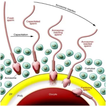
Fertilization is a multistep and complex process culminating in the merge of gamete membranes, cytoplasmic unity and fusion of genome, initiating the development of a new individual. This merge is achieved through gamete interactions, specifically cell adhesion and subsequent fusion of the gametes’ plasma membranes (PM). Although a key event in this process, there is still very little information about the mechanism or the molecules involved in membrane fusion1,2. Fertilization begins when free-swimming sperm approach oocyte within the oviduct. Mammalian sperm cannot fertilize the oocyte immediately when ejaculated and must first undergo a physiological change called capacitation (in the reproductive tract) and a subsequent morphological alteration known as the acrosome reaction when they contact with the zona pellucida (ZP) of the oocyte. After completion of the acrosome reaction, sperm penetrates the ZP, finally adhering and fusing with the plasma membrane of the oocyte. Gamete fusion results in egg activation, pronuclear formation and syngamy3,4 (Figure 1).
Figure 1. Fertilization process: mechanism of sperm-oocyte interactions. PVS: Perivetelline space; ZP, Zona Pellucida Reproduced from24.
Sperm are remarkable and atypical cells with a peculiar functionality: they are produced in the testis of one organism, and upon release they invade the female tract and deliver their genetic material to an oocyte (fertilization). During spermatogenesis, spermatozoa became terminally differentiated. These highly specialized cells are composed of three main regions: the head, the midpiece, and the tail (Figure 2). The head comprises the condensed nucleus (in which DNA condensing histones are removed and replaced by protamines, resulting in a super condensed DNA), redundant nuclear envelope, and the acrosome vesicle. The acrosome is a large secretory vesicle holding hydrolytic enzymes that assist the sperm penetration though the ZP at fertilization. The sperm tail consists of a flagellum, a motile cilium that comprises an axoneme that consists of an array of microtubules in a typical 9+2 arrangement (two central microtubules surrounded by a ring of nine microtubules pairs). Other proteins structures are arranged around the axoneme, such as the outer dense fibres and fibrous sheath, adding strength and resistance to the tail. This fibrous sheath functions as a scaffold for proteins in signalling pathways that regulate sperm maturation, motility, capacitation and acrosome reaction. The sperm midpiece connects the head and the tail and is composed by a large number of mitochondria (where ATP production occurs through oxidative phosphorylation) that are spiral wrapped around the central flagellum5–11. During spermatogenesis most of the cytosol and redundant organelles such as the Golgi apparatus, endoplasmic reticulum (ER), lysosomes, peroxisomes and ribosomes are removed. The sperm cells are transcriptionally silent and it is generally accepted that they are translationally silent too. There are some findings indicating that mammalian spermatozoa contains nuclear-encoded mRNAs in addition to their ability to synthesize mitochondrial-encoded RNA and proteins. According to Gur
et al. (2005)12, the ability of sperm cells to synthesize proteins is critical for the maturation step leading to successful fertilization. The sperm PM, tightly attached to the underlying cellular structures along the whole sperm body to provide rigidity, is highly polarized and dynamically reorganizes itself during capacitation and other fertilization processes, such as sperm-oocyte adhesion and fusion processes7,8,13 .
Figure 2. Sperm geometry: a mature sperm cell consists of head, midpiece and tail. The head is essentially divided into condensed DNA and acrosome. The miedpiece contains the mitochondrial sheath. The tail or flagellum is composed by microtubules that allow sperm motility. Reproduced from14.
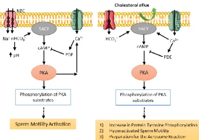
As soon as sperm cells are deposited in the female reproductive tract and pass the cervical mucus, they undergo many biochemical and physiological changes in a process called capacitation; this includes a change in the pattern of sperm motility known as hyperactivation, and the sperm becomes competent to fertilize an oocyte15,16. The biochemical changes that occur in this process include an efflux of cholesterol from the PM leading to an increase in membrane fluidity and permeability to bicarbonate and calcium ions. This results in increased bicarbonate (HCO3-) concentration, intracellular pH, and increased Ca2+ and cyclic adenosine monophosphate (cAMP) levels4,17. The process of capacitation can be divided into two signalling events: a) fast events such as the initiation of sperm motility, occurring as soon as the spermatozoa is released from the epididymis, and b) slow events such as changes in the motility pattern and the acquisition of the sperm capacity to undergo agonist-stimulated acrosome reaction (Figure 3)17,18.
Capacitation is regulated by two parallel pathways, one requiring activation of PKA and another involving activation of Ser/Thr protein phosphatases, such as PP2A. Taken together, sperm capacitation results in the induction of glycolysis in the sperm tail required for the hyperativated motility. In the sperm head it causes the redistribution of surface molecules, aggregation of lipid rafts and formation of a functional ZP binding protein complex20.
Once capacitated, sperm cells demonstrate a vigorous swimming pattern considered to give sperm the strong thrusting power that allows them to penetrate the ZP4,21 .
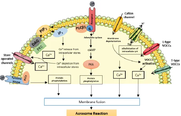
After capacitation sperm undergo a morphological change called acrosome reaction, initiating the process of fertilization. The sperm acrosome is a Golgi-derived organelle localized in the apical region of sperm head. The acrosome exocytosis is a multipoint membrane fusion event since the outer acrosome membrane and the PM fuse at multiples sites, and as a consequence, only the inner acrosome membrane becomes a new plasma membrane20,22. During the process of acrosome reaction this vesicle undergoes exocytosis and releases its contents including hydrolytic enzymes such as hyaluronidase (PH-20), which are required for the penetration of cumulus cell layers and the ZP for completing the sperm-oocyte fusion4,23. The sperm surface binds to at least three of the four human ZP proteins (ZP1, ZP3 and ZP4) by multiple proteins, most likely organized into ‘zona binding protein complexes’20,24. Binding of a capacitated sperm to the zona matrix activates two prominent signalling pathways (Figure 4). One is the pertussis toxin-sensitive Gi protein-coupled receptor linked to PLCβ1. The other is a putative tyrosine kinase receptor coupled to PLCγ, which leads to activation of components involved in fusion of the outer acrosomal membrane with the sperm plasma membrane, resulting in the acrosome reaction23,24.
The acrosome exocytosis enables the sperm to reach the perivitelline space and exposes the inner acrosomal membrane and the equatorial segment of the head, which is required for the sperm to fuse with the oocyte membrane20.
Although fascinating, the interaction between sperm and oocyte is yet poorly understood25. Currently, the term sperm-oocyte interaction includes two cellular events: sperm-oocyte adhesion and sperm-oocyte fusion (Figure 5). For cell-cell fusion, cell adhesion is a precursor step, mediated by adhesion molecules on the sperm and oocyte surfaces, which brings the membranes in close apposition. Ultimately, this membrane interaction leads to the actual fusion event, with mixing of the lipid bilayers, formation of the fusion pores, and establishment of cytoplasmic continuity between the two cells2.
Figure 4. Schematic representation of various pathways involved in ZP-mediated acrosomal exocytosis; cAMP, cyclic adenosine monophosphate; DAG, 1,2-diacylglycerol; PKA, protein kinase A; IP3, 1,4,5-inositol triphosphate; PIP2, phosphatidylinositol 4,5-bisphosphate; PLC, phospholipase C; SOC, store-operated channel; VOCC, voltage-operated calcium channel; ZP, zona pellucida. Adapted from23.
Figure 5. Schematic diagram illustrating the distinct steps of sperm binding and fusion with the oocyte plasma membrane. Purple, equatorial segment; orange, inner acrosomal membrane. Reproduced from2.
During recent years, efforts have been made towards the identification of the molecular players in sperm-oocyte interactions and their function. Several molecules on the oocyte or the sperm side were classified essential, highly relevant, or associated with essential molecules. Although the concept of multiprotein complexes on both membranes has been accepted in recent years, the first known molecules directly implicated in mammalian fertilization have been only recently discovered1. The proteins involved in sperm-oocyte interactions are summarized in Table 1 (sperm proteins) and Table 2 (oocyte proteins).
Table 1. Sperm proteins involved in sperm-oocyte interaction1,2,20,25–29.
Protein Summary of Data Role in
fertilization Binding partner
Izumo 1
Belongs to an immunoglobulin superfamily of type I membrane proteins; Izumo1 relocates during
acrosome reaction from the anterior part of the sperm head
to the sites where the fusion would take place.
Izumo1-/- mice are
completely infertile; essential for fertilization Juno ADAMs (A desintegrin and metalloprotease) Fertilin (ADAM 1 and ADAM2)
Fertilin is composed of two glycosylated transmembrane subunits (ADAM1B and ADAM2)
that make a heterodimer. It is distributed on the plasma membrane over the entire sperm
head.
Adam-/- knockout
has serious defects in gamete membrane interaction
ADAMs are binding partners for several members of integrin
family expressed in oocyte (α9β1 integrin)
ADAM3
ADAM3 may have a role in regulating aspects of sperm function. ADAM3 has been proposed to be a sperm protein
that mediates sperm-ZP3 interaction
Adam3-null sperm
bind poorly to the ZP. ADAM3 is not essential for fusion
ADAM3 binds a ZP component(s) directly
SPESP 1 (Sperm equatorial segment protein)
Belong to the SPESP1 family. Localized in the equatorial region
before acrosome reaction and a part of the SPESP1 fragment may remain in the equatorial segment
after the acrosome reaction.
Spesp1-/- males
have sperm with reduced ability to
undergo sperm-oocyte fusion.
Protein Summary of Data Role in
fertilization Binding partner
SLLP1 ( Sperm Lyzozyme-Like Acrosomal Protein)
Belong to the glycosyl hydrolase 22 family; SLLP1 relocates into the equatorial segment after the
acrosome reaction. Specific antibody against SLLP1 blocked both fertilization and binding. SAS1B
Table 2. Oocyte proteins involved in sperm-oocyte interaction1,2,20,25–28 .
Protein Summary of data Role in fertilization Binding partner
Juno (Folate receptor 4)
GPI-anchored extracellular protein on the oocyte surface. The exact molecule structure has not been fully examined.
Juno-/- female mice are
absolute infertile. IZUMO1 This interaction seems to represent a necessary and essential adhesion step rather that the
exact fusogenic
action.
Tetraspanins CD9
Members of the tetraspanin family. Through interactions with other proteins in cis,
tetraspanins create tetraspanin-enriched microdomains, which are a distinct type of membrane
microdomain.
Cd9-/- oocytes failed to fuse
with sperm. Recent
observations suggest that the CD9 molecule is secreted from the oocytes in vesicle, translocating to the sperm surface and promoting in this way the fusion process. CD9 is likely to function in conjunction with CD81.
There is no evidence for an exact binding
partner, as the
interaction with
IZUMO1, however
tempting, has not been proved.
CD81
Cd81-/- mouse shows defects
in female fertility and sperm-egg interaction. Cd9-/-/ Cd81 -/- female mice are completely
infertile
Protein Summary of data Role in fertilization Binding partner
Integrins
Integrins are heterodimeric membrane proteins, made up of an α and a β subunit.
Oocytes deficient in β1 integrin show defects in sperm-oocyte binding and
oocytes with reduced
amounts of α9 support sperm binding and fusion less
well than the control
oocytes.
α9β1 integrin
interact with several ADAMs.
GPI-anchored proteins
GPI-anchored proteins possess a covalently linked glycosylated
phosphatidylinositol moiety which serves to attach the protein portion of the molecule to the cell surface lipid bilayer.
The absence of all the oocyte GPI-anchored proteins may alter the normal lipid domain composition and may change the repertoire of protein-protein interaction in lipid domains.
Because tetraspanins localize in raft-like microdomains, it is possible that exists an association between these molecules. SAS1B (Sperm Acrosomal SLLP1 Binding) SAS1B is an oolemal metalloprotease that is concentrated in a dome corresponding to the microvilli region and in the perivetelline space.
Sas1b-/- mice showed a
significantly lowered fertilization rate
SLLP1 is believed to be one of the binding factors in the attachment-fusion machinery on the oocyte surface.
Gamete’s proteins that participate in sperm-oocyte interactions not only may act as a binding partner for a molecule on the opposite gamete or as a fusogen, but they also may interact in cis with other membrane proteins and regulate those proteins function, organization or overall membrane order. Figure 6 shows a schematic diagram illustrating the molecules implicated in gamete membrane binding and fusion.
Upon fusion, sperm activates the egg by inducing calcium oscillations and leading to the completion of the second meiotic cell division. This activation leads to exocytosis from peripherally located cortical granules, such as ovastacin-containing granules, which results in cleavage of ZP2 and by its turn in a decrease of the affinity to sperm. This phenomenon is called the ‘zona reaction’ and prevents polyspermy4.
1.2 CD81: a member of the protein tetraspanin superfamily
One of the oocyte proteins involved in sperm-oocyte interactions is CD81 (also known as a target of antiproliferative antibody 1, TAPA-1). CD81 is a 26 kDa integral membrane protein member of the tetraspanin family, an evolutionarily conserved family of membrane proteins, with at least 32 distinct family members in mammals30. Tetraspanins are defined by four transmembrane (TM) domains delimiting two extracytoplasmic regions of unequal size, a small extracellular loop (EC1) and a large extracellular loop (EC2), and by the presence of several conserved amino acids, including an absolutely conserved CCG motif and two other cysteine residues in the EC2 30. This family of proteins have been implicated in a multitude of biological processes, including fertilization of oocytes, susceptibility to infection, metastasis of cancer, and cell-cell interactions in the central nervous and immune systems. These activities are due to the association of different tetraspanins with each other and with other tissue type-specific proteins in the cell membrane. These associations take the form of tetraspanin-enriched microdomains (TEM) that form a dynamic membrane network known as the ‘tetraspanin web’31.
CD81 was originally discovered in 1990 as a target of an antiproliferative antibody on human B cells, and subsequent studies have shown that it is involved in a surprising range of physiological responses32. This molecule is highly conserved in mammals and is expressed on most human tissues with notable exceptions of red blood cells and platelets33. CD81 is abundant on various types of endocytic membranes and has been widely used as an exosomal marker. The CD81 gene has been mapped to chromosome 11p15.5 in human and in the syntenic region of chromosome 7 in mouse. Several other tetraspanin genes are located on chromosome 11 and 12 and the genomic structure is shared by each of the tetraspanin genes, indicating that they derive from a common ancestral gene34.
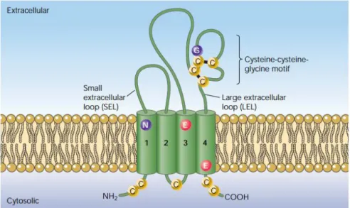
The molecular model of the complete CD81 structure was proposed in 2006 by Seigneuret35. These studies on the protein topology demonstrated that the two long hydrophilic domains of the molecule are extracellular (Figure 7). The EC1 domain, also referred to as the small cellular loop (SEL), is located between TM1 and TM2, and EC2 or LEL (large extracellular domain) is located between TM3 and TM4. The SEL is thought not to be involved in protein binding. The LEL domain is the most variable between the tetraspanin family members. The sequence variability within LEL is clustered in one hypervariable region that is coded by exon 6. This exon also codes for
the short CCG motif, conserved in all tetraspanins. The LEL region is folded into a mushroom-like structure as a result of two disulphide bridges. The crystal CD81 structure shows helical regions (a, b and e) that are conserved among the tetraspanins and form a tetraspanin-homodimerization interface. The mushroom-like patch is formed by helices c and d, and mediates specific lateral protein-protein interactions and ligand binding32,33,36.
Figure 7. Structural features of tetraspanins. Reproduced from37 .
CD81 consists of four TM domains: TM1 and TM2 flank the SEL, whereas TM3 and TM4 flank the LEL. Each TM domain contains conserved polar and non-polar aminoacid residues. These conserved residues in TM 1, 3 and 4 are responsible for a proper tetraspanin biosynthesis and maturation via intramolecular interactions, and contribute to the formation of the tetraspanin web through hydrophobic interactions between tetraspanins. Despite their short length, the cytoplasmic regions of tetraspanins (the cytoplasmic tails and short inner loop), can participate in various functions. These domains contain highly conserved cysteine residues that provide sites for palmitoylation, which contributes to the clustering of tetraspanin microdomains. The N-terminal intracellular CD81 contains two palmitoylatable cysteines, and the cytoplasmic C-terminal domain is involved in the sorting and targeting of CD81 to a determined intracellular region, and can mediate interactions with cytoskeletal or signalling proteins33,36,38.
CD81 is a nonglycosylated protein, differing from other tetraspanin proteins that are glycosylated in either one of two extracellular domains. The CD81 protein undergoes posttranslational modifications by acylation with a fatty acid, not necessarily myristic acid33.
As mentioned above, one of the most striking features of tetraspanins is their ability to form lateral associations with multiple partner proteins, including each other, in a dynamic assembly described as the ‘tetraspanin web’. The composition of the web is cell type-specific, where partnerships are formed with cell surface receptors, adhesion molecules, and transmembrane signalling proteins (Figure 8). The tetraspanin web mediates the cross talk between cell surface stimuli and intracellular signaling pathways, allowing cells to respond to a constantly changing environment in a specific and highly regulated manner31.
Through the use of different detergents in tetraspanin co-immunoprecipitations, three levels of interactions in the tetraspanin web have been proposed: primary, secondary and tertiary. Primary interactions have been referred to direct associations within the tetraspanin web with their non-tetraspanin partner molecules. Some associations with partners are highly stoichiometric, are highly avid and retained after lysis in harsh detergents such as 1% NP-40 or 1% Triton X-100. The most common primary partners of tetraspanins are specific members of the integrin and of the immunoglobulin superfamilies and these partnership can be formed with the extracellular domains of the interacting molecules. The secondary interactions have been referred to the ‘tetraspanin-tetraspanin associations’ and are the weaker interactions between direct complexes. These interactions are not disrupted by mild detergents (as 1% Brij 96 or 1% Brij 97), and are not Figure 8. Membrane clustering of tetraspanin-enriched microdomains. A. Aerial view of tetraspanin-partner and tetraspanin-tetraspanin interactions. B. Side view of lateral associations of tetraspanin with partner molecules. Clustering is facilitated by palmitoylation. Tetraspanins are shown in shades of green; partners of the immunoglobulin superfamily are shown in red and brown; and partners of the integrin are in blue. Reproduced from39.
stoichiometric. Recent studies have demonstrated that palmitoylation is necessary for the maintenance of these secondary tetraspanin-tetraspanin interactions. The tertiary interactions refer to the indirect associations of tetraspanins with additional proteins. These interactions are not disrupted in mild detergents, such as 1% CHAPS or 1% Brij 98. Functionally, these interactions cluster with cholesterol in tetraspanin-enriched microdomains, enabling lateral dynamic organization in the membrane and cross-talk with intracellular signalling and cytoskeletal structures39,40.
The tetraspanin-partner associations occur during the biosynthesis of tetraspanins. The web assembly may occur in multiple steps in conjunction with trafficking within the different cellular compartments in which web subunits are formed. Early in biosynthesis, tetraspanins can form specific interactions with their associated partner proteins in the ER, serving as building blocks for the assembly of the tetraspanin web. Tetraspanin-tetraspanin interactions are likely to form in the Golgi upon palmitoylation, serving as building blocks for the assembly of large multicomponent cell-surface complexes39.
A major difficulty in the study of tetraspanins is to identify functions that are specific for a given tetraspanin, and to determine how this function relates to specific tetraspanin-associated proteins. CD81 has been implicated in immune cell functions and can directly associate with EWI-2 and EWI-F, a pair of cell surface proteins of the immunoglobulin superfamily. CD81 supports maturation and surface expression of EWI-2, which modulates integrin-dependent cell motility and spreading. Antibody crosslinking of CD81 can co-stimulate T cells, suggesting positive effector roles. On B cells CD81 is required for molecular organization and efficient collaborations between the B cell receptors and its partners (CD21, CD19 and various signalling enzymes). Also, CD81 associates with and facilitates the biosynthesis of CD19, a molecule that plays a role in the B-cell receptor signalling. CD81 also closely associates with the α4β1 integrin, regulating α4β1 adhesion under shear flow conditions. CD81 plays a specific role in various human infection diseases: it is essential for the entry of malaria parasite into hepatocytes; its expression in immune cells is associated with HCV-induced pathogenesis; CD81 has been identified as a cell-surface receptor for hepatitis C virus (HCV)30,40.
CD81 modulates cell signalling events, affecting processes such as cell proliferation, apoptosis and tumour metastasis. CD81 interacts with different cytoplasmic molecules, facilitating
the activation of signalling cascades. The association of CD81 with type II phosphatylinositol 4-kinase (PI4K) may play a role in cell migration, bacterial infection and tumour cell proliferation. Recent studies strongly suggest that CD81 control cell migration due to its interaction with the small GTPase Rac. Further CD81 and CD9 positively modulate the activation of the extracellular signal-regulated kinase 1/2 (ERK1/2)/MAPK pathway to induce proliferation. CD81 and CD9 are closely related tetraspanins that have been linked to several processes, including apoptosis, and cell-cell fusion processes, such as like sperm-oocyte fusion. Few recent studies have started to implicate CD81 in nervous system physiology30,40.
1.2.3.1 CD81 in fertility
From the membrane molecular players involved in mouse fertilization, only three so far have been shown to be essential: Izumo1 on the sperm surface, its oocyte-based receptor Juno, and the tetraspanin CD9 on the oocyte surface. These molecules are also present on human oocyte and sperm cells. CD9 is not the only tetraspanin expressed by human and mouse oocytes, which also express CD81, shown to associate with tetraspanin-enriched membrane structures on the surface of oocytes27,41.
The lack of CD81 in the mouse female induces fertility defects, and the use of an anti-CD81 mAb partially inhibited sperm-oocyte binding and fusion42. The role of CD81 in sperm-oocyte fusion was demonstrated by in vitro insemination using Cd81 -/- knockout mice. Deletion of the Cd81 gene in mice results in a 40% reduction of female fertility. Cd9-/- Cd81-/- double knockout mice were completely infertile41.
Tanigawa et al. (2008) demonstrated that CD81 is continuously expressed in the oocyte and in cumulus cells surrounding ovulated oocytes. These results indicate that the sperm may encounter CD81 on the somatic cells surrounding oocytes before sperm and oocyte directly interact. When Cd81-/- oocytes are incubated with sperm, some of the sperm that penetrated into
the Cd81-/- oocytes perivitelline space fail to undergo the acrosome reaction. The impaired fusibility of CD81-/- oocytes may thus be partially caused by failure of the acrosome reaction43. A recent study performed by Ohnami et al. (2012) shows that oocyte CD81 was mainly localized at the inner portion of ZP, and may be involved in any step of fusion-related events prior to membrane fusion (Figure 9). Before the sperm-oocyte fusion, CD9 inside the PVS is transferred to the sperm membrane44. The CD9-containing membrane fragments are transferred from the oocyte to sperm via “exosome-like” vesicles. This interaction between membranes could be involved in the activation of sperm molecular complexes involved in binding and/or fusion with the oocyte
membrane45. Ohnami et al. (2012) suggest that CD81 may help to transfer CD9 of the oocyte into the sperm membrane through the formation of a complex between them44.
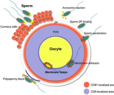
Figura 9. Schematic representation of the distribution of the CD81 tetraspanin in oocytes and sperm upon fertilization. Before fertilization, CD81 localizes in the inner region of the ZP. When the sperm penetrates the PVS, CD81 and CD9 appears to adhere to the sperm surface via exosomes. Orange area, CD81-localized area; light blue area, CD9-localized area; ZP, zona pellucida; PVS, perivitelline space. Reproduced from44.
Interestingly, CD81 was one of the most abundant interactors that appeared in a Yeast Two-Hybrid screen of human testis cDNA library previously performed46. The protein used as bait, and whose interactors we were studying, was the Amyloid Precursor Protein, APP.
1.3 Amyloid Precursor Protein (APP)
Since the molecular cloning of the APP gene in 1987, the functions of APP and its cleavage products have been subject of intense investigation. APP, located in chromosome 21, is the most well studied member of APP family due its role in Alzheimer´s disease(AD)47–50. AD is the most prevalent neurodegenerative disorder and it is characterized by the presence of neurofibrillary tangles and amyloid plaques in the brain. Neurofibrillary tangles consist largely of hyperphosphorylated twisted filaments of the microtubule-associated protein tau. Extracellular amyloid plaques are deposits of differently sized small peptides, ranging in size from 38 to 44 aminoacids and called β-amyloid (Aβ), derived from sequential proteolytic cleavages of its APP precursor51.
APP is a member of an evolutionary conserved gene family that also includes the mammalian APP-like protein-1 and -2 (APLP1 and APLP2). These proteins are highly homologous, sharing ~38-51% amino acid sequence identity52,53. The nearest APP-relative is APLP2 in which a segment of the deduced polypeptide (position 694-763) has 70.6% homology with the transmembrane cytoplasmic domain of the APP54,55. This family is conserved across a variety of species, and members can be found in invertebrates, including C. elegans (APP-like, APL-1) and D.
melanogaster (APP-like, APPL)56,57.
All APP family members are type 1 integral membrane proteins with a single membrane-spanning domain, a large ectoplasmic N-terminal regions and a shorter cytoplasmic C-terminal region. The APP sequence can be divided into multiple distinct domains, as demonstrated in Figure 10. The ectoplasmatic region of APP, which constitutes the major part of the protein, can be divided into E1 and E2 domains. The E1 domain can be further subdivided into a heparin-binding/growth factor-like domain (HFBD/GFLD) and a metal binding domain (cooper-binding domain and zinc-binding domain). The E1 domain is followed by an acidic region (Ac) and a Kunitz-type protease inhibitor (KPI) domain (that is subject to alternative splicing in both APP and APLP2). The E2 region consists of another HFBD/GFLD and a RERMS motif. The cytoplasmic region of APP contains a protein interaction motif, namely the YENPTY sequence, which is conserved in all APP homologues and participated in APP/APLPs endocytosis. The Aβ domain derived from the region of the protein encoded by parts of exons 16 and 17 is unique to the APP protein (Figure 10)58,59.
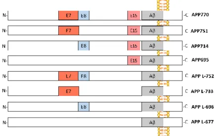
The mammalian APP gene contains 18 exons (exons 1-13, 13a, and 14-18), of which exons 7, 8 and 15 can be alternatively spliced and generate multiple isoforms that are named according to their length in amino acids: L-APP 677, APP695, L-APP696, APP714, L-APP733, APP751, L-APP752, APP77060–62 (Figure 11). APP is ubiquitously expressed, but the relative abundance of each isoform varies according to the cell type. The predominant transcripts are APP695, APP751 and APP770, in which APP695 isoform is mainly found in neuronal tissue, whereas APP751 is the predominant variant elsewhere62. The APP transcripts excluding exon 15 were first discovered in peripheral leukocytes and immunocompetent cells of the brain and where named leukocyte-derived APP (L-APP); these are ubiquitously expressed with the exception of neurons63.
Figure 10. Schematic illustration of the domain organization of the members of APP protein family. All members share conserved E1 and E2 extracellular domains, an acid domain (Ac) and the internalization YENPTY motif in the carboxyl terminus. Note that Aβ is unique for APP. HFBD/GFLD, Heparin-binding/growth factor-like domain; CuBD, Copper binding domain; ZnBD, Zinc binding domain; KPI, Kunitz-type protease inhibitor domain. Adapted from58,59.
Figure 11. Exon structure of APP isoforms. The alternatively spliced exons are indicated in colours (E7, E8 and E15). E7, exon 7 ( KPI domains); E8, exon 8; E15, exon 15; Aβ, beta-amyloid.
Like APP, APLP2 contains three exons that can be alternatively spliced. Four APLP2 isoforms are denoted as L-APLP2 isoforms in analogy to alternative splicing of APP exon 15. APLP1 gene does not give rise to alternatively spliced exons, producing a single transcript60. APLP2, similarly to APP, is ubiquitous, whereas APLP1 is found primarily in cells of the nervous system64.
APP is produced in large quantities in neurons and is very rapidly metabolized. Initially targeted to the secretory pathway, it is proteolytic processed into various fragments during its intracellular trafficking65 (Figure 12).
APP is synthetized in the endoplasmic reticulum (ER) and then transported though the Golgi apparatus to the trans-Golgi-network (TGN). From the TGN, APP can be transported in TGN-derived secretory vesicles to the PM (Figure 12, step 1). During the transport from the ER to the PM, APP can undergo post-translational modifications by N- and O-linked glycosylation, ectodomain and cytoplasmic phosphorylation, and tyrosine sulphation. Only a small fraction of APP remains in the plasma membrane, whereas the majority of APP localizes in the TGN. On PM, APP undergoes proteolytic processing or is internalized due to the presence of its YENPTY internalization motif (Figure 12, step 2). Following endocytosis, APP is delivered to endosomes, and a fraction of the endocytosed molecules is recycled to the cell surface. Significant amounts of internalized APP also undergo degradation in lysosomes (Figure 12, step 3)66–68.
The proteolytic processing of APP can be divided into two different pathways: the amyloidogenic pathway, which leads to generation of Aβ, and the nonamyloidogenic pathway (Figure 13). Both pathways are mediated by at least three cleavage events. In the
non-Figure 12. Intracellular trafficking of APP. Reproduced from66.
amyloidogenic pathway APP is cleaved approximately in the middle of the Aβ region by α-secretase, which generates a soluble N-terminal ectodomain termed sAPPα, that is release from the cell surface, and a truncated APP C-terminal fragment (CTF) (αCTF or C83). This fragment is subsequently cleaved by γ-secretase, leading to the secretion of a small non-pathogenic peptide p3 and a free APP intracellular domain (AICD) which is released into the cytosol. In the amyloidogenic pathway, the β-secretase activity initiates Aβ generation by shedding a large part of the ectodomain of APP (sAPPβ) and generating an APP CTF (βCTF or C99). This fragment is then cleaved by γ-secretase, generating Aβ and AICD. This final cleavage is not precise and under physiological conditions can occur at least between aminoacids 37 and 43 of the Aβ domain, generating Aβ1-40, Aβ1-42, carboxyl-terminal-truncated and amino-terminal-truncated Aβ peptides59,65,66,69,70.
Although APP processing has been widely studied, the location of the secretase activities within the cell, and the identification of the enzymes involved have not yet been fully elucidated. α-secretase is a zinc metalloproteinase and is a member of the ADAM (A Desintegrin and Metalloprotease) family. Three of this family members have been suggested to be α-secretase: Figure 13. APP processing involves proteolytic cleavage by several secretases. The amyloidogenic pathway releases Aβ peptides through cleavage by β- and γ-secretases. The non-amyloidogenic pathway is initiated by α-secretase cleavage, which occurs in the middle of the Aβ domain, and results in the release of several soluble APP fragments. Aβ, β-amyloid; APP-CTF, APP C-terminal fragment; AICD, APP intracellular domain. Adapted from70.
ADAM9, ADAM10, and ADAM17. α-cleavage occurs at the PM and in intracellular post-Golgi compartments. Beta-site amyloid precursor protein cleaving enzymes 1 and 2 (BACE1 and BACE2) were identified as the major β-secretases. BACE is a membrane-bound aspartyl proteases with a characteristic type I transmembrane domain. The optimal pH of BACE activity agrees with the β-site cleavage of APP taking place in more acidic cellular compartments, such as the endosomes. BACE cleavage has also been suggested to occur in the PM lipid rafts. γ-secretase is an aspartyl protease with low sequence specificity that cleaves the substrate within its transmembrane domain. This enzyme is a protein complex that consists of anterior pharynx defective 1 (APH-1), nicastrin, presenilin 1 or 2 (PS1 or PS2), and presenilin enhancer-2 (PEN-2). The existing in vivo and in vitro data on presenilin point it as the responsible for the aspartyl γ-secretase activity. Noteworthy, there is some evidence that Aβ1-40 and Aβ1-42 are generated in different cellular compartments59,65,66,69. Of note, although the sequences where the various secretases cleave are very different in the two mammalian homologous, both APLP1 And APLP2 are believed to be proteolytically processed in a similar way as APP59.
Although APP and its homologues have been the subject of several studies, the physiological functions remains largely unknown. Several in vitro and in vivo studies have yielded strong evidence for the roles of APP, APLP1 and APLP2 in the developing and adult nervous system, in cell adhesion, neuronal survival, neurite outgrowth, synaptogenesis, vesicular transport, neuronal migration, modulation of synaptic plasticity, calcium metabolism, and insulin and glucose homeostasis. APP-derived proteolytic fragments mediate various and sometimes antagonist functions. Therefore, the net effect of full-length APP in cellular activity may be a combination of its metabolites´ functions. The N-terminal fragment, sAPPα, is neuroprotective, promotes neurite outgrowth and synaptogenesis, acts as a growth factor, and regulates cell adhesion. On the other hand, sAPPβ fragment can stimulate axonal pruning and neuronal cell death. The Aβ peptide has neurotoxic effects resulting in a neurodegenerative cascade leading to synaptic dysfunction and formation of intraneuronal fibrillary tangles. Although excessive Aβ causes neurotoxicity, low levels can positively modulate synaptic plasticity, revealing a novel physiological function for Aβ. The AICD fragment has been shown to possess transactivation activity and can regulate transcription of multiple genes, induce apoptosis, and may play a role in sensitizing neurons to toxic stimuli. Full length APP has been suggested to function as a cell surface receptor contributing to cell adhesion
and cell-cell interactions via its extracellular domain and possibly though trans-dimerization (reviewed in59,65,71).
1.3.3.1 APP and fertility
Although APP has been extensively investigated in association with AD pathology of the brain, APP biological functions are not restricted to the nervous system. Based on findings using the double App and Aplp2 knockout mice, APP and APLP2 have been implicated in fertilization and may play a role in mammalian fertility. Knockout (KO) mice, homozygotes to either App /-) or Aplp2 (-/-) were healthy and fertile. Contrary to this data, 80% double KO mice to App (-(-/-) and Aplp2 (-(-/-) die within the first week and the surviving mice are infertile (9 of 10 females and all males). This findings suggests that APP and APLP2 can substitute each other functionally and are required for early postnatal development, normal growth, strength/balance, and fertility72.
In 1990, Shoji and Kawarabayashi73 demonstrated that native APP is present in acrosomes and in growing flagella, suggesting that APP appears in particular phases of spermatogenesis, especially the head and tail formation phase in spermatids.
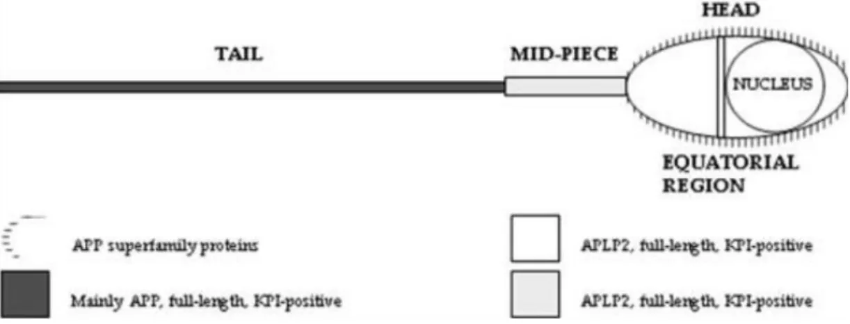
A human sperm membrane protein termed YWK-II (latter shown to be homologous with APLP2) was immunolocated in male and female gametes. YWK-II/APLP2 is expressed in the mouse PM that envelops the acrosome, suggesting its participation in sperm-egg interactions. YWK-II/APLP2 may be a Gαo-coupled receptor and was shown to interact with Müllerian-inhibiting substance (MIS), which is well known to be required for sex determination. These interactions suggest that YWK-II/APLP2 may participate on germ cell differentiation and sperm metabolism74–76. Given the high degree of homology between APP and APLP2, and taking into account the Shoji and Kawarabayashi (1990) findings, da Cruz e Silva group performed a study in order to characterize the subcellular distribution of the APP superfamily members in human sperm. This study was carry out using a battery of antibodies that either recognize APP-specific epitopes or epitopes shared with other APP family members77. The results of this study show that in human sperm, the tail mainly expresses full-length KPI-positive APP, the mid-piece mainly expresses KPI-positive APLP2 variants, the equatorial region mainly contain KPI-containing ALPL2 variants, and the surface of the entire spermatozoa contains APP and/or APLPs, with particular abundance on the head (Figure 14). These results prove the presence of APP itself in human sperm. According to the authors the presence of APP family members along the entire length of the tail may be related to signalling events involved in sperm motility, whereas their presence on the surface, especially in the equatorial region,
suggests their involvement in sperm-oocyte interactions77. However, no binding partner has been described for APP in sperm-oocyte interactors or many other sperm functions.
The identification of the protein networks interacting with APP can be very useful to approach APP functions. To date, most proteins interacting with APP have been identified in the brain, but less is known about the pathways involved in testis and sperm functions.
In order to identify tissue-specific interacting proteins to approach APP functions in sperm and testis, our lab performed a Yeast Two-Hybrid (YTH) screen of a human testis cDNA library, using full-length human APP as bait, followed by a comprehensive bioinformatics analysis. From the total of the clones screened, 147 were positive and encoded 37 known proteins. Only one protein had previously been identified as an APP interacting protein (RANBP9)46. The analysis of the YTH screen revealed that one of the most abundant interactions was with CD81 (5 out of the 147 positive clones). The study included a network structure and bioinformatics analyses to determine the biologically relevant APP protein-protein interactions and revealed prominent topological properties for CD8146.
Aims of the Thesis
CD81 is involved in reproduction, namely in sperm-oocyte interaction, but few is known about the molecular mechanisms involved in this CD81 function. APP and APLPs are expressed in the plasma membrane of the sperm head, a region involved in sperm-oocyte interaction, and APP has revealed to be a putative CD81 interacting protein. Taking these data into account, and the facts that APP is a very important protein of the nervous system and the CD81 biological functions in neuronal cells are also still far from clear, CD81 has emerged as a very interesting target of study for us.
The main general aim of this thesis was to expand the knowledge of CD81 in sperm cells and in a neuronal cell line, though the study of some of its interactions with proteins such as APP. The specific objectives set to attain this goal were to:
- Perform a bioinformatics analysis of CD81 interacting proteins;
- Characterize the expression of CD81 in sperm cells and in different cell lines, including a neuronal-like cell line;
- Determine and characterize the CD81-APP physical and functional interaction;
- Evaluate the co-localization of CD81 with some of its interacting proteins, in sperm-cells and in a neuronal cell line.
Materials and Methods
3.1 Bioinformatics analyses
The identification of proteins that interact with human CD81 was carried out using the public database ‘Biological General Repository for Interaction Dataset’ (BioGrid)78. Only the interactions that were curated in Homo sapiens have been included. In addition, a literature survey was conducted to complement data on other interactions of CD81 absent from BioGrid by searching these in PubMed. The proteins involved in this study were collected with their own ID: gene symbol (www.genecards.org/) and UniProtKB (www.uniprot.org/uniprot/?query=&sort=score).The gene symbol of all collected proteins were loaded into the GeneMANIA79 tool to generate a protein-protein interaction network. The network was generated using only data on physical interactions and using the “Biological process based” as the network weighting method.
The Gene Ontology (GO)-term enrichment analysis was carried out using PANTHER - Protein Analysis Through Evolutionary Relationships80 according to GO annotation (‘biological processes’, ‘molecular functions’ and ‘cellular component’). The gene symbol of all collected proteins were loaded into PANTHER and the enrichment of specific GO-terms was tested using the “statistical overrepresentation test”, followed by the Bonferroni multiple testing correction to control for the false discovery rate.
Similar to the GO-term enrichment analysis, cell-signalling pathways were also analysed using the “statistical overrepresentation test” implemented in PANTHER.
3.2 Preparation of CD81 cDNAs
The mApple-CD81 (Plasmid #54875) and pCDM8 hCD81 (Plasmid #11588) plasmids were purchased from Addgene (Cambridge, UK). Both plasmids encoded CD81, but mApple-CD81 has an orange-fluorescing tag (mApple) on N-terminal. The mApple-CD81 plasmid has bacterial resistance to kanamycin and pCDM8 hCD81 to ampicillin.
The plasmids were acquired as transformed bacteria in stab culture format. After being received, the bacteria were streaked from the stab onto a Luria Broth (LB) agar plate to isolate single colonies, further used to amplify and purified the plasmidic DNA by “Midiprep” using NucleoBond® Xtra Midi (Macherey-Nagel, Germany). One isolated colony of streaked LB agar plate was used to inoclute 3 ml of LB medium containing the appropriate antibiotic, and incubated for 16-18 h at 37oC with shaking (200 rpm). An aliquot of 150 µl was used to inoculate 150 ml of LB medium, also with the appropriate antibiotic, and the culture incubated overnight (ON) at 37oC with shaking (200 rpm). Cells were centrifuged at 6000 g, for 10 min at 4oC, the supernatant discarded, and 8 ml of resuspension Buffer RES added. After resuspension, 8 ml of lysis Buffer LYS was added, gently mixed by inverted the tube 5 times and incubated at RT for 5 min. Once the solution became clear and viscous, 8 ml of neutralization Buffer NEU was added to the lysate cells and then centrifuged at 12000 g for 10 min. Following, a NucleoBond® Xtra column filter was equilibrated with 12 ml of equilibration buffer EQU, placed into a 50 ml disposable plastic tube and the supernatant was poured into the column. The column filter was washed with 5 ml of equilibration buffer EQU and then discarded. The column was then washed with 8 ml of washing buffer WASH. To elute the DNA, the column was transferred to a new 15 ml plastic tube and 5 ml of elution buffer EDU was added. The precipitation of eluted DNA was carried out by adding 3.5 ml RT isopropanol and the DNA recovered by centrifugation (15000 g, 30 min at 4oC). The supernatant was discarded, and the pellets rinsed with 2 ml of 70% ethanol RT. The solutions were centrifuged at (15000 g, 5 min) the supernatant again removed, and the pellet allowed to dry. After all traces of ethanol have disappeared, the DNA was resuspended in Milli Q water and stored at -20oC. DNA concentration and its 260/280 nm purity ratio were calculated upon spectrophotometer measurement.
3.3 Mammalian cell assays
In the impossibility of performing sperm-oocyte interaction assays in our lab, immortalized cell lines were used to study the putative CD81 and APP interaction. The Chinese hamster ovary (CHO) cell line, of ovary nature, and the CG-1 spg cell line, derived from postnatal day 10 mouse testis and described as an intermediate spermatogenic cell type between type B spermatogonia and primary spermatocytes, were used. SH-SY5Y cell line was also used, due to its neuronal nature and its good expression of endogenous APP. The SH-SY5Y human neuroblastoma cells are originally derived from the cell line SK-N-SH, established from a bone marrow biopsy of a neuroblastoma patient.
CHO cells were maintained in Ham’s F-12 nutrient mixture (F-12; Sigma-Aldrich) containing 2 mM L-glutamine, 10% heat inactivated Foetal Bovine Serum (FBS; Gibco) and 1% antibiotic/antimycotic (AA) mix (Gibco). GC-1 cells were maintained in Dulbeco’s Minimum Essential Media (DMEM; Gibco) supplemented with 10% FBS (Gibco) and 1% AAs mix (Gibco). SH-SY5Y were maintained in 10%FBS/1% AA mix-supplemented Minimum Essential Media (MEM):F12 (1:1). All cell lines were maintained in a 5% CO2 humidified incubator at 37oC, and subcultured when a 70-80% confluence was reached.
Fresh sperm samples were obtained from patients undergoing routine semen analysis or fertility treatments in the Human Reproduction Service at University Hospitals of Coimbra. Patients signed informed consent forms and the samples were used in accordance with the appropriate Ethical and Internal Review Board of the participating institution. Sperm samples were obtained by masturbation after 3-5 days of sexual abstinence and routine seminal samples analysis was performed according to the World Health Organization (WHO) guidelines81. All the samples used in this study had no detectable leukocytes (or any other round cells). After liquefaction, sperm cells were prepared by direct swim-up and allowed to capacitate in a supplemented Earl’s balance salt solution (sEBSS) for at least 3 h at 37oC under 5% CO2/95% air before starting the experiments.
Since the CHO line was a recent cell line used in our laboratory, we first characterize its growth. A CHO cells growth curve was determined by using two different methods of cell viability analysis, the Trypan blue (TB) exclusion dye method and the Resazurin metabolic colorimetric assay. CHO cells suspended in growth medium were seeded (1x10⁴ cells/well) of 6-well plate (Corning) and incubated at 37oC in a humidified 5% CO2. At a daily basis, media was substituted with fresh medium supplemented 10% of a resazurin solution [0.1 mg/ml resazurin salt solution (Sigma- Aldrich) in phosphate buffer solution (PBS; Pierce, Thermo Scientific)] with an end-volume of 1 ml per well. The cultures were incubated for 4h at 37oC in 5% CO2. Following this incubation period, the resazurin-containing growth medium was collected, the cells washed with PBS, suspended in PBS (volume depending on the average cell number), an aliquot mixed 9:1 with Trypan Blue solution (Sigma-Aldrich) was loaded in a hemocytometer, and cells were scored. In parallel, the rate of resazurin conversion to resofurin by metabolically active cells was evaluated via
spectrophotometric analysis at 570 and 600 nm (Infinite M200 microplate reader; Tecan Männedorf, Switzerland) of the resazurin-containing medium. A final optical density (O.D.f) was determined for each sample, as follows: (O.D. 570/O.D.600)-(O.D.570c/O.D. c 600), where ‘OD.c’ are the O.D.s of control samples (fresh medium supplemented with resazurin, never in contact with cells). This procedure was carried out during 10 days, at the same hour, in triplicate.
In order to study the physical and functional interactions between APP and CD81, transient transfections of their cDNAs were performed in CHO and SH-SY5Y cells. Cells were plated and growth on plastic culture dishes (6-well or 100 mm) (Corning), 24 h before transfection, to be at ~70-80% confluence at the time of transfection. The growth medium was changed immediately prior to the transfection procedure. Cells were transient transfected with CD81 cDNAs (mApple-CD81 and untagged pCDM8 h(mApple-CD81), alone or together with APP cDNAs (Wt APP-GFP and untagged pRC/CMV APP).The empty vectors (mApple, pCDM8, pEGFP-N1 and pRC/CMV) were single- or co-transfected as experimental controls.
All the transfection procedures were performed using the TurboFect™ reagent (Fermentas Life Science), a cationic polymer that forms a positively charged complex with DNA that is easily endocytosed by eukaryotic cells. For each well of 6-well plate, a certain amount of different cDNA was diluted in 100 µl of serum-free growth medium. After gently vortexing, a ratio of 1:2 µl TurboFect™ reagent was added to the DNA, mixed and incubated during 20 min at RT. This solution was added to each well and mixed by gently agitating the plate. The cells were incubated at 37oC in a 5% CO2 incubator, the cell medium changed after 6 h of transfection and cells and conditioned media (in some experiments) were collected after 24h of total transfection time. Table 3 presents details on the transient transfections performed in each assay.