Faculdade de Engenharia da Universidade do Porto
Development of Hydroxyapatite-based
Nanoparticles for Medical Imaging Diagnosis
Ana Azevedo Moço
Dissertação realizada no âmbito do Mestrado Integrado em Bioengenharia
para candidatura à obtenção do grau de Mestre em Engenharia Biomédica
Orientador:____________________________
Co-Orientador:_________________________
Porto, Junho 2013
ii
This thesis was supervised by:
Supervisor
Dr. Marta Laranjeira
INEB – Instituto de Engenharia Biomédica, Universidade do Porto
FEUP – Faculdade de Engenharia da Universidade do Porto
Co-supervisor
Professor Dr. Fernando Jorge Monteiro
INEB – Instituto de Engenharia Biomédica, Universidade do Porto
FEUP – Faculdade de Engenharia da Universidade do Porto
The research described in this thesis was conducted at:
INEB – Instituto de Engenharia Biomédica, Divisão de Biomateriais,
Universidade do Porto, Portugal;
Resumo
O cancro é ainda uma das principais causas de morte e é facto que um diagnóstico precoce aumenta significativamente as possibilidades de sobrevivência do paciente, otimizando a eficácia do tratamento. Este objectivo pode ser atingido através de um equipamento adequado de detecção e um agente de contraste.
A Ressonância Magnética (RM) é uma técnica clinica de imagem com alta resolução espacial e contraste dos tecidos moles que permite uma visualização não-invasiva e tomográfica de estruturas anatómicas. É hoje aceite que agentes de contraste magnéticos de dimensão nanométrica são capazes de maximizar a sensibilidade da RM encurtando os tempos de relaxividade dos protões da água presentes nas áreas dos tecidos onde o agente se acumula. Estes agentes caracterizam-se por apresentarem vantagens distintas em relação aos agentes de contraste convencionais, nomeadamente pela sua elevada área de superfície, pela possibilidade de entrega específica a uma localização tumoral-alvo e pela possibilidade de projetar o seu tempo de circulação sanguíneo ou outras características relevantes.
A citotoxicidade é ainda uma questão importante que limita a aplicação clínica de muitos nano-agentes de contraste propostos até agora. A hidroxiapatite é um composto biocerâmico que não só incorpora iões facilmente, mas também confere biocompatibilidade ao sistema, o que faz a sua conjugação com iões magnéticos uma abordagem apelativa na imagem localizada de cancro. Tentativas consideráveis têm sido realizadas no âmbito deste tema, mas existe ainda uma necessidade para estandardizar procedimentos e comparar sistemas nano capazes de realçar o contraste tumoral e potencialmente poderiam permitir uma detecção específica de cancro.
No presente trabalho, nanoparticulas de hidroxiapatite dopadas com iões magnéticos foram produzidas com êxito por um método otimizado e reproduzível de precipitação química. Apurou-se que todas as partículas de nanohidroxiapatite dopadas de Gd, Fe(II), Fe(III) e Co possuíam a forma de bastonete e um tamanho médio entre 30 e 60nm.
A estrutura principal de hidroxiapatite não foi alterada pela percentagem de dopagem embora esta se tenha provado como diretamente proporcional aos níveis de momento magnético da amostra. Hidroxiapatite dopada com 10% Gd apresentou os níveis mais altos de
vi
momento magnético entre todos os iões dopantes e também os valores mais próximos dos de um agente de contraste já comercializado.
A viabilidade e a morfologia de células humanas microvasculares endoteliais da derme não sugerem uma alteração significativa após incubação com os materiais sintetizados.
Com efeito, os resultados mostram que as nanoparticulas de hidroxiapatite dopadas com 10% Gd são as mais promissoras dentro do grupo testado e, portanto, podem tornar-se um valioso biomaterial para agente de contraste de RM.
Abstract
Cancer is still one of the main causes of death and it is known that an early diagnostic significantly increases chances of survival of the patient, optimizing the efficacy of the treatment. This can be achieved with an appropriate detection device and contrast agent.
Magnetic Resonance Imaging (MRI) is a clinical imaging technique that allows for noninvasive tomographic visualization of anatomic structures with high spatial resolution and soft tissue contrast. It is well accepted that nanosized magnetic contrast agents can maximize MRI sensitivity shortening the relaxation times of bulk water protons in tissue where the agent accumulates. They are characterized by distinct advantages over conventional contrast agents namely their high surface area, possibility of delivery to a specific cancer site by active targeting and the possibility of engineering their blood circulation half-life, among others.
Cytotoxicity is still a major issue that limits the clinical application of many nano-contrast agents proposed so far. Bioceramic hydroxyapatite not only easily incorporates ions but also confers biocompatibility to the system which makes its conjugation with magnetically responsive ions an appealing approach in targeted cancer imaging. Considerable attempts have been made within this subject but there is a need to standardize procedures and compare nanosized systems capable of enhancing tumor contrast that would hereafter allow targeted cancer detection.
In the present work, magnetic ion doped hydroxyapatite nanoparticles were successfully produced by an optimized and reproducible wet chemical precipitation method. It was found that all Gd, Fe(II), Fe(III) and Co doped nanohydroxyapatite particles were rod-like shaped and of average size between 30 and 60nm.
Main hydroxyapatite structure was not altered by the percentage of doping although this percentage was proven to be directly proportional to the magnetic moment levels of the sample. 10% Gd doped HAP presented the highest magnetic moment levels between all doping ions and also the closest values to the ones of a commercialized contrast agent.
Viability and morphology of human dermal microvascular endothelial cells did not seem to be significantly altered after incubation with the synthesized materials.
viii
In effect, the results show that 10% Gd doped nanohydroxyapatite is the most promising from the group of nanoparticles tested and therefore can be a valuable biomaterial contrast agent for MR imaging.
Expect problems and
eat them for breakfast.
Acknowledgements
First of all, I have to thank my family and friends for all the support provided during the last 5 years, and especially during the elaboration of this dissertation. Thank you for the best season of my life so far.
Next, I would like to acknowledge my supervisors for this project, Professor Fernando Jorge Monteiro and Doctor Marta Laranjeira for all the patience, inspiration, and friendship. Thank you also to the Biocomposites team for welcoming me and making me feel a part of the team.
I also express gratitude to IFIMUP, especially to Doctor Maria Paz Garcia for her help and availability with all the measurements carried at this center.
Following, thank you to Doctor Raquel Temudo from Hospital Pedro Hispano for providing me the commercial contrast agent.
I am also very grateful to Ricardo Vidal (INEB), Susana Carrilho (INEB), Susana de Almeida (FEUP-DEMM) and Luciana Gomes (FEUP-DEQ) for all the support throughout the project.
Contents
Resumo ... v
Abstract ... vii
Acknowledgements ... xi
Contents ... xiii
List of figures ... xv
List of tables ... xvii
Abbreviations ... xix
... 21
Chapter 1
Introduction ... 211.1 - Cancer Disease ... 21
1.1.1 - Physiological characteristics of benign and malignant solid tumors ... 21
1.1.2 - Current tumor imaging perspective ... 24
1.1.2.1 - Magnetic resonance imaging ... 24
1.2 - Nanoparticles in tumor detection ... 25
1.2.1 - Biodistribution of the nanoparticles ... 25
1.2.2 - Magnetically responsive nanoparticles ... 26
1.2.2.1 - Hydroxyapatite-based nanoparticles as MRI contrast agents ... 28
1.3 - Next generation multifunctional nanoparticles for cancer imaging and therapy ... 31
1.4 - Aim and outline of the thesis... 31
... 33
Chapter 2
Materials and Methods ... 33xiv
2.2 - Physicochemical Characterization ... 34
2.2.1 - Size, morphology and zeta potential ... 34
2.2.2 - Chemical profile ... 35
2.2.3 - Crystal phase analysis ... 35
2.2.4 - Magnetic properties ... 35
2.3 - Biological response ... 35
2.3.1 - Cell culture and maintenance ... 35
2.3.2 - Cell metabolic activity evaluation ... 36
2.3.3 - Cellular morphology ... 36
2.3.4 - Statistical analysis ... 36
... 37
Chapter 3
Results and Discussion ... 373.1 - Physicochemical Characterization ... 37
3.1.1 - Size, morphology and zeta potential ... 37
3.1.2 - Chemical profile ... 41
3.1.3 - Crystal phase analysis ... 43
3.1.4 - Magnetic properties ... 44
3.2 - Nanotoxicity analysis ... 46
... 51
Chapter 4
Conclusions and future perspectives ... 51List of figures
Figure 1.1 The primary tumor microenvironment (adapted from [9]) ... 23
Figure 1.2 Illustration of longitudinal relaxation time T1 and transverse relaxation time T2. RF excitations are applied with a TR (top) with each excitation producing a magnetic resonance signal (Mxy) that exponentially decays with a time constant T2 (bottom right). Between excitations, the magnetization also exponentially returns to its equilibrium state, pointing along the direction of the main magnetic field, with a time constant T1 (bottom left). ... 25
Figure 1.3 Transmission Electron Microscopy image of the HAp nanoparticles loaded with CoFe2O4, highlighting their core-shell structure. ... 29
Figure 1.4 Nuclear Magnetic Resonance images of doped nHAp in comparison to Gd-DTPA (Magnevist®) showing T1-weighted bright contrast. ... 30
Figure 2.1 Representation of the nanoparticles fabrication conditions. a-Peristaltic pump; b-temperature controller and heating plate with magnetic stirrer; c-pH meter. ... 34
Figure 3.2 TEM images of the undoped and doped nanoHAP ... 38
Figure 3.1 NanoHAP dimensions obtained with TEM images (Figure 3.1) observation through Image J® software ... 38
Figure 3.3 ZetaSizer results displaying (A) the average particle sizes and (B) the average zeta potential of the doped and undoped nanoHAP ... 39
Figure 3.4 FTIR spectra of nanoHAP samples with different percentages of (A) Gd (B) Fe(II) (C) Fe(III) (D) Co doping and undoped HAP ... 42
Figure 3.5 X-ray diffraction pattern of commercialized pure HAP, Gd5_HAP, Co5_HAP and Fe(II)HAP. Blue symbols indicate the presence of whitlockite peaks on Fe(II)10_HAP among the hydroxyapatite composition ... 43
Figure 3.6 Variation of magnetization with the applied field of ±50kOe for synthesized doped and undoped nanoHAP: (A) Gd doping (B) Fe(II) doping (C) Fe(III) doping (D) Co doping (E) highest doping percentage in contrast with undoped nanoHAP and Dotarem® ... 45
Figure 3.7 Cell metabolic activity assay (resazurin) of hDMECs cultured for 4 and 24h with doped and undoped nanoHAP at a concentration of 500µg/mL. * indicates a significant statistical difference between TCPS 24h and all the other groups (p<0.05; n=5) ... 46
Figure 3.8 Morphology observation of hDMECs cultured for 4 and 24h with doped and undoped nanoHAP. Double nuclei characteristic is highlighted in picture A (Fe(II)10_HAP after 4h of culture). F-Actin is shown in green and cells nuclei in blue. Magnification 10x. ... 49
List of tables
Table 1.1 Summary of distinct features of benign and malignant tumors (adapted from [5]) ... 22
Table 1.2 Non-exhaustive list of MRI approved contrast agents used in clinical practice (Adapted from [16], [28] and [29]) ... 27
Table 1.3 A summary of hydroxyapatite-based magnetic nanoparticles non-commercialized
Abbreviations
List of abbreviations
BMA Bismethylammide
BMDC Bone Marrow Derived Cells
CT Computed Tomography
DLS Dynamic Light Scattering
DO3A [4,7-bis-carboxymethyl-10-(2-aminoethyl)-1,4,7,10-tetraaza-cyclododec-1-yl]-acetic acid
DOTA 1,4,7,10-tetraazacyclododecane-1,4,7,10-tetraacetic acids DPDP Dipyridoxyl Diphosphate
DTPA Diethylenetriaminepentaacetic Acid
EPR Enhanced Penetration and Retention effect FBS Foetal Bovine Serum
FDA U.S. Food and Administration FTIR Fourrier Transform Infrared Gd-BOPTA Gadobentate-dimeglumine
HAp Hydroxyapatite
hDMEC Human dermal microvascular endothelial cell hFOB Human fetal osteoblast cell line
hUVEC Human umbilical vein endothelial cell
HP Hydroxypropyl
KB Human nasopharyngeal carcinoma L929 Normal mouse lung fibroblast cell line MDSC Myeloid Cell-Derived Suppressor Cells MGC-803 Human gastric carcinoma cells
MMP Matrix Metalloproteinases MRI Magnetic Resonance Imaging MSC Mesenchymal Stem Cells nanoHAP Nano-hydroxyapatite NIRF Near-infrared Fluorescence
NPs Nanoparticles
OECD Organization for Economic Co-Operation and Development PAI Photoacoustic Imaging
xx PET Positron Emission Tomography
R3-IGF-1 Recombinant long R insulin-like growth factor-1 RES Reticuloendothelial System
RF Radio-frequency
rhEGF Human recombinant epidermal growth factor rhFGF-β Human recombinant fibroblast growth factor-β
RT Room temperature
Saos-2 Osteoblast-like cells
SPECT Single-Photon Emission Computed Tomography SPIO Superparamagnetic Iron Oxides
SQUID Superconducting quantum interference device SWNTs Single Walled Nantotubes
TEM TIE2-expressing monocytes
TR Repetition Time
UE Electrophoretic mobility USI Ultrasound Imaging
VEGF Vascular Endothelial Growth Factor XRD X-ray diffraction
Chapter 1
Introduction
1.1 - Cancer Disease
1.1.1 -
Physiological characteristics of benign and malignant solid
tumors
Not only cancer is a major public health problem, with estimated 5.2 million new cases of cancer diagnosed in OECD countries[1] and an average of 83 million years of “healthy life” lost due to death and disability in 2008, but this also constituted a total economic impact of $895 billion worldwide in the same year[2]. So, even though research efforts to improve current cancer therapies over the past three decades have led to an improvement in patient survival, there is still a need for new and better strategies.
But, in order to develop new therapies for cancer treatment, it is crucial to get a better insight of tumors.
In common medical usage, a neoplasm, a mass of tissue that continuously replicates which is not influenced by the regulatory processes that control normal cell growth, is often referred to as a tumor.
In general, all tumors have two basic components: the parenchyma, which is constituted by transformed or neoplastic cells and determines the neoplasm behavior, and the non-neoplastic stroma, which, in turn, consists of connective tissue, blood vessels and host-derived inflammatory cells, therefore providing support for the growth of parenchymal cells. However, neoplasms or tumors can be categorized into benign or malign, regarding their potential clinical behavior and performance according to some parameters: differentiation and anaplasia (lack of differentiation), rate of growth, local invasion, and metastasis.
A benign tumor will remain localized, with no chance of spreading to other sites, can be surgically removed and it is considered to be relatively innocent. Nevertheless, benign tumors can evolve to a malignant state requiring regular follow-up.
22
Malignant tumors are referred to as cancers and most of them have the ability to invade and destroy adjacent structures and spread to distant sites (metastasize) to possibly cause death.
In a more detailed perspective, as illustrated in Table 1, malignant tumors have distinct parenchymal cells, from well differentiated to completely undifferentiated states. It is well known that the more differentiated a cell is, the more likely it performs its normal functional activities. That said, anaplasia is a common feature in malignant tumors and it involves dedifferentiation, loss of the structural and functional differentiation of specialized cells or failure of differentiation in stem cells. Anaplastic nuclei are variable and peculiar in size and shape and mitoses are often numerous and distinctly atypical. Therefore, generally, the more rapidly growing and the more anaplastic a tumor, the less likely it is to have specialized functional activity.
Malignant tumors also do not present an often feature in the benign ones: an enclosing fibrous capsule probably derived from the host stroma that isolates them from the hosting environment. Thus, most cancers grow progressively penetrating and destroying surrounding tissues without developing well-defined capsules. Metastasizing and local invasion constitute the most reliable features that distinguish malignant from benign tumors.
However, not all cancers have the same ability to metastasize. Examples like basal cell carcinomas or primary brain tumors are highly invasive in their primary sites of origin but rarely metastasize[3, 4].
Table 1.1 Summary of distinct features of benign and malignant tumors (adapted from [5])
Because of their rapidly growing feature, most cancers tend to spread locally and to distant organism sites, process called metastasizing. Angiogenesis, known as the formation of new blood vessels from existing ones, is essential to the transformation of a small, often dormant cluster of cancer cells to a solid tumor and also to the metastasizing process, hypothesis stated for the first time in 1971 by Judah Folkman[6]. For solid tumors of 1-2mm3, simple diffusion is the way oxygen and nutrients reach the center of the tumor, but for solid tumors bigger than 2mm3, a state of cellular hypoxia begins, which initiates signaling events that upregulate the production of pro-angiogenic factors, such as vascular endothelial growth
B
ENIGNT
UMORM
ALIGNANTT
UMOR Differentiationand anaplasia
Composed of well-differentiated cells that closely resemble their normal counterparts; mitoses are limited in number and of normal configuration.
Characterized by a wide range of parenchymal cell differentiation; anaplasia is common; mitoses are often numerous and distinctly atypical.
Rate of growth Most benign tumors increase in size slowly over the span of months to years.
Most cancers progressively enlarge over time, some slowly, others rapidly.
Local invasion Remains localized at its site of origin.
Most cancers infiltrate, invade, or metastasize to distant sites.
Metastasis Nonmetastatic Most cancers develop secondary
implants (metastases)
discontinuous with the primary tumor, in remote tissues.
factor. In turn, VEGF regulates endothelial cell proliferation and enhances permeability of the microvessels, giving rise to neovasculatures different from those of normal tissues with an irregular shape, of high density and heterogeneity and poorly oxygenated, resulting in regions of hypoxia. The lack of oxygen stimulates the tumor cells to have an anaerobic metabolism that causes an extracellular acidosis in response to excessive production of lactic acid and CO2. It has been reported that extracellular normal tissue pH is around 7.4, whereas extracellular tumor pH is generally between 6.0 and 7.0 and this characteristic has been a source of interest especially in targeted cancer therapies investigation[7]. The initiation of the tumor angiogenesis is crucial to the exponential tumor growth and is called angiogenic switch. It can occur at different stages in the tumor-progression pathway and it is defined by several distinct phases such as vessel dilation, angiogenic sprouting, new vessel formation and maturation and recruitment of perivascular cells.
In addition, cancer cells tend to compress lymphatic vessels forcing them to collapse, therefore creating lack of functional lymphatic system mostly within the tumor central area.
This characteristic along with a leaky tumor vasculature induces an accumulation of vascular contents inside the tumor and an interstitial hypertension which hampers the transport of macromolecules from the capillaries to the interstitial fluid[8].
Also, the tumor microenvironment plays a major role in the modulation of the metastatic behavior of most cancers. It is now recognized that primary and metastatic tumors induce the mobilization of bone marrow-derived cells that help the tumor to progress, survive and grow at a secondary site. As portrayed in Figure 1.1, a plethora of cell types interact with each
24
other, responsible for the secretion of, for example, VEGF and matrix metalloproteinases, enabling angiogenesis malignant progression. Endothelial cells in the blood and lymphatic vessels, pericytes, fibroblasts, macrophages, neutrophils, mast cells, myeloid cell-derived suppressor cells, mesenchymal stem cells and TIE2-expressing monocytes are among the main cell types that interact with cancer cells[9].
1.1.2 -
Current tumor imaging perspective
Early detection of cancer disease allows the maximum likelihood of successful treatment and recovery, since early localization of malignant solid tumors may allow a directed therapy to its site, optimizing the efficacy of the treatment. This can be achieved with an appropriate detection device.
The type of information required or the characteristics of the biological target condition the performance of the in-vivo imaging technique used and the choice of the contrast agent or probe. For instance, MRI is better suited for clinical use than fluorescence imaging, since it is more powerful and safer on humans[10].
Imaging modalities play an important role in the clinical management of cancer, including screening, diagnosis, treatment planning and therapy monitoring. Table 2 summarizes the most commonly used bioimaging modalities in tumor diagnosis, paying special attention to current NP-based contrast agents for each technique (MRI, computed tomography, positron emission tomography, single-photon emission computed tomography, ultrasound imaging, photoacoustic imaging and near-infrared fluorescence).
These most commonly used imaging modalities in clinical detection of cancer are all nonspecific, therefore they cannot distinguish between malignant or benign tumors and none leads to 100% accurate detection. Besides, most of the presented technologies and their contrast agents involve exposure to radiation or heavy metals, which present potential health risks to patients and limit the ways they can be applied.
The goal of cancer imaging should be to detect and/or image the smallest possible number of tumor cells, since there are currently no reliable methods for in vivo imaging of tumors smaller than 1cm3, constituted with approximately 109 cells[11].
Also, there are certain types of cancer that are still currently hard to detect and image such as ovarian cancer because of its extra thin earliest proliferative lesion or pediatric cancer, since it is difficult to preform clinical trials in young ages and because they constitute a small market size[11].
1.1.2.1 - Magnetic resonance imaging
MRI is a non-invasive FDA approved for clinical use imaging technique (since 1985) that does not expose the patient to ionizing radiation and offers a high spatial resolution of approximately 50μm. It is also accepted that magnetic fields are not contraindicated for humans and MR scanners are safe with a static magnetic field of 8 Tesla, except for patients whose body contains magnetizable material such as implanted metallic medical devices, pacemakers, artificial limbs or tattoos[12, 13].
MRI is based on the detection of nuclear magnetic resonance signals that are emitted by hydrogen protons in body tissues, membrane lipids, proteins, etc., under the effect of a magnetic field and it is used for the diagnosis and monitoring of human diseases, since it enables whole-organisms anatomical imaging. MRI can quickly produce pictures of wide range
of bodily tissues, though the clarity of these pictures is sometimes insufficient for diagnoses, which can be improved through the appliance of a contrast agent that enhances the visibility of the tissues it is in contact with.
Technically, the application of a transverse, or perpendicular to the main magnetic field, radiofrequency-field induces the transversal magnetization of the hydrogen protons. When these try to realign again with the longitudinal axis of the magnetic field, returning to a state of equilibrium (relaxation process), an electric field is created, generating the resonance signal (Mxy).
In addition, three tissue parameters influence MRI image contrast, which are sensitivity to proton density, longitudinal relaxation times T1 (return of longitudinal magnetization to align with the external magnetic field) and transverse relaxation times T2 (vanishing of transverse magnetization), which are illustrated in Figure 1.2 (Adapted from [14]). However, here remains a possibility of poor contrast between healthy and diseased tissue due to small variation in their relaxation times.
A method to improve image contrast and diagnostic value of MRI is to introduce contrast enhancer agents that increase relaxation rates of water protons in tissue where the agent accumulates. By manipulating TR and time delay after excitation before the signal is measured, the relative contributions of T1 and T2 can be manipulated to produce T1- and T2 -weighted images. More specifically, the agents that increase T1 signal produce a bright contrast and those that reduce the T2 signal create a dark contrast. Therefore, agents that reduce T1 or T2 relaxation times are said to have individual relaxivities values [15], [16], [10].
1.2 - Nanoparticles in tumor detection
1.2.1 -
Biodistribution of the nanoparticles
Nanoparticles constitute a very powerful tool in cancer therapy and diagnosis due to some key characteristics. Primarily, their size range, 10-100nm, allows NPs use in several Figure 1.2 Illustration of longitudinal relaxation time T1 and transverse relaxation time T2. RF excitations are applied with a TR (top) with each excitation producing a magnetic resonance signal (Mxy) that exponentially decays with a time constant T2 (bottom right). Between excitations, the magnetization also exponentially returns to its equilibrium state, pointing along the direction of the main magnetic field, with a time constant T1 (bottom left).
26
modifications at the molecular level such as cell-receptor binding for site-selective imaging and targeting[17] or for localization of encapsulated therapeutics for delivery[18].
Notwithstanding, tumor tissues targeting occurs after injection of the NPs into the systemic blood circulation, process which depends on the blood capillaries characteristics, administration site and surface properties of the NPs. Their biodistribution in the circulation blood stream is often associated with a RES uptake, which makes the strategies to stealth for long circulation times with minimal nonspecific binding an important area of focus.
In tumor and inflammatory tissues, or in RES-rich organs (liver, spleen, bone marrow), 100nm particles can easily penetrate since these tissues present discontinuous basal lamina caused by degradation during the abnormal angiogenesis process of tumor growth[8], therefore creating discontinuous capillary walls that also have a large number of approximately 100nm pores.[19]
Also, due to specific characteristics of their irrigating system, namely hypervascularization, extensive production of vascular permeability factors, lack of smooth muscle layer, no constant blood flow and direction and inefficient lymphatic drainage, solid tumors exhibit an EPR, which can be an important feature in the design of selective cancer diagnosis and therapies techniques. Briefly, once the nano-sized particles coupled with macromolecular anticancer/cancer detection drugs are intravenously administered, they cannot infiltrate the tight endothelial junctions of normal blood vessels, so they penetrate the tumor tissues and become trapped in the interstitium of tumor with no access to the lymphatic system and slow venus return[20].
With an increasing number of nanoparticle formulations under review by the FDA, growing government funding, and exponentially increasing submission of patents for novel formulations, the outlook for nanoparticle systems in cancer imaging and therapeutics is promising.
1.2.2 -
Magnetically responsive nanoparticles
Magnetic nanoparticles are successful in biomedical applications, particularly as contrast agents for MRI, largely because of their enhanced proton relaxation capabilities. It was actually in 1978 that Oghushi was one of the firsts to demonstrate that magnetic nanoparticles, specifically dextran-magnetite, can reduce the T2 relaxation times in a controlled and useful way[21].
Most commonly used magnetic materials to produce contrast are paramagnetic or superparamagnetic substances, mainly superparamagnetic iron oxide particles and soluble paramagnetic metal chelates, and this characteristic means that they present net positive magnetic susceptibilities, becoming magnetized by an external magnetic field. Iron oxide particles consist of specific iron oxide cores and coating materials including dextran. Regarding their main application and biodistribution, contrast agents can also be categorized into extracellular agents that distribute non-specifically throughout the body and are rapidly excreted by the kidneys (portrayed by contrast agents like Magnevist® and Dotarem®, in Table 3), into blood pool agents that are larger than extracellular ones so they cannot leak into the interstitium and finally into tissue/organ-specific contrast agents which have a special affinity for a specific type of cell or receptor and tend to accumulate in the target sites.
Concerning superparamagnetic contrast agents, first generation magnetic nanoparticles were polydisperse SPIO, heterogeneous in size, which were quickly cleared from the blood of the liver RES, serving as contrast enhancing agents to detect primary and metastatic hepatic tumors[22]. As described in Table 3, MRI approved contrast agents that are currently on the market and are specific for liver, such as Feridex IV® or Teslascan®, work in the same way. Later, smaller monodisperse iron oxide nanoparticles started to be produced since it was shown that smaller particle sizes increased blood circulation time and consequently induced a high uptake by the macrophages of the lymph nodes[23].
However, iron oxide particles without a surface coating are not stable in aqueous media, therefore tending to aggregate and precipitate, being sequestered by macrophages of the RES in response to the nonspecific adsorption of plasma proteins to the aggregates. Simultaneously, intrinsic superparamagnetic properties are weakened[24]. Concerning the structure of this kind of contrast agents, they usually consist of an iron oxide core (Fe3O4) along with an incorporator material such as dextran (Feridex IV®), carboxydextran (Resovist®), chitosan[25], starch[26], lipid[27], among others. Superparamagnetic substances have mutually aligned magnetic ions, which allow them to have higher relaxivities compared to the ones of paramagnetic substances.
Table 1.2 Non-exhaustive list of MRI approved contrast agents used in clinical practice (Adapted from [16], [28] and [29])
Contrast agent Composition Classification Target
Feridex IV® SPIO+dextran T2-agent Suspected liver metastases
Resovist® SPIO+carboxydextran T2-agent Suspected liver metastases
Gastromark® SPIO+silane T2-agent Bowel marker
Magnevist® Gd+DTPA T1-agent Extracellular agent
Omniscan® Gd+DTPA+BMA T1-agent Extracellular agent
ProHance® Gd+DO3A+HP T1-agent Extracellular agent
Dotarem® Gd+DOTA T1-agent Extracellular agent
Gadovist® Gd+DO3A+Butriol T1-agent Extracellular agent
Multihance® Gd+BOPTA T1-agent Liver specific agent
Teslascan® Mn+DPDP T1-agent Liver specific agent MR contrast may also be enhanced by the presence in vivo of paramagnetic substances, commonly metal ions with unpaired electrons such as gadolinium (Gd3+), manganese (Mn2+, Mn3+), iron (Fe2+, Fe3+), copper (Cu2+)[30], cobalt (Co2+)[31] lanthanide ion series (La3+) and dysprosium (Dy3+)[32]. The first generation of such contrast agents consists on T
1-agents, namely Gd complexes. Gadolinium has seven unpaired electrons and a large magnetic moment making it close to ideal to use as an MRI contrast agent. However, Gd and other metal ions cannot be used in the ionic form due to undesirable biodistribution and moderately high toxicity. For this reason, paramagnetic metal ions have been complexed with ligands to form chelates that are excreted intact from the body, reducing toxicity. The mentioned ligands include DTPA (Magnevist®), DOTA (Dotarem®) and other derivatives.
Even though iron oxide nanoparticles seem the most promising in MRI contrast agent applications, there are still many challenges to overcome in the formulation of these structures. One of those is to develop a coating material capable of stabilizing the iron oxide particle and also provide controllable functional active groups to allow bioconjugation of specific ligands, which, for example, dextran is not capable of. Considering its in vivo use,
28
iron oxide particles of small size (5-150nm) and high mass magnetization are desired in addition to the proposed target specificity.
1.2.2.1 - Hydroxyapatite-based nanoparticles as MRI contrast agents
Hydroxyapatite Ca10(PO4)6(OH)2 is a bioceramic material with a calcium-to-phosphorus ratio similar to that of natural bone and teeth, being biocompatible, bioactive and biodegradable, therefore, with great clinical interest. It has been used for medical purposes over the last three decades, firstly in the form of implants for bone reconstruction [33-35]. HAp is also produced in form of powders, scaffolds and coatings over metallic implants or composites, which was referred to increase the osteoconductive properties of the implant, facilitating new bone formation and reducing recovery time after surgery[36, 37]. Nanosized HAp possessing uniform size and morphology has many applications in different fields of medicine and it its currently being tested as delivery vehicle of growth factors[38], antibiotics, anticancer drugs[18], enzymes, genes[39], among others.
Also, some physicochemical properties of the apatite structure allow it to be dominated by different compositions, therefore, allowing an easy incorporation of ions in the crystal lattice. The calcium ions can be exchanged with most cations without affecting the stability of the phosphate [40]. All facts considered build the assumption that this biomaterial is a plausible candidate for its integration with a magnetically responsive nanoparticle in order to form a contrast agent compound.
So far, several methods of preparing HAp nanoparticles have been exploited in the literature, namely sol-gel, biomimetic deposition, electrodeposition[41], ultrasonic spray freeze-drying, spray dry, combustion synthesis[42], being wet precipitation the easiest and more widely used technique. Briefly, this method can be translated into the following popular reaction scheme:
Equation 1 is the reaction between calcium hydroxide and ortho-phosphoric acidis usually preferred because it has a simple formulation with the advantage of water being the only byproduct. However, hydroxyapatite precipitation is still a relatively complex process which depends on several parameters (calcium and phosphate ion concentration, pH, temperature, etc.) that influence final morphology, size and even chemical formulation[43].
Concerning the formulation of a MRI contrast agent consisting of HAp nanoparticle with incorporated magnetic substances, efforts have been made in this direction (Table 1.3).
The workgroup of Feng-Huei Lin produced Fe2+ loaded Hap nanoparticles by the wet chemical precipitation method and tested them in vitro(2007)[44] and in vivo(2009)[45], for hyperthermia cancer therapy or image enhancer for MRI applications. These NPs were characterized by a size distribution between 20 and 50nm with aggregated sphere shape. Their individual morphology was categorized as short-rod and sphere. Concerning in vitro cytotoxicity results in a mouse embryonic fibroblast cell line (3T3), magnetic NPs were not harmful to the cultured cells. When injected subcutaneously in mice, the newly-formed NPs powders showed signs of biocompatibility, with bone metabolism not disturbed. However, their liver metabolism was affected, showing signs of hepatitis, but with no seriously damaged or fatal outcome.
Petchsang et at.[46] presented some interesting results on studying magnetic properties of monodisperse Cobalt-ferrite-doped hydroxyapatite nanoparticles of approximately 90nm of 5Ca(OH)2 + 3H3PO4 Ca10(PO4)6(OH)2 + 9H2O (1)
diameter, generated by a microwave irradiation technique. The team targets MRI contrast agent as a main focus for the synthesized particles, but also including other biomedical applications such as hyperthermia treatment of cancer or magnetically target drug delivery. The produced nanoparticles presented a clearly distinct core consisting of Co-ferrite and a shell constituted by HAp with a spheroidal-like shape, as pictured in Figure 1.3. Cytotoxicity was not assessed in this work.
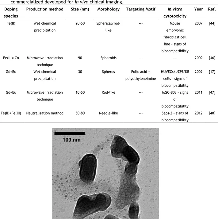
Table 1.3 A summary of hydroxyapatite-based magnetic nanoparticles non-commercialized developed for in vivo clinical imaging.
Doping species
Production method Size (nm) Morphology Targeting Motif In vitro
cytotoxicity
Year Ref. Fe(II) Wet chemical
precipitation 20-50 Spherical/rod-like --- Mouse embryonic fibroblast cell line – signs of biocompatibility 2007 [44]
Fe(III)+Co Microwave irradiation technique
90 Spheroids --- --- 2009 [46] Gd+Eu Wet chemical
precipitation
30 Spheres Folic acid + polyethyleneimine
HUVECs/L929/KB cells – signs of biocompatibility
2009 [17]
Gd+Eu Microwave irradiation technique
10-50 Rod-like --- MGC-803 – signs of biocompatibility
2011 [47]
Fe(II)+Fe(III) Neutralization method 50-80 Needle-like --- Saos-2 – signs of biocompatibility
2012 [48]
Figure 1.3 Transmission Electron Microscopy image of the HAp nanoparticles loaded with CoFe2O4, highlighting their core-shell structure.
30
On the other hand, Ashokan[17] tested the potential of multi-modal HAp nanoparticles doped with paramagnetic Gd and Europium and functionalized with folic acid (nHAp) and polyethyleneimine, in order to specifically target cancer cells and serve as a multi-modal contrast agent for three clinical imaging modalities (MRI, X-ray imaging and NIRF). These were produced by wet chemical precipitation method which originated nearly 80% of monodisperse non-functionalized particles with ~30nm diameter and spherical shape. In addition, the new particles showed better magnetic properties with brighter contrast than the clinically used Gd-DTPA (Magnevist®), in Figure 1.4. In vitro cytotoxicity studies using human umbilical vein endothelial cells and a cancer cell line revealed no apparent toxicity even up to relatively higher doses of 500 mg/mL.
Feng Cheng et al. [47] also focused work on multifunctional Eu/Gd dual-doped hydroxyapatite nanorods for drug delivery applications with image guidance. Using a rapid microwave-assisted hydrothermal method, nanorods with diameter ranging between 10 and 50nm were created. In addition, the synthesized NPs showed no appreciable toxicity in human gastric carcinoma cells.
Silvia Panseri et al.[48] produced Fe(II)/Fe(III) doped HAP NPs by a neutralization method optimized by the group. Suggesting applications in bone tissue regeneration as well in diagnostic imaging or magnetic drug delivery, the proposed NPs with needle-like structure registered sizes of 5-20nm in width and up to 50-80nm in length. In vitro results showed that these NPs did not reduce osteoblast-like cell viability.
As described above, magnetic nanoparticles can provide excellent performance as contrast agents and once injected in the blood stream they can be monitored by MRI. However, the distribution kinetics of current commercially available nano-contrast agents is described as passive or non-targeted, which restricts their clinical application. Consequently, considerable efforts are focused on the preparation of targeted magnetic nanoparticles to expand the range of pathologies to which these contrast agents could be applied. In addition, Figure 1.4 Nuclear Magnetic Resonance images of doped nHAp in comparison to Gd-DTPA (Magnevist®) showing T1-weighted bright contrast.
magnetic NPs can be regarded as 3D platforms onto which different molecules can be assembled, such as drugs for cancer theranostics (imaging and diagnostics).
1.3 - Next generation multifunctional nanoparticles for
cancer imaging and therapy
It is believed that the future of clinical oncology relies on the detection and treatment of malignant solid tumors in the earliest stage possible, increasing the chances to eradicate it. For this reason, it is highly anticipated that third generation magnetic nanoparticles can not only provide sensitive imaging information in cancer patients but also selectively deliver anticancer drugs to tumor sites. Besides early cancer diagnostic, this strategy would also be able to surpass the poor specificity and dose-limiting toxicities of pharmacologically active cancer drugs during current chemotherapy.
Nevertheless, this goal faces many challenges, namely the delivery of the probe to the targeted tumor along with its biocompatibility and toxicity, the stability of the probe onto the nanoparticle and the effectiveness of the signal enhancement in vivo.
Concerning tissue/organ-specific contrast agents, they usually consist of a coated paramagnetic or superparamagnetic label and a target-group molecule – probe – having a characteristic affinity for a specific cell or receptor. One of these probes believed to be highly specific against some cancers are monoclonal antibodies, such as an antibody against the endothelial integrin αvβ3 expressed on growing angiogenic vessels in some tumors (still not commercialized)[49]. Other target ligands involve soluble metalloporphyrins since they seem to have preferential selective uptake and retention by tumor tissues, folic acid once it binds to the high affinity folate receptors that exist in the serum of cancerous patients and on the cell surface of several human cancers of epithelial origin, bioactive peptides, sialic acids, among others[16].
For tumor-targeted therapy, methods to increase the loading capacity of anticancer drugs in the nanoparticles and control their release at target cells remain quite challenging.
1.4 - Aim and outline of the thesis
Most cancers have the ability to invade and destroy adjacent structures, developing permissive niches that promote tumor cell survival and proliferation and possibly cause death. Not only cancer is a major public health problem registering an exponential increase in new cases every year, but it also constitutes a great economic impact worldwide. So, even though research efforts to improve current cancer therapies over the past three decades have led to an improvement in patient survival, there is still a need for new and better strategies.
Early detection of cancer disease allows the maximum likelihood of successful treatment and recovery, since early localization of malignant solid tumors may allow a directed therapy to its site, optimizing the efficacy of the treatment. However, the most commonly used imaging modalities in clinical detection of cancer are all nonspecific, therefore they cannot distinguish between malignant or benign tumors and none leads to 100% accurate detection.
32
MRI is a non-invasive and commonly used imaging modality that can maximize its diagnostic value with the combined use of magnetic contrast agents. In addition, MRI contrast agents with nanometric dimensions constitute a very powerful tool in cancer therapy and diagnosis possessing distinct advantages over conventional ones due to some key characteristics such as their small size and high surface area, the possibility of delivery to a specific cancer site by active targeting and the possibility of engineering their blood circulation half-life, among other features.
Although some nano-contrast agents have been proposed so far, cytotoxicity is still a major issue that endangers their clinical use. Hydroxyapatite is a bioceramic material that enables the inclusion of ions in its crystal lattice and also confers biocompatibility to the system which makes its conjugation with magnetically responsive ions an appealing approach in targeted cancer imaging.
As previously described, some efforts to develop hydroxyapatite-based MRI contrast agents have been made. Nevertheless, there is a need to standardize the procedures of fabrication of the magnetic NPs. Also, it is not included in the literature a study that compares different doping agents produced by the same method and tested on the same parameters.
This project main goal is to produce, characterize and compare hydroxyapatite nanoparticles doped with particular magnetic species (Gd, Fe(II), Fe(III) and Co) and ultimately infer about their potential as MRI contrast agents. This main goal can be divided into smaller tasks that consist on:
Choose the right method a optimizing the available conditions in order to produce doped nanoparticles with sizes between 10 and 100nm;
Characterize the produced NPs in terms of their size, magnetic properties and also characterize the process of ion doping and its influence on the HAP structure;
Evaluate any toxic effects of the doped NPs in a common cell line in a certain time frame.
Chapter 2
Materials and Methods
2.1 - Synthesis of the Hydroxyapatite-based nanoparticles
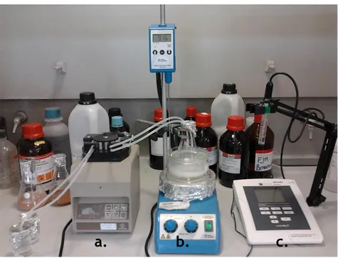
The hydroxyapatite-based nanoparticles were prepared by the wet chemical precipitation method. In a typical synthesis procedure of pure hydroxyapatite nanoparticles, 40mL of 0.5M Ca(OH)2 (98% extra pure, Acros Organics) was prepared and heated to 100ºC to which 40mL of 0.3M H3PO4 (85 wt% solution in water, Acros Organics) was added at a rate of 500µL/min. At this point, the magnetic dopants Gadolinium chloride hexahydrate Cl3Gd.6H2O (99%, Sigma Aldrich), Cobalt nitrate hexahydrate Co(NO3)2.6H2O (98+%, Acros Organics), Iron nitrate nonahydrate Fe(NO3)3 9H2O (98+%, Acros Organics) and Iron chloride tetrahydrate FeCl2.4H2O (99+%, Acros Organics) were added simultaneously with the phosphate precursor H3PO4 dropwise to the Ca(OH)2 solution at the rate of500µL/min to produce doped hydroxyapatite nanoparticles. The amount of dopant ions added depended on the required percentage of doping (2.5%, 5% and 10%) with respect to the atomic percentage of Ca2+. The applied nomenclature to the nanoparticles is formatted as XY_HAP, where X is the doped magnetic ion and Y represents the percentage of doping. All solutions were prepared with deionized water and the (Ca+doping ion)/P molar ratio was maintained at 1.67 in all procedures.The reaction was carried out at 100ºC in a water bath with constant stirring of 650rpm, as portrayed in figure 2.1. Throughout the reaction, pH was maintained at ~7.4 using ammonium hydroxide (25% solution in water, Acros Organics). After the reaction, the mixture was kept in these conditions for 2 hours and then incubated at room temperature overnight. Prior to the drying step at 60ºC, samples were centrifuged (5 min, ~2070g) and washed 3 times with hot deionized water. Finally, all nanoparticles were powdered using mortar and pestle and stored in a desiccator for further use. This protocol was adapted from the one described by Ashokan et al [17].
34
2.2 - Physicochemical Characterization
2.2.1 -
Size, morphology and zeta potential
Transmission electron microscope (TEM) was employed for high resolution imaging of pure and doped nanoHAP. A Jeol JEM 1400 microscope was used to observe nanoparticle size and morphology. To prepare samples for observation, the powders were suspended in deionized water using a concentration of 0.05g/L and the dispersant sodium dodecyl sulfate (SDS, 20% solution in water, Sigma Aldrich) was added in a concentration of 10x10-3 mol/L. Solutions were then sonicated for 15 seconds (20x) (Sonics&Materials, VC50) and applied to a copper grid. Images were examined using ImageJ® software to quantify nanoparticles dimensions.
The zeta potential and size of the synthesized nanoparticles were measured with a ZetaSizer Nano ZS. This device uses Dynamic Light Scattering (DLS) and calculates the particle size distribution from their diffusion under Brownian motion and applying the Stokes-Einstein relationship, D=kT/3πηDH, where D is the translational diffusion coefficient of a spherical particle in a fluid, k is the Boltzmann’s constant, T is the absolute temperature, η is the viscosity and DH is the hydrodynamic diameter.
On the other hand, zeta potential is calculated through the particles electrophoretic mobility which is the movement of a charged particle relative to the liquid when an electric field is applied. A laser beam hits the moving particles and induces a change of frequency by the Doppler Effect. This difference is proportional to the electrophoretic mobility (UE) and zeta potential (z) is analyzed according to Henry’s equation, UE=2εzf(Ka)/3η, where ε is the dielectric constant, f(Ka) is Henry’s function and η is the viscosity[50].
For this characterization, the powders were suspended in deionized water and prepared the same way as for TEM observations.
Figure 2.1 Representation of the nanoparticles fabrication conditions. a-Peristaltic pump;
b-temperature controller and heating plate with magnetic stirrer; c-pH meter.
2.2.2 -
Chemical profile
The infrared spectra that contain the species characteristic chemical bonds were recorded with a Fourrier Transform Infrared (FTIR) spectrometer Perkin-Elmer 2000 with a 4cm-1 spectral resolution and 100 scans per sample. In order to perform the analysis, ~2mg of each powdered sample was mixed with ~200mg of potassium bromide KBr to maintain low levels of humidity and discs were produced in a uniaxial press (Grasedy Specac).
2.2.3 -
Crystal phase analysis
In order to infer about the particles crystallinity before and after doping, X-ray diffraction (XRD) measurements were made. For this technique, samples were sintered to improve the obtained signal. The nanoHAP powders were heated up to 1000ºC at a rate of 4ºC/min and then heat-treated for 1h at this temperature, being the appropriate sintering temperature for nanosized HAP prepared by this method[41].
The x-ray diffraction patterns were obtained in a PANalytical X’Pert PRO TCU 1000 diffractometer, using monochromatic Cu radiation (λ = 1.541874Å). Data were acquired for 2Θ values between 8º and 100º with steps of 0,008º. Commercialized undoped nanoHAP was analyzed by other team member and was used as a control after heat-treatment.
2.2.4 -
Magnetic properties
The magnetic properties of the doped and undoped nanoparticles were studied by superconducting quantum interference device (SQUID, Quantum design Ref: MPMS-5S) in an applied magnetic field of•±50 kOe at 300K. The strength of the magnetic moment is presented as a mass magnetization (M) – magnetic moment per total mass of each sample.
For these measurements, the previously prepared powders were weighed and inserted in a see-through capsule and, in turn, the capsule was inserted in a see-through straw that is introduced into the SQUID magnetometer for further analysis.
2.3 - Biological response
2.3.1 -
Cell culture and maintenance
Human Dermal Microvascular Endothelial Cells (hDMECs) were purchased from ScienCell Research Laboratories (Catalog number: 2000) and cultured in Endothelial Cell Basal medium (EBM®-2, Clonetics Lonza) supplemented with 2% foetal bovine serum (FBS), 0.04% hydrocortisone, 0.4% human recombinant fibroblast growth factor-β (rhFGF-β), 0.1% vascular endothelial growth factor (VEGF), 0.1% ascorbic acid, 0.1% Gentamicin Sulfate and Amphotericin-B, 0.1% heparin, 0.1% human recombinant epidermal growth factor (rhEGF) and 0.1% recombinant long R insulin-like growth factor-1 (R3-IGF-1) . The medium was renewed every 2-3 days until confluence was reached and cells were used until passage 7. All the procedures described with live cells were performed in sterile conditions on a laminar flow chamber and incubation was carried out in a 5% CO2 and 37 °C atmosphere.
36
2.3.2 -
Cell metabolic activity evaluation
In order to estimate the in vitro cell viability of the nanoHAP with the highest doping concentration (10%) of Gd, Fe(II), Fe(III) and Co, a resazurin (Sigma) assay was performed. This method is based on a cell viability indicator that uses natural reducing power of living cells to convert resazurin to the fluorescent product, resofurin. Fluorescence acts as a proportional response of cell metabolic activity[51].
Each well of the 96-well plate was seeded with 5000 hDME cells. After 24h of culture, the medium in the wells was refreshed with the medium containing autoclaved nanoparticles at a concentration of 500µg/mL. Finally, cells were incubated with new medium and resazurin assay was performed at the time points of 4h and 24h. Briefly, after previously removing the culture medium form the wells, resazurin was added in fresh medium at a final concentration of 10% (v/v) and incubated for 4h, at 37ºC. 100μL of medium was then extracted from each sample and fluorescence was measured at λex=540nm and λem=590nm in a microplate reader.
2.3.3 -
Cellular morphology
After 4 and 24 hours of culture, cell morphology was observed by staining the F-actin with Alexa Fluor ® 488 phalloidin (Invitrogen, Molecular Probes) and counterstaining the nuclei with DAPI (4',6-diamidino-2-phenylindole, Merck). Culture medium was removed from the wells and samples were washed 2x with PBS and fixated with 4% formaldehyde at room temperature (RT) for 15 minutes. After washing with PBS twice, cells were permeabilized for 30 minutes with 0.1% (v/v) Triton X-100 and washed again 3x with PBS. The samples were then incubated with the phalloidin diluted 1:100 in PBS for 30 minutes in the dark at RT and washed 3x with PBS. To stain the nuclei of the cells, samples were incubated with DAPI at a concentration of 0.1µg/mL in PBS for 10 min and washed with PBS 3x. Two drops of FluoromountTMwere added and the samples were stored at 4ºC in the dark until observation under an inverted fluorescence microscope with proper light filters.
2.3.4 -
Statistical analysis
Data from the resazurin assay were analyzed using Scheffe test for multiple comparisons. Results were considered significant when p<0.05. Calculations and descriptive statistic were performed using SPSS-Software for Windows® (version 17.0) and Microsoft Office Excel 2010®. All data are presented as average values ± standard deviation.
Chapter 3
Results and Discussion
3.1 - Physicochemical Characterization
3.1.1 -
Size, morphology and zeta potential
Both size and morphology of nanoparticles synthesized as MRI contrast agents influence their interaction not only with each other in a suspension, but also with the cells they are in contact with.
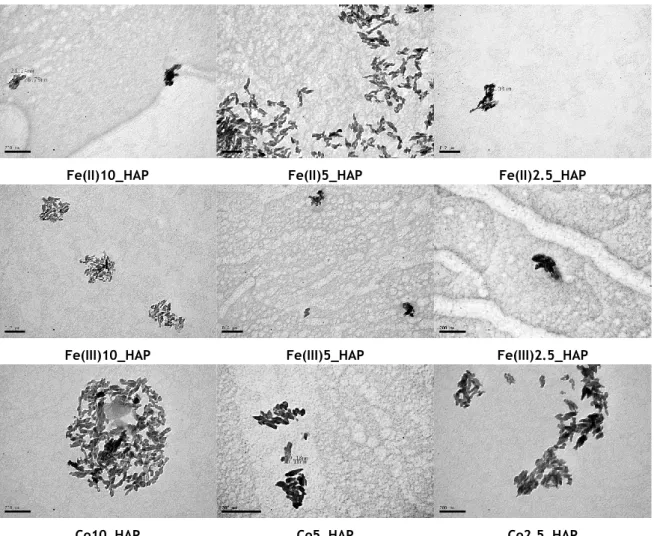
Undoped HAP
38
Fe(II)10_HAP Fe(II)5_HAP Fe(II)2.5_HAP
Fe(III)10_HAP Fe(III)5_HAP Fe(III)2.5_HAP
Co10_HAP Co5_HAP Co2.5_HAP
Figure 3.2 TEM images of the undoped and doped nanoHAP
Figure 3.1 NanoHAP dimensions obtained with TEM images (Figure 3.1) observation through Image J® software 0,0 10,0 20,0 30,0 40,0 50,0 60,0 70,0 80,0 Si ze (n m )
The TEM image (Figure 3.1) of undoped hydroxyapatite indicates the formation of spherical nanoHAP of average size ~11 nm. On the other hand, all images of doped nanoparticles show a predominant more elongated shape that can be identified as a rod-like shape. In addition, these species present slightly higher dimensions, as also shown in Figure 3.2. The studied doped nanoparticles present longitudinal dimensions between ~34 and ~59nm and cross sectional dimensions between ~15 and ~29nm. These differences in size and morphology from the doped and undoped species might have been induced by differences in electric charge and ionic radius between the Ca2+ ions and the Gd3+/Fe2+/Fe3+/Co2+ doping ions. This may be influencing the crystal growth of nanoHAp, leading to altered size and shape of doped NPs compared to undoped NPs, as suggested by other authors in similar studies[47].
Considering the main application of the synthesized nanoparticles in tumor cells imaging and future applications in tumor cell specific targeting, ligand coated NPs can be efficiently directed toward and subsequently internalized by tumor cells. However, cellular uptake of
0 50 100 150 200 250 300 Si ze (n m ) -60 -50 -40 -30 -20 -10 0 Zeta Po ten ti al (m V )
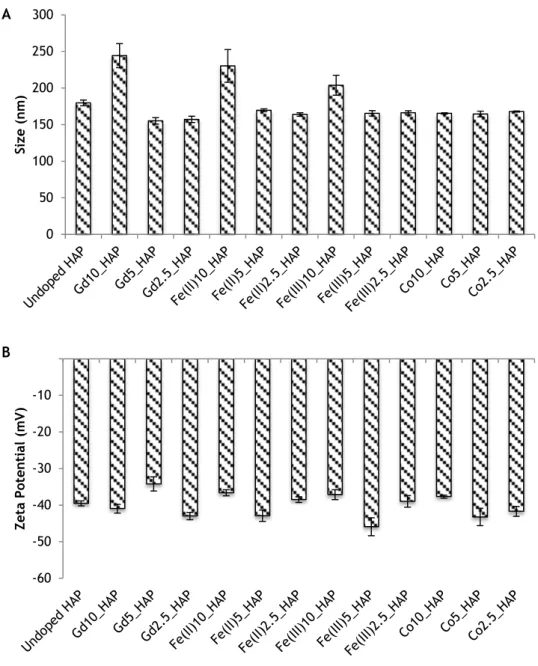
Figure 3.3 ZetaSizer results displaying (A) the average particle sizes and (B) the average zeta potential of the doped and undoped nanoHAP
A
B A
40
NPs is size-dependent and some authors have found 40-60nm to be the optimal NP size range for endocytosis and mediation of other signaling processes [52, 53]. Consequently, the results obtained through TEM observation are in agreement with those described in the literature.
In relation to the shape of the particles, although there is a lot of unconformity among authors, recent findings indicate that rod-like shaped nanoparticles are significantly better in cancer targeting, exhibiting higher specific uptake and lower nonspecific uptake in breast cancer cells compared with spherical nanoparticles [54]. This also constitutes a positive indicator that the doped nanoHAP produced in this work is suitable for targeted cancer imaging.
Comparing the TEM results to the ones obtained with the ZetaSizer, size measurements differ. As portrayed in Figure 3.3A, the studied doped and undoped nanoparticles present sizes between a range of 155 and 244nm. Size between both methods differs due to the fact that ZetaSizer uses DLS technology whose approach recognizes each particle as a perfect sphere that does not represent the actual shape of the synthesized NPs. Also, the ZetaSizer measurements most likely represent the size of NPs aggregates that were not effectively dispersed after sonication.
Concerning the measured zeta potential of the samples, it fluctuates between 45 and -34mV with small deviations (Figure 3.3B). All preparations consisted on stable colloidal suspensions either at or above critical micelle concentration for the negatively charged surfactant SDS which explains the strongly negative zeta potential obtained in all suspensions[55].
3.1.2 -
Chemical profile
4000 3700 3400 3100 2800 2500 2200 1900 1600 1300 1000 700 400 Trans m it tanc e (% ) Wavenumber (cm-1)Undoped HAP Gd10_HAP Gd5_HAP Gd2.5_HAP
4000 3700 3400 3100 2800 2500 2200 1900 1600 1300 1000 700 400 Trans m it tanc e (% ) Wavenumber (cm-1)
Undoped HAP Fe(II)10_HAP Fe(II)5_HAP Fe(II)2.5_HAP
4000 3700 3400 3100 2800 2500 2200 1900 1600 1300 1000 700 400 Trans m it tanc e (% ) Wavenumber (cm-1)
Undoped HAP Fe(III)10_HAP Fe(III)5_HAP Fe(III)2.5_HAP
A B C OH -H2O HPO4 2-CO3 2-PO4
3-42
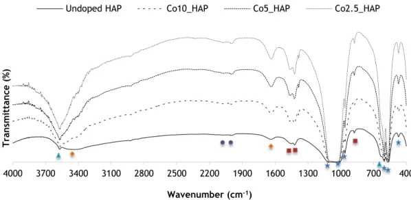
In order to chemically characterize the process of ion doping in terms of changes in the nanoHAP profile, a FTIR analysis was performed (Figure 3.4).
Considering all the represented species, the FTIR spectra are characterized by lattice (~3573cm-1) and vibrational (~634cm-1) OH bands, phosphate bands (ʋ1~962cm-1, ʋ2 ~470cm-1, ʋ3 ~1090-~1110 and ~1035cm-1, ʋ4 ~603 and ~565cm-1), residual carbonates C-O resulting from the atmospheric CO2 adsorbed by the apatite (~1457, ~1421 and ʋ2 ~876cm-1) and lattice water (3417-3495cm-1 and ~1637cm-1) which are typical in a nanoHAP spectra [17, 44, 56-58]. All species also present a few weak peaks 2500-2000cm-1 that might correspond to HPO
4 2-groups as previously reported in the literature[59].
In a more close analysis to each type of ion doping into nanoHAP, the addition of Gd increases the intensity of the band assigned to moisture in the samples (2500-3500cm-1)[60] and also increases the intensity of the peak assigned to the hydroxyl groups (3570 cm-1) but does not significantly affect the rest of the spectra(Figure 3.4A). Fe(II) doping also produces a similar effect to the referred water band and hydroxyl peak (Figure 3.4B) and Fe(III) shows only a major increase in intensity of the hydroxyl peak, compared to pure HAP(Figure 3.4C). Cobalt insertion in the HAP matrix does not significantly alter the spectra compared to the nanoHAP one. A sharp peak is also found in Co10_HAP ~800cm-1 which might correspond to the presence of some remaining nitrate from the used reagents [61, 62].
4000 3700 3400 3100 2800 2500 2200 1900 1600 1300 1000 700 400 Trans m it tanc e (% ) Wavenumber (cm-1)
Undoped HAP Co10_HAP Co5_HAP Co2.5_HAP
Figure 3.4 FTIR spectra of nanoHAP samples with different percentages of (A) Gd (B) Fe(II) (C) Fe(III) (D) Co doping and undoped HAP
3.1.3 -
Crystal phase analysis
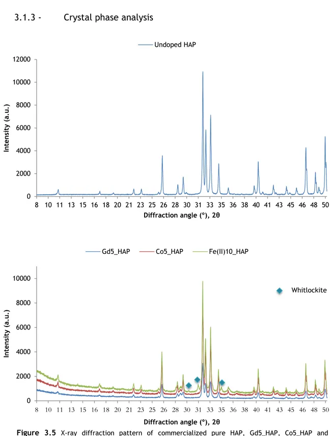
XRD technique was used to infer about phase composition and crystallinity of the HAP nanoparticles after magnetic ion doping. Figure 3.5 shows the XRD patterns of some heat-treated samples that were successfully analyzed and compared to commercialized undoped nanoHAP.
In Gd5_HAP and Co5_HAP, no unexpected phase was formed and all peaks correspond solely to hydroxyapatite phase, accordingly to Centre for Diffraction Data (ICDD) standard
0 2000 4000 6000 8000 10000 8 10 11 13 15 16 18 20 21 23 25 26 28 30 31 33 35 36 38 40 41 43 45 46 48 50 In ten si ty (a. u .) Diffraction angle (º), 2θ
Gd5_HAP Co5_HAP Fe(II)10_HAP
Figure 3.5 X-ray diffraction pattern of commercialized pure HAP, Gd5_HAP, Co5_HAP and Fe(II)HAP. Blue symbols indicate the presence of whitlockite peaks on Fe(II)10_HAP among the hydroxyapatite composition 0 2000 4000 6000 8000 10000 12000 8 10 11 13 15 16 18 20 21 23 25 26 28 30 31 33 35 36 38 40 41 43 45 46 48 50 In ten si ty (a. u .) Diffraction angle (º), 2θ Undoped HAP Whitlockite
44
data (PDF file #9-432). However, XRD analysis of Fe(II)10_HAP indicated the presence of whitlockite, a form of calcium phosphate, between the hydroxyapatite composition. This could have formed during heat-treatment and propitiated by the Fe(II) content of the sample, which might be modifying the apatite structure and producing secondary phases[63]. Further sample analysis is needed.
3.1.4 -
Magnetic properties
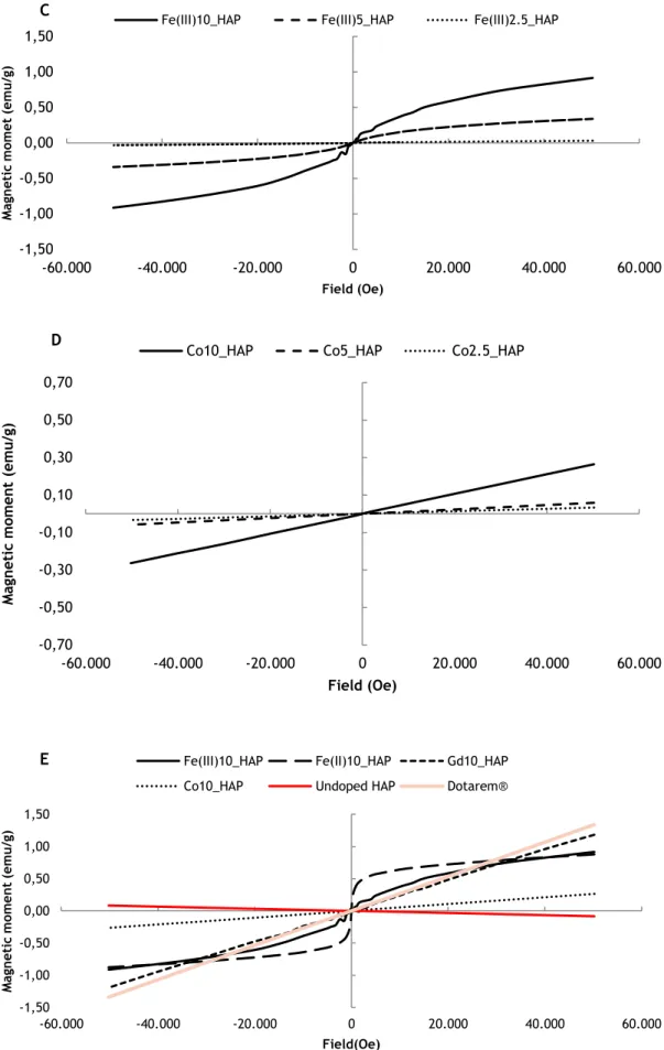
According to the main objective of this work towards developing a robust and reproducible contrast agent system capable of enhancing tumor contrast, it was important to illustrate the magnetism of the synthesized doped nanoHAP. The relationship of magnetization to applied field (±50kOe) at room temperature of the nanoHAP with different doping ions is plotted in figure 3.6.
In all doped nanoparticles (Gd_HAP, Fe(II)_HAP, Fe(III)HAP and Co_HAP), the magnetization level increased with the doping concentration of each ion, confirming their predominant role in making these nanoparticles magnetically functional.
-1,50 -1,00 -0,50 0,00 0,50 1,00 1,50 -60.000 -40.000 -20.000 0 20.000 40.000 60.000 Ma gn e tic m om e n t (a m u /g ) Field(Oe)
Gd10_HAP Gd5_HAP Gd2,5_HAP
-1,50 -1,00 -0,50 0,00 0,50 1,00 -60.000 -40.000 -20.000 0 20.000 40.000 60.000 Ma gn e tic m om e n t (e m u /g ) Field (Oe)
Fe(II)10_HAP Fe(II)5_HAP Fe(II)2.5_HAP
A
-0,70 -0,50 -0,30 -0,10 0,10 0,30 0,50 0,70 -60.000 -40.000 -20.000 0 20.000 40.000 60.000 Mag n etic m om en t (em u /g ) Field (Oe)
Co10_HAP Co5_HAP Co2.5_HAP
-1,50 -1,00 -0,50 0,00 0,50 1,00 1,50 -60.000 -40.000 -20.000 0 20.000 40.000 60.000 Ma gn e tic m om e t (e m u /g ) Field (Oe)
Fe(III)10_HAP Fe(III)5_HAP Fe(III)2.5_HAP
-1,50 -1,00 -0,50 0,00 0,50 1,00 1,50 -60.000 -40.000 -20.000 0 20.000 40.000 60.000 Ma gn e tic m om e n t (e m u /g ) Field(Oe)
Fe(III)10_HAP Fe(II)10_HAP Gd10_HAP Co10_HAP Undoped HAP Dotarem®
Figure 3.6 Variation of magnetization with the applied field of ±50kOe for synthesized doped and undoped nanoHAP: (A) Gd doping (B) Fe(II) doping (C) Fe(III) doping (D) Co doping (E) highest doping percentage in contrast with undoped nanoHAP and Dotarem®
C
D
![Table 1.1 Summary of distinct features of benign and malignant tumors (adapted from [5])](https://thumb-eu.123doks.com/thumbv2/123dok_br/15719462.1070299/22.892.95.754.657.974/table-summary-distinct-features-benign-malignant-tumors-adapted.webp)
![Figure 1.1 The primary tumor microenvironment (adapted from [9])](https://thumb-eu.123doks.com/thumbv2/123dok_br/15719462.1070299/23.892.159.771.440.948/figure-primary-tumor-microenvironment-adapted.webp)
![Table 1.2 Non-exhaustive list of MRI approved contrast agents used in clinical practice (Adapted from [16], [28] and [29])](https://thumb-eu.123doks.com/thumbv2/123dok_br/15719462.1070299/27.892.130.802.554.807/table-exhaustive-approved-contrast-agents-clinical-practice-adapted.webp)