Susana Isabel Conde Jesus Palma
Mestre em Engenharia Biomédica
Engineered MRI nanoprobes based on
superparamagnetic iron oxide
nanoparticles
Dissertação para obtenção do Grau de Doutor em
Bioengenharia (MIT-Portugal)
Orientador: Prof. Doutora Ana Cecília Roque, FCT-UNL
III
Engineered MRI nanoprobes based on superparamagnetic iron oxide
nanoparticles
Copyright © Susana Isabel Conde Jesus Palma, Faculdade de Ciências e Tecnologia, Universidade Nova de Lisboa.
V
VII
Acknowledgements
During the four years of the PhD Adventure, I had the chance to work in a multidisciplinary environment, building bridges between different areas of expertise in bioengineering research. It has been a very interesting challenge, which was only possible with the contribution and support of several people.
I deeply thank my supervisor Prof. Ana Cecília Roque for having given me the opportunity to be part of her research group and work with her in this challenging project. I am very grateful for the rigor, exigence, brainstorming and advice but also for the patience and confidence that you had in me, Cecília. Without your support, the achievements of this “nano -adventure” were not possible.
When I met Dr. M. Puerto Morales in a conference, in Lubeck, I could not imagine the importance she would have in my PhD. I thank Puerto for my stay in her Lab at ICCM (Madrid), for giving me the opportunity to meet and/or work with Marzia Marciello, Sabino Vientemillas, Gorka Salas, Amalia Ruiz, Yurena Luengo, Fernando Herranz and Jesús Ruiz-Cabello. I am very grateful to Puerto for her availability to make VSM measurements of my nanoparticles and to discuss the results of the work, giving an important input to my PhD project. It was such a fruitful partnership.
I thank Prof. Alexandra R. Fernandes for receiving me in her Lab at UCIBIO, DCV (FCT-UNL) for the in vitro assays with HC116 cell line, and for her availability to discuss the results. I leave here also a very sincere “thank you” to Joana Silva and Pedro Martins, from Prof. Alexandra’s Lab, for having introduced me to the human cell culture techniques and for the technical support during my experiments in the cell culture lab. I express also my gratitude to Prof. Pedro Baptista and his team for sharing the Lab equipment with me.
This work would be more difficult without the collaboration of Alexandra Carvalho at CENIMAT (FCT-UNL), who I acknowledge for the support regarding the MRI experiments and availability to discuss results and share ideas. It was a nice and fruitful collaboration.
I thank Prof. Joaquim Sampaio Cabral for the opportunity to work with the cell line ReNcell VM in his Lab (SCBL at iBB (IST-UL)) and for providing the resources and facilities for the experiments.
VIII
always with a friendly and fun environment, but also very well organized and tidy. Dear colleagues Abid Hussain, Ricardo Branco, Ana Pina, Íris Batalha, Telma Barroso, Margarida Dias, Vijaykumar Dahdge, Cláudia Fernandes, Henrique Carvalho, Carina Rodrigues and, more recently, José Almeida, Aline Viecinski, Luís Silva and Arménio Barbosa: thank you for the nice environment in the Lab, and for the discussions in the group meetings and during the lab daily routine. A special acknowledgement goes to Abid, who was the first to introduce me to the Lab bench and nanoparticle synthesis. Íris Batalha, Margarida Dias and Ana Pina, the self-named “orsinhas”: thanks for the healthy discussions, for all the laughing and for being there also in the frustrating and unhappy moments of life during the PhD. A big and smiley “thank you” to all the members that passed by the Biomolecular Engineering Group during my PhD, not forgetting my “aluninhas” Rosarinho Nazaré and Patrícia Traguedo, who introduced me to the teaching side of academic research life.
I acknowledge the technical support of Dr. Carla Rodrigues, from Laboratório de Análises of REQUIMTE and also Prof. César Laia (for the collaboration with using the DLS at DQ), Prof. Luísa Ferreira (for the help with FTIR analyses), Dr. Paulo Lemos and Dr. Christophe Roca (for the help with the fluorescence microscope). I would like to acknowledge also D. Maria José Carapinha and Isabel Rodrigues for their help with bureaucratic paper work, and D. Maria da Palma Afonso and Mafalda Manita for the assistance in keeping the labware clean for the daily laboratory activities.
None of the achievements of this thesis would have been possible without the support and love of my friends and family. They helped me to relax and patiently listened to my concerns and doubts in the most stressful days. But, at the same time, they were also the ones who celebrated with me the enthusiasm of positive results and published papers!
Agradeço aos meus colegas da Banda da Sociedade Musical Sesimbrense e da Bota Big Band pela Música, pelos momentos de companheirismo e de paródia. Aos meus amigos Cristina, Ângelo (e Bia, claro), Sara, Maria da Luz, Carla, Tânia, e Ana Dionísio obrigada por me terem ouvido nos dias mais depressivos do doutoramento e por terem partilhado comigo momentos de descontracção e festa para sair da rotina do Lab!
Ao meu pai, à minha mãe e ao meu irmão, agradeço o interesse e o esforço que sempre demonstraram em perceber e acompanhar o meu trabalho, apesar ser pouco convencional e complexo de explicar… Obrigada pela enooorrrrme paciência e todo o apoio e carinho.
IX
Abstract
This project aimed to engineer new T2 MRI contrast agents for cell labeling based on formulations containing monodisperse iron oxide magnetic nanoparticles (MNP) coated with natural and synthetic polymers. Monodisperse MNP capped with hydrophobic ligands were synthesized by a thermal decomposition method, and further stabilized in aqueous media with citric acid or meso-2,3-dimercaptosuccinic acid (DMSA) through a ligand exchange reaction. Hydrophilic MNP-DMSA, with optimal hydrodynamic size distribution, colloidal stability and magnetic properties, were used for further functionalization with different coating materials. A covalent coupling strategy was devised to bind the biopolymer gum Arabic (GA) onto MNP-DMSA and produce an efficient contrast agent, which enhanced cellular uptake in human colorectal carcinoma cells (HCT116 cell line) compared to uncoated MNP-DMSA. A similar protocol was employed to coat MNP-DMSA with a novel biopolymer produced by a biotechnological process, the exopolysaccharide (EPS) Fucopol. Similar to MNP-DMSA-GA, MNP-DMSA-EPS improved cellular uptake in HCT116 cells compared to MNP-DMSA. However, MNP-DMSA-EPS were particularly efficient towards the neural stem/progenitor cell line ReNcell VM, for which a better iron dose-dependent MRI contrast enhancement was obtained at low iron concentrations and short incubation times. A combination of synthetic and biological coating materials was also explored in this project, to design a dynamic tumor-targeting nanoprobe activated by the acidic pH of tumors. The pH-dependent affinity pair neutravidin/iminobiotin, was combined in a multilayer architecture with the synthetic polymers poy-L-lysine and poly(ethylene glycol) and yielded an efficient MRI nanoprobe with ability to distinguish cells cultured in acidic pH conditions form cells cultured in physiological pH conditions.
Keywords: iron oxide magnetic nanoparticles (MNP), magnetic resonance imaging (MRI), gum
XI
Resumo
O objectivo desta tese consiste na engenharia de novos agentes de contraste T2 para marcação celular através de imagiologia por ressonância magnética (MRI), usando formulações com nanopartículas magnéticas de óxido de ferro (MNP) revestidas com polímeros naturais e com polímeros sintéticos. O método da decomposição térmica foi usado para sintetizar MNP monodispersas revestidas com ligandos hidrofóbicos. Para as estabilizar em meio aquoso, os ligandos hidrofóbicos foram substituídos por moléculas hidrofílicas, como o ácido cítrico ou o ácido meso-2,3,-dimercaptosuccínico (DMSA), através de uma reacção de intercâmbio de ligandos. As MNP-DMSA, hidrofílicas, com estabilidade coloidal e propriedades magnéticas optimizadas, foram revestidas com diferentes materiais. Para acoplar o biopolímero goma arábica às MNP-DMSA, foi usada uma ligação covalente que permitiu obter um agente de contraste eficiente e com um nível de captação celular melhorado face às MNP-DMSA em células humanas de carcinoma colorectal (linha celular HCT116). Um protocolo experimental semelhante foi usado para revestir as MNP-DMSA com um biopolímero novo produzido por via biotecnológica, o exopolisacarídeo (EPS) Fucopol. Tal como as DMSA-GA, as MNP-DMSA-EPS melhoraram o nível de captação celular nas células HCT116 face às MNP-DMSA. No entanto, foram particularmente eficientes numa linha de células estaminais/progenitoras neurais (ReNcell VM), nas quais se obteve um melhoramento mais intenso do contraste em função da dose de ferro nas imagens por MRI, para doses baixas de ferro e tempos de incubação curtos. Neste projecto foi também explorada uma combinação de materiais de revestimento sintéticos e biológicos para desenvolver uma nano-sonda dinâmica para marcação de tumores, activada pelo pH ácido tumoral. O par de afinidade dependente do pH neutravidina/iminobiotina foi combinado com poli-L-lisina e poli(etilenoglicol) através de uma arquitectura multi-camada, resultando numa nano-sonda para MRI eficiente e capaz de distinguir células cultivadas em condições ácidas de células cultivadas em condições fisiológicas.
Palavras-chave: nanopartículas magnéticas de óxido de ferro (MNP), imagiologia por
XIII
Table of Contents
XIV
XV
3.4. Conclusions _____________________________________________________________ 84 3.5. References _____________________________________________________________ 85 Chapter 4: A value-added exopolysaccharide as a coating agent for MRI nanoprobes ______ 89 4.1. Introduction _____________________________________________________________ 91 4.2. Experimental Section _____________________________________________________ 92 4.2.1. Materials ____________________________________________________________ 92 4.2.2. Synthesis and phase transfer of iron oxide magnetic nanoparticles (MNP-DMSA) ___ 92 4.2.3. Preparation of EPS-coated magnetic nanoparticles (MNP-DMSA-EPS) ___________ 93 4.2.4. Characterization of magnetic nanoparticles _________________________________ 93 4.2.5. Cell culture and labeling ________________________________________________ 94 4.2.6. Multi-lineage differentiation of ReNcell VM _________________________________ 95 4.2.7. Cytotoxicity evaluation _________________________________________________ 95 4.2.8. Identification of cellular iron by Prussian blue staining ________________________ 96 4.2.9. Intracellular localization of magnetic nanoparticles ___________________________ 96 4.2.10. Iron quantification ____________________________________________________ 97 4.2.11. In vitro MRI of cell phantoms ___________________________________________ 97 4.3 Results and Discussion ____________________________________________________ 97 4.3.1. Particle size, composition and surface chemistry ____________________________ 97 4.3.2 Magnetic properties and relaxivities measurements __________________________ 100 4.3.3 Cell-nanoparticle interactions ___________________________________________ 103 4.3.4 Differentiation of MNP labeled neural stem/progenitor cells ____________________ 108 4.3.5 In vitro MRI of MNP-DMSA-EPS labeled cells ______________________________ 109 4.4 Conclusion _____________________________________________________________ 111 4.5 References _____________________________________________________________ 112 Chapter 5: An affinity-triggered MRI nanoprobe for pH-dependent cell labeling ___________ 117 5.1. Introduction ____________________________________________________________ 119 5.2. Experimental Section ____________________________________________________ 121 5.2.1. Materials ___________________________________________________________ 121 5.2.2. Production of multi-layer functionalized magnetic nanoparticles ________________ 121
XVI
XVII
Index of Figures
Figure 1.1. Schematic representation of the effect of applying an external magnetic field on
magnetite at different size scales. ________________________________________________ 2
Figure 1.2. Superparamagnetism features. _________________________________________ 4 Figure 1.3. Schematic representation of MNP surface modification with biocompatible coating
and functional moieties. _______________________________________________________ 10
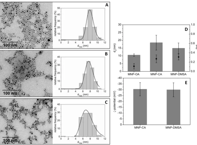
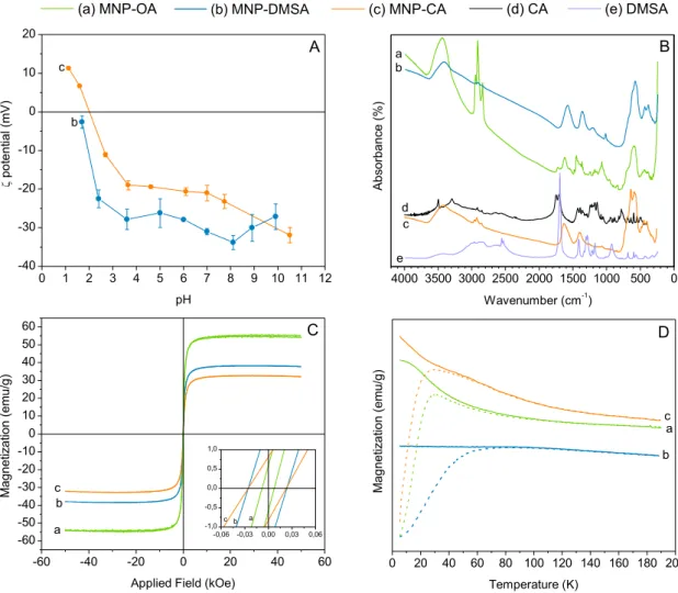
Figure 2.1. Morphology, size and zeta potential of the hydrophobic and hydrophilic
nanoparticles. _______________________________________________________________ 49
Figure 2.2. Surface and magnetic properties of the hydrophobic and hydrophilic nanoparticles.
__________________________________________________________________________ 51
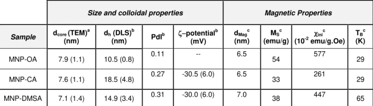
Figure 2.3. MNP-DMSA functionalization possibilities using gum Arabic as model biomolecule.
__________________________________________________________________________ 55
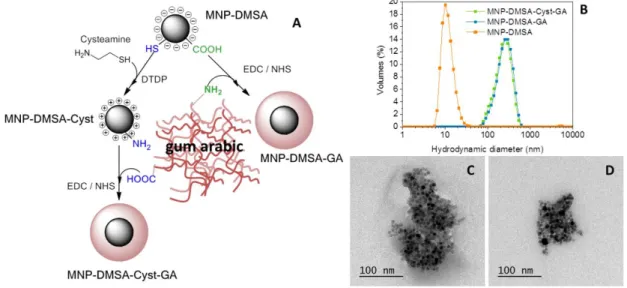
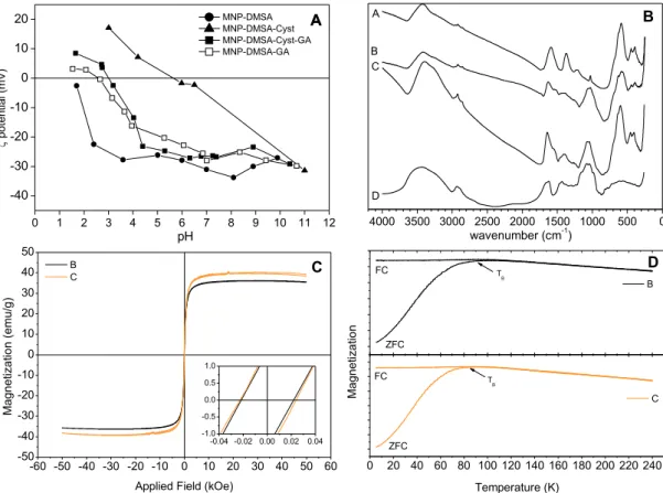
Figure 2.4. Surface and magnetic properties of the particles coated with gum Arabic. ______ 56 Figure 2.5. Determination of the relaxation rates of MNP-DMSA-Cyst-GA and MNP-DMSA-GA
as a function of iron concentration, respective linear adjustments and r2 values. ___________ 57
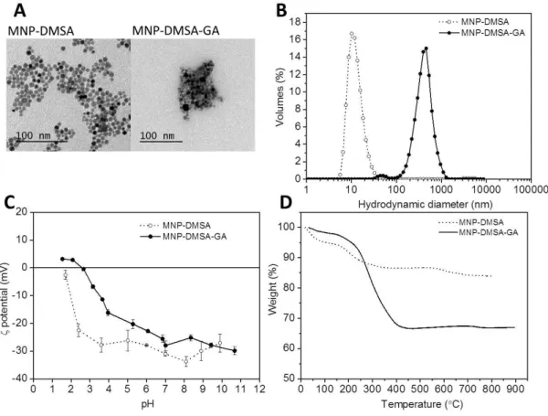
Figure 3.1. Size, colloidal stability and composition of the produced particles. ____________ 73 Figure 3.2. MNP-DMSA-GA colloidal stability over time, in different conditions. ___________ 74 Figure 3.3. Differential Thermal Analysis (DTA) of MNP-DMSA and MNP-DMSA-GA. ______ 74 Figure 3.4. FTIR spectra of MNP-DMSA-GA in comparison with MNP-DMSA and free GA. __ 75 Figure 3.5. Magnetic properties of MNP-DMSA and MNP-DMSA-GA. ___________________ 76 Figure 3.6. Relaxivities and T2-MRI phantoms of MNP-DMSA-GA. _____________________ 77 Figure 3.7. Determination of the nanoparticles and GA effects on HCT116 cell viability._____ 78 Figure 3.8. In vitro interactions of MNPs with HCT116 cells observed by microscopy and iron
uptake quantification. _________________________________________________________ 79
Figure 3.9. Effect of particle incubation time on the amount of iron per cell, quantified by ICP. 80 Figure 3.10. Bright field and fluorescence microscopy images of cells incubated for 48 h with
MNP-DMSA-GA at IC50. _______________________________________________________ 81
Figure 3.11. Localization of MNP-DMSA-GA within HCT116 cells after 48h incubation at IC50.
__________________________________________________________________________ 81
XVIII
Figure 3.13. Fold changes in pro-apoptotic (p21 and BAX) and anti-apoptotic (BCL-2) genes
expression at 3.5h, 6h, 12h, and 48h after cell incubation with MNP-DMSA-GA at IC50. _____ 83
Figure 3.14. Evaluation of in vitro MRI cell labeling efficiency with DMSA and
MNP-DMSA-GA. _________________________________________________________________ 84
Figure 4.1 Characterization of size and composition of the nanoparticles before and after EPS
coating. ____________________________________________________________________ 98
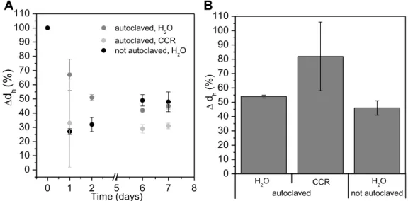
Figure 4.2. Variation of hydrodynamic diameter, polydispersity index and zeta potential of
MNP-DMSA and MNP-MNP-DMSA-EPS dispersed in different conditions. ________________________ 99
Figure 4.3. Evaluation of magnetic and relaxometric properties of DMSA and
MNP-DMSA-EPS. _______________________________________________________________ 101
Figure 4.4. Details regarding magnetic properties of MNP-DMSA and MNP-DMSA-EPS.
_________________________________________________________________________ 102
Figure 4.5. Cell cytotoxicity assay results for HCT116 and ReNcell VM cells incubated with
MNPs and EPS. ____________________________________________________________ 104
Figure 4.6. Representation of cell cytotoxicity assay results in logarithmic form (log10 [Fe] (or
log10 [EPS])). _______________________________________________________________ 104
Figure 4.7. Observation and quantification of iron in cell cultures incubated with MNP-DMSA
and MNP-DMSA-EPS. _______________________________________________________ 105
Figure 4.8. Contribution of internalized and adsorbed nanoparticles for the cellular iron found in
cells after labeling with MNP-DMSA or MNP-DMSA-EPS. ___________________________ 106
Figure 4.9. Tracking the localization of MNP-DMSA-EPS within (A) HCT116 cells and (B)
ReNcell VM cells by microscopy. _______________________________________________ 108
Figure 4.10. Immunohistochemistry of post-labeled ReNcell VM cells at day 14 of culture. _ 109 Figure 4.11. Efficacy of MNP-DMSA-EPS for in vitro MRI cell labeling. _________________ 111 Figure 5.1. Schematic representation of the multilayer pH-sensitive MNPs and concept for
achieving preferential interactions with tumoral cells. _______________________________ 121
Figure 5.2. Multi-layer MNP assembly. __________________________________________ 127 Figure 5.3. pH-dependent dissociation of Nav-bPEG layer from multilayer nanoparticles as a
result of 20 h exposition to different pH buffers. ____________________________________ 129
Figure 5.4. Effect of pH treatment on multilayer nanoparticles surface charge and size. ____ 130 Figure 5.5. Variation of multilayer nanoparticles size distributions after being exposed to PBS at
different pHs. ______________________________________________________________ 131
Figure 5.6. Bright field microscopy images of preparations stained with Prussian blue for iron
identification, obtained after exposing HCT116 cells to multilayer nanoparticles at 10 µg Fe/ml for 5 h. ___________________________________________________________________ 132
Figure 5.7. Tracking nanoparticles localization after incubation of HCT116 clels for 5 h in acidic
XIX
Figure 5.8. pH-dependent cell-nanoparticle interactions after 5 h of incubation with the
nanoprobes at 10 µg Fe/ml in acidic (pH 6.5) and physiological (pH 7.4) culture medium. __ 135
Figure 5.9. Contribution of internalized and adsorbed nanoparticles for the proportion of cellular
iron found in cells after labeling. ________________________________________________ 136
Figure 6.1. Summary of the average relative iron uptake per cell upon incubation with the MNPs
XXI
Index of Tables
Table 1.1. Applications of superparamagnetic iron oxide nanoparticles in the biomedical field. 6 Table 1.2. Utilization of biopolymers extracted from natural sources in the development of
MNP-based MRI contrast agents. ____________________________________________________ 16 __________________________________________________________________________ 17
Table 1.3. Utilization of biopolymers obtained by biotechnological processes in the development
of MNP-based MRI contrast agents. _____________________________________________ 17
Table 1.4. Utilization of synthetic polymers in the development of MNP-based MRI contrast
agents. ____________________________________________________________________ 22
Table 2.1. Summary of size, zeta potential and magnetic characterization results for the
hydrophobic (MNP-OA) and hydrophilic (MNP-CA and MNP-DMSA). ___________________ 54
Table 3.1. Summary of size, magnetic and relaxometric properties of the produced MNP in
comparison with a commercial MNP-based contrast agent. ___________________________ 77
Table 3.2. Quantitative characterization of the cell-nanoparticles interactions. ____________ 80 Table 4.1. Uptake of iron by HCT116 and ReNcell VM after incubation with MNP-DMSA and
MNP-DMSA-EPS.___________________________________________________________ 105
Table 4.2. Distribution of cellular iron between internalized and adsorbed fractions _______ 106 Table 5.1. Average hydrodynamic diameter and zeta potential of multi-layer nanoparticles at
each assembly step. _________________________________________________________ 128
Table 5.2. Characterization of Nav-bPEG layer. Nav-bPEG conjugation proportion, Nav-bPEG
layering conditions and quantification after exposing the multilayer MNPs to different pH
conditions. ________________________________________________________________ 129
Table 6.1. Summary of size, colloidal and relaxometric properties of the MNP produced in this
thesis. ____________________________________________________________________ 144
Table 6.2. Overall summary of the interactions between the different MNP produced in this
XXIII
Abbreviations
bPEG Biotin-modified PEG
CA Citric acid
Cyst Cysteamine hydrochloride
DAPI (4',6-diamidino-2-phenylindole)
DLS Dynamic Light Scattering
DMEM Dulbecco’s modified Eagle’s medium DMSA Meso-2,3-dimercaptosuccinic acid
DMSO Dimethylsulfoxide
DTA Differential thermal analysis DTDP 2,2’-dithiodipyridine
EDC N-(3-dimethylaminopropyl)-N’-ethyl-carbodiimide
EPS Exopolysaccharide
FBS Fetal bovine serum
FC Field cooling
FTIR Fourier transform infrared spectroscopy
GA Gum Arabic
ib Iminobiotin
IC50 Relative half-maximal inhibitory concentration
ICP-AES Inductively coupled plasma atomic emission spectroscopy
LbL Layer-by-Layer
MNP Iron oxide Magnetic Nanoparticles
MNP-CA Iron oxide magnetic nanoparticles coated with citric acid MNP-DMSA Iron oxide magnetic nanoparticles coated with
meso-2,3-dimercaptosuccinic acid
MNP-DMSA-Cyst-GA MNP-DMSA coated with Cyst and GA
MNP-DMSA-EPS MNP-DMSA coated with the exopolysaccharide Fucopol MNP-DMSA-GA MNP-DMSA coated with gum Arabic
MNP-DMSA-PLLib-Nav-bPEG MNP-DMSA coated with iminobiotin-modified Poly-L-lysine, neutravidin and biotin-modified poly(ethyleneglycol) MNP-OA Iron oxide magnetic nanoparticles coated with oleic acid and
oleylamine
MR Magnetic resonance
MRI Magnetic Resonance Imaging
MS Saturation Magnetization
MTT 3-(4,5-dimethylthiazol-2-yl)-2,5-diphenyl tetrazolium bromide
Nav Neutravidin
NHS N-hydroxysuccinimide
NMR Nuclear magnetic resonance
PBS Phosphate buffered saline
PdI Polydispersity index
PEG Poly(ethyleneglycol)
PLL Poly-L-Lysine
XXIV
SPION Superparamagnetic Iron Oxide Nanoparticles
TE Echo time
TR Repetition time
TEM Transmission Electron Microscopy
TGA Thermogravimetric analysis
TR Repetition time
VSM Vibrating sample magnetometry
XXV
Background
Magnetic Resonance Imaging (MRI) is a noninvasive medical imaging technique with a wide range of applications in diagnostics which has been used in the clinic for more than 30 years. Among the currently available clinical imaging techniques, MRI offers important advantages, mainly because it does not use harmful radiation and, besides being noninvasive, provides excellent spatial resolution (sub-millimeter, the best among X-ray CT, PET, SPECT and ultrasound), and anatomical information of deep tissue structures. The major challenge with MRI is its relatively low sensitivity (10-3 M to 10-5 M) compared to other imaging methods, but it can be improved with the administration of better contrast agents, which augment the visibility of specific body structures by enhancing the contrast of the images. Traditionally, gadolinium-based paramagnetic compounds are used for this purpose (as T1 contrast agents) and, currently, these are the only products approved by health regulatory agencies being used in the clinic as MRI contrast agents.1–3
Superparamagnetic iron oxide nanoparticles (MNP) are a different class of MRI contrast agents (T2 contrast agents) and potential alternatives to gadolinium-based agents. They possess a superior magnetic moment than gadolinium and therefore lower doses of MNP are required to provide adequate image contrast.4 In addition, iron oxides are biocompatible and biodegradable at the doses needed for contrast enhancement.5 Since iron is a naturally occurring metal in the human body, there are specialized metabolic pathways and clearance mechanisms for regulation of iron homeostasis, unlike for gadolinium. The potential for long-term cytotoxicity of MNP is thus reduced. Some MNP-based MRI contrast agents have been approved for clinical use in the past, but were withdrawn from the market due to economical rather than safety reasons.6,7 Therefore, research continues dedicating large efforts to the development of new nanoprobes based on iron oxide nanoparticle formulations with MRI applications.
XXVI
imaging modalities. Detecting cancer and metastases, monitoring cancer treatment response, detecting inflammation or tracking the fate of transplanted stem cells are some of the current applications of MNP-based MRI nanoprobes under research.1,8
Natural and synthetic polymers are popular classes of materials used as the basis for MNP coating and engineering, with polysaccharides (e.g. dextran) and poly(ethyleneglycol) (PEG) among the most employed materials. While polymeric coatings aim primarily at stabilizing the MNP in biological fluids, they can also modulate the particles magnetic properties and be modified with biologically active molecules or responsive chemical groups towards the design of multifunctional nanoprobes, namely tissue or cell-targeted and stimuli-responsive nanoprobes.9
Continuous advances in polysaccharide production by biotechnological means are leading to greener and more sustainable processes which isolate new biocompatible and biodegradable materials. However, the exploitation of new polysaccharides for the design of MNP-based MRI nanoprobes is limited and the commercially available polymers are still preferred.10–12 On the other hand, a number of polymer-coated nanoprobes have been developed based on engineered synthetic polymers bearing environment-sensitive bonds or chemical groups that make them change properties as a response to changes in environmental characteristics.13 Yet, some biological interactions found in Nature could be used instead to provide the same type of responsiveness.
The project presented in this thesis aimed to engineer new T2 MRI contrast agents based on formulations containing monodisperse iron oxide MNP coated with natural and synthetic polymers. The novelty of the work relies on:
The development of a new strategy to couple natural polymers onto monodisperse superparamagnetic iron oxide nanoparticles;
The demonstration of the feasibility of a new exopolysaccharide, produced through a biotechnological process, as a coating material in a MNP-based MRI nanoprobe;
XXVII
References
(1) Sharifi, S.; Seyednejad, H.; Laurent, S.; Atyabi, F.; Saei, A. A.; Mahmoudi, M. Superparamagnetic Iron Oxide Nanoparticles for in Vivo Molecular and Cellular Imaging. Contrast Media Mol. Imaging2015, DOI:10.1002/cmmi.1638.
(2) Sim, N.; Parker, D. Critical Design Issues in the Targeted Molecular Imaging of Cell Surface Receptors. Chem. Soc. Rev.2015, 44, 2122–2134, DOI:10.1039/c4cs00364k.
(3) Srivastava, A. K.; Kadayakkara, D. K.; Bar-Shir, A.; Gilad, A. A.; McMahon, M. T.; Bulte, J. W. M. Advances in Using MRI Probes and Sensors for in Vivo Cell Tracking as Applied to Regenerative
Medicine. Dis. Model. Mech.2015, 8, 323–336, DOI:10.1242/dmm.018499.
(4) Wang, Y.-X. J. Superparamagnetic Iron Oxide Based MRI Contrast Agents: Current Status of Clinical Application. Quant. Imaging Med. Surg.2011, 1, 35–40,
DOI:10.3978/j.issn.2223-4292.2011.08.03.
(5) Weissleder, R.; Stark, D. D.; Engelstad, B. L.; Bacon, B. R.; Compton, C. C.; White, D. L.; Jacobs, P.; Lewis, J. Superparamagnetic Iron Oxide: Pharmacokinetics and Toxicity. AJR. Am. J. Roentgenol.
1989, 152, 167–173, DOI:10.2214/ajr.152.1.167.
(6) Corot, C.; Warlin, D. Superparamagnetic Iron Oxide Nanoparticles for MRI: Contrast Media Pharmaceutical Company R&D Perspective. Wiley Interdiscip. Rev. Nanomed. Nanobiotechnol.2013, DOI:10.1002/wnan.1225.
(7) Modo, M.; Kolosnjaj-Tabi, J.; Nicholls, F.; Ling, W.; Wilhelm, C.; Debarge, O.; Gazeau, F.; Clement, O. Considerations for the Clinical Use of Contrast Agents for Cellular MRI in Regenerative Medicine. Contrast Media Mol. Imaging2013, 8, 439–455, DOI:10.1002/cmmi.1547.
(8) Rosen, J. E.; Chan, L.; Shieh, D.-B.; Gu, F. X. Iron Oxide Nanoparticles for Targeted Cancer Imaging and Diagnostics. Nanomedicine2012, 8, 275–290, DOI:10.1016/j.nano.2011.08.017.
(9) Boyer, C.; Whittaker, M. R.; Bulmus, V.; Liu, J.; Davis, T. P. The Design and Utility of Polymer-Stabilized Iron-Oxide Nanoparticles for Nanomedicine Applications. NPG Asia Mater.2010, 2, 23–30, DOI:10.1038/asiamat.2010.6.
(10) Chang, P. R.; Yu, J.; Ma, X.; Anderson, D. P. Polysaccharides as Stabilizers for the Synthesis of Magnetic Nanoparticles. Carbohydr. Polym.2011, 83, 640–644, DOI:10.1016/j.carbpol.2010.08.027.
(11) Sivakumar, B.; Aswathy, R. G.; Sreejith, R.; Nagaoka, Y.; Iwai, S.; Suzuki, M.; Fukuda, T.; Hasumura, T.; Yoshida, Y.; Maekawa, T.; et al. Bacterial Exopolysaccharide Based Magnetic
Nanoparticles: A Versatile Nanotool for Cancer Cell Imaging, Targeted Drug Delivery and Synergistic Effect of Drug and Hyperthermia Mediated Cancer Therapy. J. Biomed. Nanotechnol.2014, 10, 885–899.
(12) Uthaman, S.; Lee, S. J.; Cherukula, K.; Cho, C.-S.; Park, I.-K. Polysaccharide-Coated Magnetic Nanoparticles for Imaging and Gene Therapy. Biomed Res. Int.2014, Article ID 959175.
Chapter 1: Hybrid magnetic-polymeric iron oxide nanoprobes for MRI: from preparation to application
1
Chapter 1
Hybrid magnetic-polymeric iron oxide nanoprobes for
MRI: from preparation to application
Chapter 1: Hybrid magnetic-polymeric iron oxide nanoprobes for MRI: from preparation to application
2
1.1. Physical properties of MNP
At the macroscale, bulk magnetite (Fe3O4) and maghemite (γ-Fe2O3) are ferrimagnetic, meaning that they exhibit permanent magnetic moment at room temperature even in the absence of an external magnetic field. Ferrimagnetic behavior arises from the combination of atomic composition and crystal structure of these materials. Bulk iron oxides consist of Fe2+ and Fe3+, which possess unpaired electrons. As a consequence, the sum of the magnetic moments generated by the unpaired electrons creates a net magnetic moment for each atom. Due to strong magnetic coupling interactions and to the organization of the atoms in the metal crystalline structure, net magnetic moments of adjacent atoms align with each other (either in parallel or antiparallel direction), thus creating a permanent magnetization within the solid, even in the absence of an external magnetic field. Due to energetic requirements, a ferrimagnetic solid is organized in regions called magnetic domains, where there is a mutual alignment of all atomic magnetic moments in the same direction. Between domains, magnetic moments are oriented in random directions. In a macroscopic piece of iron oxide there are a large number of domains, and all may have different magnetization orientations (Figure 1.1 A).
Figure 1.1. Schematic representation of the effect of applying an external magnetic field on magnetite at
Chapter 1: Hybrid magnetic-polymeric iron oxide nanoprobes for MRI: from preparation to application
3
When the volume of the solid iron oxides is reduced until a critical diameter, as in the case of MNP, each particle will consist of a single magnetic domain with ferromagnetic behavior (Figure 1.1 B). The critical diameter corresponds to the size at which domain boundaries are no longer energetically favorable and varies for differing materials.2 In the case of spherical magnetite (Fe3O4) the critical diameter is between 70 – 100 nm.2,3 If the size of spherical magnetite nanoparticles is further reduced to below approximately 20 nm,2 the nanoparticles become superparamagnetic (Figure 1.1 C): in the absence of an external magnetic field, the thermal energy available at room temperature is sufficient to make the magnetization of the particle as a whole to change, despite the individual atomic moments maintaining their ordered state relative to each other (Figure 1.2 A). Therefore, in a system containing superparamagnetic nanoparticles, due to the random fluctuations of the magnetic moment of each particle, the net magnetization of the system will be zero. However, when a magnetic field is applied, there will be a net statistical alignment of particles’ magnetic moments. This behavior is similar to what happens with paramagnetic materials except that the magnetic moment is not that of a single atom but of the MNP containing various atoms (can be up to 104 times larger than for a paramagnetic material);1,3 this being the reason for the designation of superparamagnetism. (Figure 1.1)
At a high enough magnetic field all the MNP magnetic moments in the system will be aligned and a maximum magnetization will be reached (the saturation magnetization), which can be very close to the bulk Ms. The evolution of the magnetization with the intensity of the externally applied magnetic field in superparamagnetic nanoparticles is described by a non-hysteretic sigmoidal M-H curve (Figure 1.2 B) proportional to the Langevin function, which takes into account a Boltzman distribution of the energy levels corresponding to all of the possible orientations of the particle magnetization moment:4
𝑚(𝐵0) = 𝑚(∞)𝐿(𝑥) (1.1)
where m(B0) is the magnetization of the suspension at a field B0, m() is the magnetization at saturation and L(x) is the Langevin function:
𝐿(x) = coth(𝑥) −1 𝑥⁄ (1.2)
with 𝑥 =𝑀𝑆(𝑇)𝑉𝐵0
𝑘𝐵𝑇 (1.3)
where MS(T) is the saturation magnetization of the bulk at temperature T, V is the volume of the
MNP core, B0 is the applied magnetic field and kB is the Boltzman constant.
Chapter 1: Hybrid magnetic-polymeric iron oxide nanoprobes for MRI: from preparation to application
4
volume is small enough at a given temperature, Ea is equal or inferior to the thermal energy available (kBT, where kB is the Boltzman constant and T is the temperature) and therefore, the magnetic moment is able to fluctuate just by thermal effect. This relation shows that superparamagnetism itself depends on the size of the MNP. In general, the smaller the MNP, the lower the transition temperature from ferrimagnetic to superparamagnetic behavior.2,3 Another effect of size reduction is the enhancement of the relative contribution of surface effects to the saturation magnetization of the particles, due to surface disorder.3 Besides size, shape also affects the magnetic properties of superparamagnetic MNP since it is known to strongly affect the magnetic anisotropy constant K and consequently the anisotropy energy barrier.
Figure 1.2. Superparamagnetism features. (A) Schematic representation of a superparamagnetic
nanoparticle in the absence of an external magnetic field. (B) Magnetization curve of a superparamagnetic fluid.
1.2. MNP synthesis methods
In general, biological applications require magnetic particle cores with a number of well-defined and reproducible structural, physio-chemical and toxicity properties. While some intrinsic properties, such as MNP core size, shape, surface chemistry and core magnetic properties can be tuned through the choice of appropriate synthesis procedures, the application of appropriate surface coatings tailors other features like colloidal stability, functionality and biocompatibility.
Chapter 1: Hybrid magnetic-polymeric iron oxide nanoprobes for MRI: from preparation to application
5
firstly described in 1981.5 It consists in the co-precipitation of a stoichiometric mixture (2:1) of ferrous (Fe2+) and ferric ions (Fe3+) salts (usually chlorides) in alkaline conditions. Although the co-precipitation method produces large amounts of MNP and permits in situ functionalization of the particles using additives (e.g. polymers), it usually yields a mixture of magnetite and maghemite, due to uncontrolled oxidation, which minimizes the magnetic properties of the ferrofluid. Tight control over synthesis parameters such as pH, ionic strength, concentration of the growth solution and nature of the base is needed in order to control MNP core size and shape, ensure the formation of mostly magnetite, and make the method reproducible.3,6 Other hydrolytic methods include hydrothermal routes,7 developed in order to improve the magnetic properties of co-precipitation MNP, or microemulsion techniques, which intend to overcome the drawback of limited control over MNP size distribution in co-precipitation by confining the space for MNP growth inside emulsions or reverse micelles.8
However, none of these methods offers such control over crystallinity, core size and monodispersity as the thermal decomposition method, which is the most popular non-hydrolytic MNP synthesis method. Organic precursors of iron like Fe(Cup)3, Fe(CO)5 and Fe(acac)3 or iron oleate complexes decomposed at elevated temperatures using organic solvents (including polyols) and surfactants result in highly monodisperse and crystalline nanoparticles of magnetite coated with hydrophobic ligands.9–13 Therefore a phase transfer step is needed in order to solubilize the MNP for biological applications. The success of this synthetic strategy relies in the separate occurrence of crystal nucleation and crystal growth. Control over particle size and shape is provided by adjusting the reaction times and the temperature but also the concentration and ratios of the reactants, nature of the solvent, precursors or addition of seeds.3,4
Details about these and other synthesis methods are addressed extensively in the literature2,4 and reveal the efforts made in the last years towards the development of methods to produce biocompatible MNP with controllable physiochemical characteristics.
1.3. Biomedical applications of MNP
Chapter 1: Hybrid magnetic-polymeric iron oxide nanoprobes for MRI: from preparation to application
6
Through the application of a magnetic field gradient in the proximity of the ferrofluid, a magnetic force is generated and MNP can be manipulated to exert control over their biodistribution in order to deliver therapeutic agents to specific organs or tissues;14–16 to transfect cells with genes; or to induce mechanical actuation towards tissue engineering scaffolds.17–19
Another interesting property is the capability of MNP to generate heat when subjected to an alternating magnetic field (AMF). Under an appropriate AMF, the magnetic moments of the MNP reorient themselves and then release energy in the form of heat during the demagnetization process. This effect is explored as a therapeutic approach for cancer cells through hyperthermia. 20,21
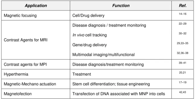
Table 1.1. Applications of superparamagnetic iron oxide nanoparticles in the biomedical field.
Application Function Ref.
Magnetic focusing Cell/Drug delivery 14–16
Contrast Agents for MRI
Disease diagnosis / treatment monitoring 22–29
In vivo cell tracking 30–32
Gene/drug delivery 29,33–35
Multimodal imaging/multifunctional 32,36–38
Contrast agents for MPI Disease diagnosis/treatment monitoring 39–41
Hyperthermia Treatment 20,21
Magnetic-Mechano actuation Stem cell differentiation; tissue engineering 17–19
Magnetofection Transfection of DNA associated with MNP into cells 42,43
Chapter 1: Hybrid magnetic-polymeric iron oxide nanoprobes for MRI: from preparation to application
7
1.4. MNP as MRI contrast agents
Magnetic Resonance Imaging (MRI) was first reported back in 1973,44 with the first contrast agent for in vivo MRI (based in manganese) being demonstrated in 1978.45 In the same year, through nuclear magnetic resonance (NMR) studies, Ohgushi et al.46 discovered the ability of iron oxide nanoparticles (crosslinked with dextran) to shorten the T2 relaxation time of water and showed that they were more efficient (by one or more orders of magnitude) than the paramagnetic ions or free radical contrast agents in relaxing neighboring nuclei.46 During the 1980’s iron oxide MNP were then demonstrated to produce contrast in in vivo MRI.47,48,49 From then on, iron oxides have been extensively used as MRI contrast agents and, in 1995, the first iron oxide MNP-based MRI contrast agent (Ferumoxides, from Guerbet, Advanced Magnetics) was approved by the American Food and Drug Administration (FDA) for human use in liver imaging.50 While many other iron oxide MNP commercial agents have appeared in the last years, some have been withdrawn from market. By now, the only approved iron oxide MNP for pharmaceutical use is Ferumoxytol,51 for the treatment of anemia, although this MNP also has properties as MRI contrast agent. In the meanwhile numerous research studies continue being reported in the literature seeking for improved MNP towards MRI applications.
MRI makes use of a strong permanent magnetic field, B0, which causes the magnetic moments of water protons in a tissue to align in its direction, precessing around B0 (at the Larmor frequency) and producing an equilibrium magnetization along the z-axis, Mz (with amplitude M0). By applying a radiofrequency (RF) magnetic field at the same frequency of the hydrogen protons precession and perpendicular to B0 (in the xy plane), the protons resonate (absorb energy form the RF pulse) and their magnetic moments start precessing coherently, such that the net magnetic moment is rotated to the transverse plate (Mxy) and precesses at the Larmor frequency. In practice, the RF transverse field is applied in a pulsed sequence. From the instant that the RF pulse is turned off, the magnetic moments of the protons relax back to equilibrium and this response is measured via induced currents in pick-up coils in the MRI scanner.1,52,53 The time required for the magnetic moments to relax to the equilibrium state (relaxation time), and therefore, MRI contrast, is tissue dependent.
MRI contrast is due to differences in proton density, spin-lattice relaxation time (T1, longitudinal relaxation time) and spin-spin relaxation time (T2, transversal relaxation time) of protons.
Chapter 1: Hybrid magnetic-polymeric iron oxide nanoprobes for MRI: from preparation to application
8
T2 is time constant of the exponential decay of transverse magnetization Mxy after a RF pulse, which corresponds to the amount of time for precessing magnetic moments to become randomly aligned (dephased) in the xy-plane after a RF pulse, eventually resulting in a net magnetic moment of zero in the xy plane. Dephasing of the magnetization of the precessing protons is due to magnetic interactions with each other and with other fluctuating moments in their surroundings.1,3
Since the natural variations of T1 and T2 in tissues are small, sometimes exogenous materials are used to enhance the contrast between tissues – contrast agents. Most contrast agents influence both T1 and T2 but usually their effect is more pronounced in either T1 or T2.
T1 contrast agents increase the MRI signal intensity, providing positive contrast enhancement in T1-weighted MR images (lighter image regions), while T2 contrast agents decrease signal intensity resulting in negative contrast in T2-weighted images (darker image regions). The current clinical contrast agents are based on paramagnetic chelates of lanthanide metals such as gadolinium, which is a T1 agent.51 The presence of paramagnetic ions near water protons shortens their T1 relaxation time through coordination with water molecules providing increased contrast. The short blood circulation times, poor detection sensitivity and toxicity concerns of gadolinium chelates had led to the continued development of superparamagnetic iron oxide-based T2 MRI contrast agents.54 Due to their larger magnetic moments, MNP-based MRI contrast agents produce higher relaxation rates at lower doses than paramagnetic ions like Gd3+.1 The low toxicity of iron, which is normally processed through various metabolic pathways, makes these agents very attractive.55
Iron oxide MNP are mainly T2 contrast agents, enhancing contrast by inducing a pronounced decrease in T2 along with a less pronounced decrease in T1. When MNP are present in the tissues and are subjected to an external magnetic field, their large magnetic moments align with it, consequently creating gradients of magnetic fields in the tissues, i.e., local inhomogeneities in the net magnetic field, through which the water protons diffuse. The dipolar coupling between magnetic moments of water protons and the magnetic moments of MNP causes dephasing of the protons magnetic moments, thereby shortening their T2 relaxation time. Due to localized differences in the uptake of the MNP by tissues, there will be regions of different MRI signal intensity, with less intensity (darkening) in the vicinity of MNP.
The addition of a MNP contrast agent causes an increase in the longitudinal (1/T1) and transversal (1/T2) relaxation rates of the water protons. The relaxation rate in the presence of MNP depends linearly on the concentration of the MNP and is given by
Chapter 1: Hybrid magnetic-polymeric iron oxide nanoprobes for MRI: from preparation to application
9
Relaxivity is defined as the slope of the above linear relation and is a measure of the efficacy of the MNP as MRI contrast agent since it defines the ability of a fixed concentration of MNP to increase the relaxation rate of the protons.
Although MNP have been used mainly as T2 contrast agents, it is possible to model their characteristics so that they have an effect on T1. For example, reduction of MNP core size to diameters of less than 10 nm, are capable of producing positive contrast in T1-weighted images. However, under these conditions their T2 effects are reduced.56,57 Nonetheless, MNP-based T1-contrast agents could be an alternative to gadolinium chelates to produce contrast enhancement in tissue regions where MR signal is naturally low.
1.4.1. Structure of MNP-based nanoprobes for MRI
The quality of a MNP MRI contrast agent in vivo depends on the physiochemical properties of magnetic core but also on the MNP ability to be stealth and escape from the reticuloendothelial system so that they can circulate in blood for sufficient time to reach the target tissues and be taken up by target cells. The stability of the MNP in biological fluids (like blood) is therefore of uttermost importance.
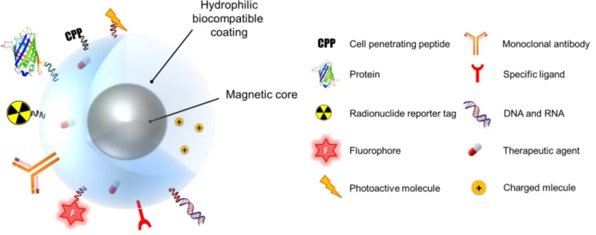
Bare iron oxide nanoparticles do not present colloidal stability at physiological pH due to the proximity of their isoelectric point (pH 6.8),58 tending to agglomerate and flocculate rapidly. Besides having neutral pH, biological fluids are complex and contain various macromolecules that readily interact with iron oxide surface and can cause colloidal instability of the nanoparticles. Also, the high surface–to-volume ratio of nanoparticles yields high surface energies which make the surface prone to oxidation, with consequences on the magnetic and relaxometric properties.1 To overcome these limitations, MNP-based MRI contrast agents are usually composed of magnetic core(s) involved in biocompatible and hydrophilic coating materials (Figure 1.3) that prevent MNP agglomeration through electrostatic and/or steric interparticle repulsions and enhance biocompatibility of the system in biological media.
MNP coatings also play significant roles in tuning MNP physiochemical properties like hydrodynamic size, magnetic core aggregation, surface charge and surface chemistry and, in particular, the magnetic59,60 and relaxometric properties61–65 of the nanoprobes. Importantly, free functional groups on the MNP wrapping molecules can be used to bind other compounds to the MNPs, such as reporter molecules for different imaging modalities,32 therapeutic agents (such as drugs, peptides, proteins, DNA/RNA),34 photoactive moieties37,66,67 and targeting moieties specific for certain cell types23 (Figure 1.3 and Table 1.1).
Chapter 1: Hybrid magnetic-polymeric iron oxide nanoprobes for MRI: from preparation to application
10
Figure 1.3. Schematic representation of MNP surface modification with biocompatible coating and
functional moieties.
1.4.2. MNP coating materials towards stabilization and functionalization of MRI nanoprobes
Depending on the chemistry of the ligand molecules present at the surface of particles after synthesis and on the purpose intended for the final particles, different coating materials and methods can be chosen. For example, it may be necessary to add new coating layers, to exchange the ligand, crosslink it or modify it with functional groups to provide particles’ stabilization and/or functionality. The most common for MNP include small organic molecules (e.g. citric acid, phosphonic acid), inorganic materials (e.g. silica) and natural/synthetic polymers natural and synthetic (e.g. dextran, poly(ethylene glycol)).
1.4.2.1. Small organic molecules
Chapter 1: Hybrid magnetic-polymeric iron oxide nanoprobes for MRI: from preparation to application
11
dimercaptosuccinic acid (DMSA),75 or methylene diphosphonic acid,76 were also used to provide increased stability to MNP synthesized in aqueous medium. Another example of the utilization of organic small molecules is the phase transfer of MNP synthesized in organic solvents. The carboxylic acid group functionality (in oleic acid) is first employed for MNP synthesis and stabilization in hydrophobic medium. Then, to render the resulting particles hydrophilic, the oleic moieties are replaced by hydrophilic small molecules with higher affinity towards iron oxide due to the presence of a larger number of iron oxide reactive groups or of groups with higher reactivity. Citric acid10,77 and DMSA78–80 are commonly employed for this purpose, leaving free carboxylic acid (and thiol groups, in the case of DMSA) at the surface of the particles to provide stability in aqueous media. The free functional groups are also useful as reactive groups for conjugation with other molecules such as targeting ligands,78 fluorophores81 or more complex polymeric constructs to engineer hybrid efficient MRI nanoprobes.82 Finally, these small organic molecules with affinity for iron oxide surfaces can be conjugated first with other molecules (e.g. PEG) to serve as anchoring moieties onto MNP. This strategy was employed to PEGylate initially hydrophobic MNPs using catechol (dopamine), dihydroxybenzamide, phosphonic or carboxylic acid moieties.61,83
1.4.2.2. Silica
Chapter 1: Hybrid magnetic-polymeric iron oxide nanoprobes for MRI: from preparation to application
12
nanoparticulate structures, such as mesopourous silica-coated MNP96,97 useful to combine imaging and drug delivery; and ultrasmall core-shell MNP with T1-weighted MRI contrast agent properties.56 Also, exploitation of the photoacoustic properties of core-shell silica-coated MNP demonstrated their potential for imaging through photoacoustic Radar imaging.98
1.4.2.3. Polymers
Along with silica-based coatings, hydrophilic polymeric coatings are preferred over small organic molecules for MNP functionalization. The main reasons for this preference are their colloidal and chemical stabilization properties and higher versatility for chemical modifications. Polymers provide colloidal stability through steric interactions established between the polymeric chains or through a combination of steric and electrostatic interactions, when charged moieties are present in the polymer. Importantly, polymeric shells also offer protection to iron oxide magnetic cores at physiological pH, which contributes for the chemical stability of the constructs. In biological terms, the polymer coating mediates the interface between the iron oxide surface and the biological medium. Therefore it dictates the way the cells “see” the nanoprobes (overall size, surface chemistry) and contributes to biodistribution and pharmacokinetics of MNP upon administration. Polymers provide MNPs with surface functionality, making possible to tailor their biological and physio-chemical properties, namely to design hybrid nanoprobes with ability to provide multimodal imaging, specific targeting, delivery and stimulated release of therapeutic agents. The MRI properties of the hybrid nanoparticles are intrinsically dependent on the interactions between the magnetic dipole created by the iron oxide core and the water protons in the vicinity. In particular, the magnitude of MRI relaxivity depends on the number of water molecules disturbed by the magnetic field generated by the MNPs. The presence of a hydrophilic polymer is of uttermost importance as it mediates the access of water molecules to the magnetic core. Manipulation of parameters such as the hydrophilicity65 and the thickness99 of the coating and the aggregation degree of magnetic cores surrounded by the polymer100 can affect the MRI properties of a hybrid MNP.
Chapter 1: Hybrid magnetic-polymeric iron oxide nanoprobes for MRI: from preparation to application
13
using a linker ligand between the MNP surface and the polymer or (b) replacement of the ligands initially at the MNP surface by a polymer bearing iron oxide anchoring ligands. The later strategy can be employed when the MNP as-synthesized are hydrophobic and solubilization into aqueous medium is performed by ligand-exchange reaction.65
1.4.3. Types of polymers used to coat MNP-based MRI nanoprobes
From the large number of polymeric materials described in the literature to produce hybrid MRI nanoprobes, two main groups can be identified: polymers with biological origin (biopolymers, in particular polysaccharides) and synthetic polymers. Table 1.2, Table 1.3 and Table 1.4 present examples of the utilization of polysaccharides and synthetic polymers to produce MNP-based MRI nanoprobes.
Polysaccharides are one of the three types of biopolymers found in Nature (polynucleotides, polypeptides and polysaccharides). The abundance of polysaccharides in Nature allied with the advances towards low cost and greener extraction/production processes have increased the interest in exploitation of polysaccharide materials for a range of applications, including nanotechnology. These biopolymers present favorable characteristics and biological properties that make them versatile materials to employ as coating materials for MNP to be used in biomedical applications. Polysaccharides are water soluble, biocompatible and biodegradable, which is crucial for clinical application in humans because health regulatory agencies demand that, besides being biocompatible, materials shall be biodegradable upon administration.50 In addition to these advantages, polysaccharides generally have biological activity, as most of them are present in structural tissues of living organisms, and can be involved in molecular recognition mechanisms.101–104 Also, they naturally present a large number of functional groups in their chains, which can serve as anchoring points onto MNP and as reactive groups for modification.
However, polysaccharides can have high degradation rates and sometimes need to be combined with other polymers or crosslinked to reduce degradation rates and enhance stability in biological environment.105 That is the case of CLIOs (cross-linked iron oxide particles), for which carboxymethyl groups were added to the dextran coating and cross-linked to epichlorohydrin to increase stability of the MNP.106 Optimal performance of polysaccharides is a challenge because synthesis of natural polymers is carried out in living organisms and thus is not strictly controlled. The alteration in structural properties of the polymers during production is difficult as well as the strict reproducibility of the polymer structure from batch to batch.
Chapter 1: Hybrid magnetic-polymeric iron oxide nanoprobes for MRI: from preparation to application
14
synthesis and modifiable properties, which potentially facilitates reproducibility and production scale-up. Indeed, polymer synthesis methodologies are well studied and are controllable, making possible to systematically add chemical modifications and functionalities on the polymer during its synthesis or to combine different polymers to obtain new materials (e.g. block copolymers) with tailor-made properties.108–113 Synthetic methods such as living radical polymerization (atom-transfer radical-polymerization (ATRP) and reversible addition– fragmentation chain-transfer polymerization (RAFT)),110 ring opening polymerization (ROP)111 and polymerization induced self-assembled approach (PISA)114 can be used to produce a virtually infinite number of tailor-made polymers with specific properties, including well defined molecular weights, polydispersity, and engineered modes of attachment to the MNPs115 that may facilitate the control of iron oxide cores aggregation to form single or multi-core MNPs. With these strategies, innovative hybrid multifunctional nanoprobes are emerging but there is still lack of in vitro and in vivo studies compared to polysaccharide-coated MNPs.
To get the best of both worlds, there is also the possibility to employ mixed coatings that combine natural and synthetic polymers, for example by covalent coupling116 or by formation of copolymers.35
1.4.3.1. MNP-polysaccharide hybrid MRI nanoprobes
Polysaccharides consist of repeating units of mono or disaccharides linked by glycosidic bonds to form linear or branched chain structures. Due to the high variability of building block composition, type of branching, molecular weight of the polymer and eventual combination with proteins (peptidoglycans) or lipids (glycolypids), polysaccharides have diverse biological and physico-chemical properties that are interesting from a biomedical perspective.
Chapter 1: Hybrid magnetic-polymeric iron oxide nanoprobes for MRI: from preparation to application
15
magnetic iron oxide nanoparticles, or after the particles are synthesized, through addition of the polymer and conjugation to the MNP by adsorption or chemical bonding.
16 Cha pte r 1: Hy bri d m ag ne tic -p oly m eri c i ro n o xid e n an op ro be s fo r M RI: fro m p re pa ra tio n t o a pp lic ati on
Table 1.2. Utilization of biopolymers extracted from natural sources in the development of MNP-based MRI contrast agents. (continued)
Biopolymer Biological source
MNP synthesis method
MNP-biopolymer assembly strategy
Biopolymer or MNP modification /
functionalization Intended purpose Ref.
Gum Arabic Acacia senegal and seyal trees
Co-precipitation
Post-synthesis
adsorption Citrate-modified cyclodextrin Magnetic drug targeting for hydrophobic drugs 123 Post-synthesis
adsorption (ultrasounds
and vortex) Rhodamine B
Simultaneous magnetic targeting and in vivo imaging
of brain tumor cells
124
Post synthesis covalent
conjugation -
MNP stabilization for biomedical and
biotechnological purposes
125,126
Fucoidan Brown seaweed Co-precipitation Adsorption onto carboxymethyldextran-coated MNP
Fucoidan amination with diaminopropane
In vivo imaging of activated
platelets for detection of intraluminal thrombus and aneurisms
102,127
Chitosan Crustaceans shell Co-precipitation
Post-synthesis
adsorption (ultrasounds and vortex)
Carboxymethylation of chitosan; rhodamine isothiocyanate (RITC); folic acid
Cancer-specific targeting, detection and imaging (fluorescence and MRI)
128
Sonochemical Post-synthesis adsorption (ultrasounds) MNPs mixed with Poly-L-Lysine solution prior to administration. In vivomesenchymal stem cells tracking of human 129
Heparin
Animal tissues (extracellular matrix)
Co-precipitation Post-synthesis adsorption (stirring,
ultrasounds) Fluorescein isothiocyanate (FITC)
In vivo imaging of human
mesenchymal stem cells 103
Co-precipitation Post-synthesis adsorption onto APTES-coated MNP
Photosensitizer pheophorbide-A (PheoA) covalently conjugated to heparin
Simultaneous in vitro
photodynamic therapy and dual-mode fluorescence/MRI imaging of cancer cells
66
Starch Green plants
Co-precipitation under high pressure homogenization conditions Post-synthesis adsorption
Red fluorescent dye DY-555– N-hydroxysuccinimide ester covalently attached to the MNP’s starch coating followed by poly-D-lysine adsorption
MRI tracking of adipose tissue-derived progenitor cells
130
Co-precipitation In situ coating
Human hepatocellular carcinoma cell line homing peptide (A54) labeled with 5-carboxyl-fluorescein
Biomolecular-targeted diagnostics and therapeutics of human tumor
17 C ha pte r 1 : H yb rid m ag ne tic -p oly m eri c i ro n o xid e n an op ro be s f or M RI: fr om p re pa ra tio n t o a pp lic ati on
Table 1.2. Utilization of biopolymers extracted from natural sources in the development of MNP-based MRI contrast agents. (continued)
Biopolymer Biological source
MNP synthesis method
MNP-biopolymer assembly strategy
Biopolymer or MNP modification /
functionalization Intended purpose Ref.
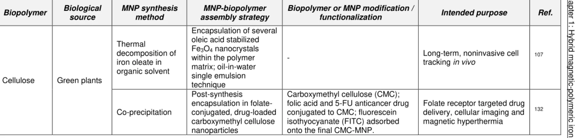
Cellulose Green plants
Thermal
decomposition of iron oleate in organic solvent
Encapsulation of several oleic acid stabilized Fe3O4 nanocrystals
within the polymer matrix; oil-in-water single emulsion technique
- Long-term, noninvasive cell tracking in vivo 107
Co-precipitation
Post-synthesis encapsulation in folate-conjugated, drug-loaded carboxymethyl cellulose nanoparticles
Carboxymethyl cellulose (CMC); folic acid and 5-FU anticancer drug conjugated to CMC; fluorescein isothyocyanate (FITC) adsorbed onto the final CMC-MNP.
Folate receptor targeted drug delivery, cellular imaging and magnetic hyperthermia
132
Table 1.3. Utilization of biopolymers obtained by biotechnological processes in the development of MNP-based MRI contrast agents. (continued)
Biopolymer Biological source MNP synthesis method
MNP-biopolymer assembly strategy
Biopolymer or MNP modification
/ functionalization Intended purpose Ref.
Dextran
Lactic acid bacteria, such as
L. mesenteroides, L. brevis and S. mutants
Co-precipitation
In situ coating
Carboxylmethylation of dextran on MNP; bombesin peptide covalently conjugated onto carboxymethyl dextran-coated MNP
Targeting and imaging of
breast cancer cells 121
Co-precipitation in presence of glucose
Folic acid covalently bond to glu-dex-MNP
Diagnosis and monitoring of treatment response of rheumatoid arthritis
28
Co-precipitation Diethylamino ethyl-modified dextran was employed to tune
MNPs charge; FITC Stem cell tracking
122
18 Cha pte r 1: Hy bri d m ag ne tic -p oly m eri c i ro n o xid e n an op ro be s fo r M RI: fro m p re pa ra tio n t o a pp lic ati on
Table 1.3. Utilization of biopolymers obtained by biotechnological processes in the development of MNP-based MRI contrast agents. (continued)
Biopolymer Biological source MNP synthesis method
MNP-biopolymer assembly strategy
Biopolymer or MNP modification
/ functionalization Intended purpose Ref.
Co-precipitation
In situ coating;
alginate was cross-linked with ferrous ions, precipitated with sodium hydroxide and oxidized to iron oxide
-
Tracking of implanted alginate microcapsules with
encapsulated rat myoblast recombinant cells.
135
Pullulan Aureobasidium pullulans (fungus) Co-precipitation
In situ coating Ethylenediamine and succinic anhydride. In vitrobone marrow-derived rat magnetic labeling of mesenchymal stem cells.
136
Co-precipitation Post synthesis coating by adsorption Cross-linked pullulan chains, with glutaraldehyde In vitrofibroblasts labeling of human 105
Hyaluronan (or
hyaluronic acid)137
Streptococci group
A and C;138
Bacilus subtilis
Co-precipitation in presence of dextran
Post-synthesis covalent coupling onto aminated MNP
Dextran coating was cross-linked and aminated with NH4OH;
Doxorubicin bound to hyaluronan-coated MNP through hydrazone linkage
Targeted drug delivery and bimodal imaging (MRI and fluorescence) of ovarian cancer cells expressing CD44 cell surface marker.
101 Commercial oleic-acid stabilized hydrophobic MNP (Sigma-Aldrich) Post-synthesis; encapsulation in hyaluronan micelles by probe-type ultrasonication method
Acylation (oleil-modification) of hyaluronan
Selective in-vitro cytotoxicity
towards cancer cells and imaging of tumor tissues
139
One pot hydrothermal synthesis in the presence of PEI
Post-synthesis covalent conjugation onto PEI-stabilized MNP
PEI stabilized-MNP previously
labeled with FITC. Imaging of surgically induced endometriosis model in rats 140,141
Thermal decomposition in of iron precursor in organic solvent
Post-synthesis electrostatic interactions with the ligand a t the surface of the MNP and coordination with MNP surface
Hyaluronan conjugated with dopamine; MNP solubilized with cetyltrimethylammonium bromide (CTAB)
Imaging of CD44 overexpressing in cancer-associated angiogenesis
19 C ha pte r 1 : H yb rid m ag ne tic -p oly m eri c i ro n o xid e n an op ro be s f or M RI: fr om p re pa ra tio n t o a pp lic ati on
Table 1.3. Utilization of biopolymers obtained by biotechnological processes in the development of MNP-based MRI contrast agents. (continued)
Biopolymer Biological source MNP synthesis method
MNP-biopolymer assembly strategy
Biopolymer or MNP modification
/ functionalization Intended purpose Ref.
Mannan Saccharomyces cerevisae Co-precipitation
Post-synthesis
adsorption -
In vitro and in vivo
mannose-mediated targeted imaging of macrophages
143
Post-synthesis
adsorption Carboxylation of mannan
Imaging of lymph node through MNP targeting to immune cells.
144,145
In situ coating - Imaging of rabbit atherosclerotic aortic wall 146