Universidade do Minho
Escola de Engenharia
Luis Manuel Fonseca Amaral
UMinho|20
14
Luis Manuel F
onseca Amar
al
Mesenchymal Stem Cell-based Differentiation
of Smooth Muscle Cells
Mesench ymal Stem Cell-based Dif ferentiation of Smoo th Muscle Cells
Dissertação de Mestrado
Mestrado Integrado em Engenharia Biomédica
Ramo de Engenharia Clinica
Trabalho efetuado sob a orientação do
Prof. Dr. Willi Jahnen-Dechent
e co-orientação do
Prof. Miguel Gama
Universidade do Minho
Escola de Engenharia
Luis Manuel Fonseca Amaral
Mesenchymal Stem Cell-based Differentiation
of Smooth Muscle Cells
Declaration
Nome: Luis Manuel Fonseca Amaral
Endereço eletrónico: [email protected] Número do Bilhete de Identidade: 13559292
Título dissertação: Mesenchymal Stem Cell-based Differentiation of Smooth Muscle Cells Ano de conclusão: 2014
Orientador: Prof. Dr. Willi Jahnen-Dechent Co-Orientador: Prof. Miguel Gama
Designação do Mestrado: Mestrado Integrado em Engenharia Biomédica Ramo: Engenharia Clínica
É AUTORIZADA A REPRODUÇÃO INTEGRAL DESTA DISSERTAÇÃO APENAS PARA
EFEITOS DE INVESTIGAÇÃO, MEDIANTE DECLARAÇÃO ESCRITA DO INTERESSADO,
QUE A TAL SE COMPROMETE;
Braga, ____/____/________
Acknowledgements
This work represents the end of my academic journey. And finally, it is time to express my sincere thanks to all of those that somehow have contributed throughout this journey, especially in this thesis.
I would like to express my gratitude to Prof. Dr. Willi Jahnen-Dechent for give me the opportunity to develop my master project in Aachen Uniklinik and for being always available for me.
I would like to acknowledge Pr. Dr. Sabine Nuess-Stein for all the help, support, knowledge and availability during my time in Aachen. Her enthusiasm about my project and all the ideas and suggestions were a great incitement during all the steps of this work.
I would like to thank Prof. Miguel Gama for always supported me to do my master thesis in Aachen, for his encouraging advices and supervising.
To Norina Labude, for the guidance, for all the help during this project and for the patient and availability to explain all my doubts. Thank you for giving me strength and encouragement.
I want also to thank Dr. Ing. Petra Mela and Ricardo for all the support, advices and ideas. To all researchers and staff I met in Aachen Uniklinik, I would like to thanks the good environment and help they provided throughout this work.
To my friends, which start this journey with me and since the beginning walked by my side. For all the experiences we shared together, talks, laughs and cries. You turn these five years the best years of my life and I couldn't have done without you.
To Cristina, for all the support and patient she has given to me. You never let me put my arms down and given me strength when I was most weak. You were the pillar that prevented me to fall. You know how important you are to me.
To my family, especially to my grandparents that always invested in me and wanted to see me graduated and finally, I almost can give them that present. They always had spoiled me with homemade sweets and motivation. To Bruno and my brothers thank you for always been there for me. Everything I am I owe to them. To my parents that always have believed in me, and never let me give up of anything. Thank you so much for everything you have done for me. I don’t have words to describe how grateful I am.
Abstract
Valvular heart disease is a major health and socioeconomic problem worldwide with approximately 300 000 valve replacements performed annually. Tissue-engineered heart valves with repair and remodelling capabilities could overcome the limitations of today’s valvular prostheses. One major limitation to this approach has been finding a reliable source of smooth muscle cells (SMC) because biopsies of these cells can be impractical and morbid as also present limited replicative capacity. The ideal cell source should be harvested in a non-or minimally invasive way and should deliver an initial high number of cells in order to drastically reduce the time needed for cell expansion. For these reasons there are many studies endeavoured to explore whether functional SMC could be generated from various types of adult mesenchymal stem cells (MSC): Umbilical cord (UC-MSC), bone marrow (BM-MSC), adipose derived (AD-MSC) and chorionic villi (CV-MSC) as possible sources for heart valve therapy. The MSC from different sources were isolated and expanded of healthy and different donor. In this study an isolation protocol was established for the CV-MSC, because it was never performed on this research group. Since the CV-MSC are barely reported in studies, this MSC source was characterized by flow cytometry to compare with other sources that are well-characterized in literature. After that, the MSC were differentiated by culture them in a culture medium containing transforming growth factor-β1 (TGF-β1) and bone morphogenetic protein 4 (BMP4), based on a published 7-day differentiation protocol. The differentiation was analysed using 4 different smooth muscle markers (α-SMA, SM22α, Calponin and SM-MHC) by immunofluorescence (IF) and immunohistochemistry (IHC). A more extensively analysis was performed by flow cytometry using the smooth muscle markers and the other markers used on different studies to characterize the MSC population for a complete phenotype characterization before and after differentiation. The IF showed promising results as the smooth muscle markers stained positive for the differentiated MSC and negative for the undifferentiated MSC. On the other hand, the IHC and flow cytometry show some contradicting results for differentiation, since the expression is not consistent within the undifferentiated and differentiated MSC. These results highlight the concept that MSC represent an easily accessible, novel cell source for heart valve therapy, but despite of the wide experiments and results in this work, it is necessary further research in this field due to the conflicting evidence and inadequate information about several cell surface markers.
Resumo
As doenças valvulares cardíacas são um problema social, económico e de saúde grave no mundo, com a realização de aproximadamente 300 000 cirurgias para substituição de válvulas cardíacas. A Engenharia de Tecidos na investigação e desenvolvimento de células cardíacas com capacidades para reparar e substituir válvulas cardíacas, pode vir a ultrapassar as limitações das atuais próteses valvulares. Contudo uma das maiores dificuldades desta investigação, tem sido encontrar uma fonte válida de células do músculo liso. A fonte ideal de colheita de células deve resultar de uma técnica minimamente invasiva e deve disponibilizar inicialmente um número elevado de células de modo a reduzir drasticamente o tempo necessário para a proliferação. Por estas razões há muitos estudos em curso de modo a investigar se as células do músculo liso podem ser obtidas a partir de vários tipos de células mesenquimatosas adultas (MSC): células do cordão umbilical (UC-MSC), medula óssea (“BM-MSC), tecido adiposo (AD-MSC) e vilosidades coriónicas (CV-MSC), como possíveis fontes para a terapêutica de válvulas cardíacas patológicas. As MSC de diferentes fontes, foram isoladas e cultivadas a partir de diferentes dadores saudáveis. Neste estudo foi estabelecido um protocolo independente para as células CV-MSC porque nunca tinha sido realizado neste grupo de investigação. Uma vez que as células CV-MSC são raramente descritas em estudos, esta fonte de MSC foi caracterizada por citometria de fluxo, para possível comparação com outro tipo de MSC que estão bem caracterizadas na literatura. Posteriormente, as MSC foram diferenciadas, isoladas e cultivadas num meio de cultura contendo o factor de crescimento TGF-β1 e BMP4 baseado num protocolo de diferenciação de 7 dias. A diferenciação foi analisada usando quatro marcadores diferentes de músculo liso (α-SMA, SM22α, Calponin and SM-MHC) através de imunofluorescência (IF) e imunohistoquímica (IHC). Para uma caracterização completa do fenótipo antes e depois da diferenciação, foi também realizada por citometria de fluxo uma análise mais extensa usando os marcadores de músculo liso e os outros marcadores utilizados por diferentes estudos na caracterização das MSC. A IF mostra bons resultados uma vez que as células diferenciadas mostraram ser positivas para os marcadores musculares enquanto que as células não diferenciadas não mostraram evidência dos marcadores. Por outro lado, a IHC e a citometria mostraram resultados contraditórios para a diferenciação, uma vez que a expressão não foi consistente nas MSC não diferenciadas e diferenciadas. Os resultados mostram que que as MSC são facilmente acessíveis e podem ser um boa alternativa para tratamento de válvulas cardíacas e apesar de se ter realizado uma análise extensiva, é necessário mais investigação para clarificar alguns pontos poucos claros que ficaram para responder em relação aos marcadores.
Table of Contents
Acknowledgements ... III Abstract ... V Resumo ... VII Table of Contents ... IX List of Abbreviations ... XIII List of Figures ... XV List of Tables ... XIX
Chapter I – General Introduction ... 1
1. Aim of Studies ...3
2. Smooth Muscle Cells ...4
3. Heart Valves ...5
4. Tissue Engineering ...6
5. Stem Cell Therapy ...7
5.1 Stem Cells Basics ...7
5.2 MSC – Definition And Sources ...10
5.3 MSC – Niche Characterization and Homing Ability ...12
5.4 MSC – Morphological and Phenotypic Characteristics ...13
6 MSC – Candidate Cells for Differentiation ...14
6.1 MSC – BM-MSC Properties and Characteristics ...15
6.2 MSC – AD-MSC Properties and Characteristics ...16
6.3 MSC – UC-MSC Properties and Characteristics ...17
6.4 MSC – CV-MSC Properties and Characteristics ...18
7. Differentiation Into Smooth Muscle Phenotype ...20
Chapter II – Materials and Methods ... 21
1. Materials ... 23
1.1 Equipment ...23
1.2 Glass Ware ...24
1.4 Chemicals ...25
1.5 Solvents ...25
1.6 Cell culture ...25
1.7 Kits ...26
1.8 Cells ...26
1.9 Primary antibodies for IHC and IF ...27
1.10 Primary antibodies for Flow cytometry ...27
1.11 Secondary Antibodies ...29
1.12 Software ...29
2. Methods ... 29
2.1 Cell Culture Mediums ...29
2.2 Solutions Preparation ...30
2.3 Cell Counting With CASY-1® ...32
2.4 Isolation And Culture Of Human Mesenchymal Stem Cells ...33
2.4.1 Isolation and Culture of Human UC-MSC ...33
2.4.2 Isolation and Culture of Human BM-MSC ...33
2.4.3 Isolation and Culture of Human CV-MSC ...34
2.5 Thawing And Freezing Cells ...35
2.5.1 Freezing ...35
2.5.2 Thawing ...36
2.6 FISH Test ...36
2.7 Induction of Smooth Muscle Differentiation ...36
2.8 IF and IHC Staining ...37
2.8.1 Immunofluorescence ...37
2.8.2 Immunohistochemistry ...38
2.9 Preparation of Cells for Flow Cytometry ...39
2.10 Flow Cytometry ...42
Chapter III – Results and Discussion ... 45
1. Isolation and Characterization of CV-MSC ... 47
1.1 CV-MSC Morphology ...47
1.2 FISH Analysis ...49
1.3 Phenotype Analysis ...50
2 Smooth Muscle Differentiation ... 53
2.1 Immunohistochemistry Analysis ...54
2.3 Flow Cytometry Analysis ...57
2.3.1 Human Umbilical Vein Endothelial Cells ...58
2.3.2 Undifferentiated and Differentiated UC-MSC ...58
2.3.3 Undifferentiated and Differentiated BM-MSC ...60
2.3.4 Undifferentiated and Differentiated CV-MSC ...61
2.3.5 Undifferentiated and Differentiated AD-MSC ...61
2.3.6 MSC Smooth Muscle Phenotype Characterization ...62
Chapter IV – Conclusions and Future Remarks ... 65
Chapter V – References ... 69
Chapter VI – Appendixes ... 79
Appendix A – IF and IHC Experiments Plan ...81
Appendix B – Flow Cytometry Experiments Plan ...83
Appendix C – Flow Cytometry Plan ...85
Appendix D – Flow Cytometry Results for a CV-MSC donor ...88
Appendix E – Surface Markers Categories ...89
Appendix F – Immunohistochemistry MSC Pictures ...90
Appendix G – Immunofluorescence MSC Pictures ...93
List of Abbreviations
AB – Antibody
AD-MSC – Adipose Derived Mesenchymal Stem Cells APC – Allophycocyanin
Aqua dest. – Distilled H2O ASC – Adult Stem Cell
BM-MSC – Bone Marrow Mesenchymal Stem Cells BMP4 – Bone Morphogenetic Protein-4
BSA – Bovine Serum Albumin C6H8O7 – Citric Acid Monohydrate
CD – Cluster Differentiation or Cluster of Determinate CH – Switzerland
CVDs – Cardiovascular Diseases
CV-MSC – Chorionic Villi Mesenchymal Stem Cells DAPI – 4',6-diamidino-2-phenylindole
DE – Germany DK – Denmark
DMEM – Dulbecco ́s Modified Eagle ́s Medium DMSO – Dimethyl Sulfoxide
ECM – Extracellular Matrix
EDTA – Ethylenediaminetetraacetic Acid ESC – Embryonic Stem Cell
FBS – Fetal Bovine Serum
FGF-Basic – Basic Fibroblast Growth Factor FISH – Fluorescence In Situ Hybridization FITC – Fluorescin
HCL – Hydrochloric Acid
HSC – Hematopoietic Stem Cells
HUASMC – Human Umbilical Artery Smooth Muscle Cells HUVEC – Human Umbilical Vein Endothelial Cells
IF – Immunofluorescence IHC – Immunohistochemistry
IL – Israel
iPSC – Induced Pluripotent Stem Cells JP – Japan
M – Molar mM – Millimolar
MSC – Mesenchymal Stem Cells NL – Netherlands
ºC – Degree Celsius
PBS – Phosphate Buffered Saline PE – R-Phycoerythrin
PE-Cy™7 – Is a tandem fluorochrome that combines PE and a cyanine dye Pen Strep – Penicillin Streptomycin
PerCP – Is a component of the photosynthetic apparatus
PerCP-Cy™5.5 – Is a tandem conjugate that combines PerCP with a cyanine dye PFA – Paraformaldehyd
RT – Room temperature
SM-MHC – Monoclonal Anti-Myosin (Smooth) antibody produced in mousse SMCs – Smooth Muscle Cells
TGF-β1 – Transforming Growth Factor-β1 UC – Umbilical Cord
UC-MSC – Umbilical Cord Mesenchymal Stem Cells UK – United Kingdom
USA – United States of America vWF – Von Willebrand factor
List of Figures
Figure I.1 - Tissue Engineering strategies, adapted from [24]. ... 6
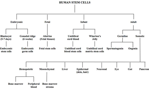
Figure I.2 – Human stem cell classification regarding their origin, adapted from [27]. ... 9
Figure I.3 – The mesengenic process: cellular transitions from MSC to differentiated cells, adapted from [43]. ... 11
Figure I.4 – Stem cells niches and their oxygen tension levels, adapted from [46]. ... 12
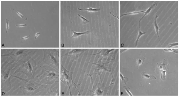
Figure I.5 - Morphologically distinct MSC in culture. A to C: Elongated spindle shaped cells. D to F: Flattened polygonal cells with short or no branches (adapted from [49]). ... 13
Figure I.6 – Bone marrow microenvironment harbouring both MSC and HSCs compartments, adapted from [29]. ... 15
Figure 7 – Example of a cross section, adapted from [59]. ... 17
Figure I.8 – Schematic Structure of a Human Placenta, adapted from [63]. ... 19
Figure II.1 – CASY-1 Electronic Cell Counter. ... 32
Figure II.2 – Schematic overview of CV-MSC isolation. In the picture 1 is visible a placenta after processing. Picture 2 and 3 shows pieces of the processed placenta, the tissue was weight and than extensively minced. The picture 4 shows the processed placenta tissue at an incubator shaker. ... 34
Figure III.1 – Pictures of a donor of CV-MSC in different passages, with a resolution of 50. From A to C represents the passage 0 of the CV-MSC during 18 days. Picture A was taken after 5 days of isolation, picture B was taken after 12 days and picture c after 18 days. The cells on picture D are on passage 2, on picture E are on passage 4 and on picture F are on passage 6. ... 48
Figure III.2 – Fluorescence Images of FISH Analysis of CV-MSC from Twin Babies. Twin 1 Is the Baby Boy and the Twin 2 is the Baby Girl. ... 49
Figure III.3 – Some results of flow cytometry analysis of the expression of surface markers for a CV-MSC donor. ... 50
Figure III.4 – Expression of SMC differentiation markers in MSC. ... 53
Figure III.5 – SMC -specific proteins. IHC staining of a BM-MSC donor under differentiation conditions during 7 days (n=3). ... 54
Figure III.6 – SMC-specific proteins. IF staining of a BM-MSC donor under differentiation conditions during 7 days (n=3). ... 56
Figure III.7 – Heat map analysis of flow cytometry data showing different expression of cell surface markers between the undifferentiated (U) MSC, differentiated (D) MSC (n=3) and HUVEC (n=2). ... 59
Figure III.8 – Heat map analysis of flow cytometry data showing different expression of smooth muscle cell markers between the undifferentiated MSC, differentiated MSC, HUVEC and HUAMSC (n=2). ... 63
Figure D.1 – Remaining flow cytometry results of the expression of surface markers for a CV-MSC donor (n=3). ... 88
Figure F.1 – SMC-specific proteins. IHC staining of an AD-MSC donor under differentiation conditions during 7 days (n=3). ... 90
Figure F.2 – SMC-specific proteins. IHC staining of a CV-MSC donor under differentiation conditions during 7 days (n=3). ... 91
Figure F.3 – SMC-specific proteins. IHC staining of an UC-MSC donor under differentiation conditions during 7 days (n=3). ... 92
Figure G.1 – SMC-specific proteins. IF staining of an AD-MSC donor under differentiation conditions during 7 days (n=3). ... 93
Figure G.2 – SMC-specific proteins. IF staining of a CV-MSC donor under differentiation conditions during 7 days (n=3). ... 94
Figure G.3 – SMC-specific proteins. IF staining of an UC-MSC donor under differentiation conditions during 7 days (n=3). Appendix H – Flow Cytometry Analysis of Different MSC sources. ... 95
List of Tables
Table I.1 – Potential US patient populations for stem cell-based therapies, adapted from [28]. ... 8
Table I.2 – Summary of the markers expression for MSC, adapted from [50, 52]. ... 14
Table I.3 – Expected results of surface markers for BM-MSC and AD-MSC, adapted from [52, 61]. . 18
Table II.1 – Interpretation of the Markers used for flow cytometry. ... 39
Table III.1 – Results of flow cytometry analysis of three different donors of each MSC source (n=3).51
Table C.1 – Flow cytometry plan for undifferentiated and differentiated AD-MSC. ... 85
Table C.2 – Flow cytometry plan for HUVEC and undifferentiated and differentiated UC-MSC, BM-MSC and CV-MSC. ... 86
Table C.3 – Flow cytometry plan for HUASMC. ... 87
Table E.1 – Commonly expressed markers on MSC. The markers in red are variably reported on literature, from being expressed to not being expressed, adapted from [51-54, 66]. ... 89
Table H.1 – Results of flow cytometry analysis for MSS phenotype characterization of the undifferentiated MSC (n=3) and HUVEC (n=2). ... 96
Table H.2 – Results of flow cytometry analysis for MSS phenotype characterization of the differentiated MSC (n=3). ... 97
Table H.3 – Results of flow cytometry analysis for the smooth muscle markers of the undifferentiated and differentiated MSC, HUVEC and HUASMC (n=2). ... 98
Chapter I
General Introduction
1. Aim of Studies
This master thesis comprises a comparative study of four different sources of mesenchymal stem cells (MSC) as potential candidates for heart valve therapy. It comprehends an interdisciplinary work merging the subjects of Biomedical Engineering and Stem Cell Biology in order to overcome, through cellular therapy, one of the major problems among the cardiovascular diseases. The main objective of this project is to study and characterise MSC from different sources with regard to the establishment of a protocol for smooth muscle differentiation. The four different human mesenchymal cells used were (i) mesenchymal stem cells from umbilical cord matrix (UC-MSC), (ii) mesenchymal stem cells from bone marrow (BM-MSC), (iii) mesenchymal stem cells from adipose tissue (AD-MSC) and (iv) mesenchymal stem cells from chorionic villi of placenta (CV-MSC). The specific tasks of this work comprise three major goals:
• To establish an isolation protocol for chorionic villi derived human mesenchymal stem cells (CM-MSC). Characterization and comparison of MSC from the four different sources listed above. All MSC types were obtained from tissue (primary cells) and during the work all cells were expanded until passage 7 up to 8.
• To characterise the phenotype of the MSC used in this work by flow cytometry. All the cells were analysed with a group of markers between different passages. As the CV-MSC are rarely reported in studies, it was very important to perform a comparison between the CV-MSC and the well-characterised MSC: the UC-MSC, BM-MSC and AD-MSC.
• To establish a smooth muscle cell differentiation protocol using MSC from different sources. The success of smooth muscle cell differentiation was analysed by immunofluorescence and immunohistochemistry through a specific set of smooth muscle cell markers. The cells were also analysed using flow cytometry with the same group of markers used to characterise the phenotype of MSC plus smooth muscle markers. Flow cytometry was very important to analyze into details the difference between the undifferentiated and differentiated cells. All the cells used were between passages 4-7.
For analysis was used a positive and negative control, human umbilical artery smooth muscle cells (HUASMC) and human umbilical vein endothelial cells (HUVEC), respectively. The project was
executed at the AG S. Neuss-Stein, Pathology/IBMT-ZMG at Uniklinik RWTH Aachen University with collaboration of AG P. Mela (AME, Helmholtz Institute) at RWTH Aachen University.
2. Smooth Muscle Cells
Smooth muscle cells (SMC) reside in various locations throughout the human body (gastrointestinal, respiratory system, reproductive system, ocular, urinary tissues and heart) and they play an essential role in maintaining the structural and functional integrity of blood vessels. SMC have different specialized functions in each tissue in which they are found. Thus, SMC are multifunctional, for instance, they maintain the viability and phenotype of endothelium by providing physical support and regulation of blood pressure by contracting and relaxing in response to a variety of stimuli; they are responsible for the alterations in the volume and diameter of hollow organs, enabling them to generate pressure and move contents; they adjust the shape of the lens and the diameter of the pupil in the eye; they produce erections of the hairs in the body; they control the passage of food through the gut and excretion of waste products; and they determine the movement of sperm cells, eggs and the delivery of a fetus [1, 2].
The fully differentiated or mature SMC proliferates at an extremely low rate and is a cell almost completely geared for contraction. For example, fully differentiated SMC in mature blood vessels proliferate at extremely low rates and produce only small amounts of extracellular matrix (ECM) proteins. They express a unique repertoire of contractile proteins, ion channels, and signalling molecules that are required for their function and that when taken in aggregate clearly distinguish it from any other cell type [1-5].
The SMC retains remarkable plasticity, such that it can undergo relatively rapid and reversible changes in its phenotype in response to changes in the local environment. However, the plasticity of the SMC has confounded efforts to understand the cellular and molecular mechanisms that control its differentiation. A given SMC can acquire a broad spectrum of different phenotypes in response to different physiological or pathological stimuli. If an artery is injured, some SMC must be recruited to repair the injury, while at the same time the contractile function of the blood vessel must be maintained for normal cardiovascular homeostasis [1, 3, 6].
There is considerable interest in understanding the cellular and molecular regulation of the differentiation of vascular smooth muscle cells for the treatment of diseases. For example, cardiovascular diseases (CVD) are the number one cause of death globally, more people die annually
from CVD than from any other cause. An estimated 17.3 million people die annually from CVD, representing 30% of all global deaths. The number of people, who die from CVD, mainly from heart disease and stroke, will increase to 23.3 million by 2030. CVD are projected to remain the single leading cause of death [7-9]. Therefore CVD are considered more than just a health care problem because of their major consequences in terms of financial and economical impact for governments, businesses and individuals. These are a global epidemic that requires a combined action from not only the individual patients but also the pharmaceutical industry and the government. Thereby the treatments for SMC therapy have been the subject of intense research in the field of cellular therapeutics [9].
3. Heart Valves
Cardiovascular diseases represent a major worldwide health care issue. A considerable amount of these diseases is represented by heart valve failures. It’s a significant cause of morbidity and mortability; over 300 000 valve replacements are performed globally per year and the number of patients requiring heart valve replacement is expected to triple by the year 2050 [10].
Heart valve disease is characterised by damage to or a defect in one of the four heart valves: the mitral, aortic, tricuspid or pulmonary. Heart valve abnormalities are caused by birth defects, age-related changes, infections, or other conditions. The valves become too hardened to open fully, or are unable to close completely hindering the flow of blood. As the tissue of the heart cannot regenerate spontaneously, valve diseases generally necessitate surgical repair or replacement of the diseased tissue in order to avoid serious and potentially fatal cardiac or systemic consequences.
Currently used heart valve prostheses can be divided into two basic groups, namely mechanical and biological prostheses. Mechanical valves are durable but suffer risks of clot formation on their prosthetic surfaces, necessitating life-long anti-coagulant drug therapy. Bioprosthetic valves, on the other hand, have minimal risk for bleeding events but are much less durable and unsuitable for pediatric applications. These disadvantages can be overcome by valve substitutes that more closely mimic their native counterparts as to adequate mechanical function and durability, as well as the absence of immunogenic and inflammatory reactions [11-14].
Tissue engineering is a promising strategy to meet these requirements by in vitro fabrication of autologous living heart valve replacements. The final goal is to create or regenerate a living valve replacement that functions well hemodynamically, repairs on-going tissue damage, and has long-term
durability and growth potential similar to those of the natural heart valves. The most immediate need for heart valve tissue engineering and regeneration technology is in the pediatric and young adult population in which the results of valve replacement are not as favourable as those in older adults [15, 16]. However, an ideal cell source enabling the fabrication or regeneration of heart valve has not been identified yet. Ideally, the cells used to seed the developing tissues should be non-immunogenic, capable of expansion in vitro to yield increased number of cells, have the ability to provide specific cell functions, easy to harvest in a non-or minimally invasive way and should deliver an initial high number of cells in order to drastically reduce the time needed for cell expansion [17-20].
4. Tissue Engineering
Tissue Engineering has emerged as a rapidly expanding approach to address the organ shortage problem. It is “an interdisciplinary field that applies the principles and methods of engineering and life sciences toward the development of biological substitutes that can restore, maintain, or improve tissue function” [21]. Currently, tissue engineering focuses mainly on associating cells with scaffolds, in order to promote cell attachment and restrict their distribution in the tissue, direct cell distribution and differentiation, sustain large tissue losses while new tissue is formed and ultimately lead to new tissue formation [22, 23].
In 1993 Langer and Vacanti described three strategies for the creation of new tissue in vitro [21]:
1. Isolated cells or substitutes. The concept of treating injured tissues with isolated cells is regarded as cell therapy.
2. Tissue inducting substances. Bioactive molecules induce cell proliferation, differentiation and metabolic activity.
3. Cells placed on or within matrices. Associated cells and substrates provide the injured tissue with continuity, and promote cell attachment and fixation.
As proposed by Langer and Vacanti, tissue engineering may be performed by several different approaches in order to obtain tissue regeneration, but the association showed on Figure I.1 (with the association of all three elements, composing bioactive constructs) is proposed to be the most promising option for tissue engineering [24, 25]. The present work is focused on the study of mesenchymal stem cells from different sources and their differentiation towards SMC.
5. Stem Cell Therapy
Stem cell research has evolved a lot in recent years due to its therapeutic potential in dealing with many diseases in the human body, which many are incurable by established therapies. The list of diseases and injuries cited as potential targets of stem cell therapy reveals, in large measure, why stem cells offer so much hope for revolutionary advances in medicine (Table I.1). These diseases are characterized by progressive cell loss, traumas and defects that become a serious health issue, because the cells that maintain the ability to divide and differentiate into more specialized cells of different tissue types are rare in adult. In contrast, the seemingly unlimited potential of the undifferentiated cells of the early embryo has made embryonic stem cells the focus of great scientific interest. The properties of stem cells need to be understood so stem cell therapy can help the body regenerate against some serious health problems [26-28].
5.1 Stem Cells Basics
Stem cells are unspecialized cells in the human body that are capable of becoming specialized cells, each with new specialized cell functions. A stem cell is uncommitted, and remains uncommitted until it receives a signal to develop into a specialized cell. They serve as a repair system by being able to divide without limit to replenish other cells. When stem cells divide, each daughter cell has the
potential to either remain as a stem cell in the stem cell pool (niche) or become mature cell type with new special functions, such as blood cells, brain cells, etc. Their self-renewal capacity combined with their differentiation capacity, makes stem cells unique [27, 29].
Table I.1 – Potential US patient populations for stem cell-based therapies, adapted from [28]. Condition Number of Patients
Cardiovascular Diseases 58 million Autoimmune diseases 30 million
Diabetes 16 million
Osteoporosis 10 million
Cancer 8.2 million
Alzheimer’s Disease 5.5 million Parkinson’s Disease 5.5 million Burns (severe) 0.3 million Spinal-cord Injuries 0.25 million
Birth Defects 0.15 million/year
Stem cells are present within most, if not all multicellular organisms and are the ultimate drivers of growth and regeneration. They are considered to be critical biological components necessary for proper growth and development during embryogenesis and they have also been demonstrated to play indispensable roles in adult species, providing a source of cellular replenishment for every mature and differentiated cell type. All stem cells originate from the fertilized egg. As a totipotent entity, the fertilized egg has the capacity to drive the formation of all intra- and extra-embryonic tissues during growth and development. It is during the process of embryonic maturation that determination occurs, a variety of more specialized stem cell types are generated with different properties that allow the development of specific tissues and organs [27, 30].
Stem cells can either be totipotent, pluripotent, multipotent or bipotent. Pluripotent stem cells have the potential to give rise to all the cells that derive from the three embryonic germ layers, more specifically the mesoderm, endoderm, and ectoderm. The germ layers are the embryonic source of all cells in the body and are originated from the blastocyst, one of the earliest stages of the embryo development. From the inner cell mass of blastocyst before it would be implanted in the uterine wall it is possible to isolate and collect one type of pluripotent cell, the embryonic stem cell (ESC). As pluripotent stem cells, ESC have the ability to give rise to any type of cell (except extra-embryonic tissue such as the trophoblast / placenta) yielding an unlimited ability to self-renewal associated to high levels
of telomerase activity. ESC have a tremendous prospective for clinical application, yet their use in therapeutics is ethically controversial due to its origin and also their unmeasured proliferation can lead to in vivo teratoma (tumour consisting of different types of tissue caused by the development of independent germ cells) formation, which make them unsafe for the therapies [29, 31-33].
The multipotent stem cell represent an undifferentiated type of cell occurring in a specialized differentiated tissue with the ability to self-renew and give rise to all types of cells from its origin tissue. Basically these stem cells have the in vitro and in vivo ability to give rise to a limited number of specialized cells, being capable of originating multiple cell lineages or more restricted progenitor populations that are able to produce precursors and finally mature cells. They also exhibit a self-renewal capacity and contribute to the tissue homeostasis by repairing and replacing the damaged cells after an injury. Despite their limited potential regarding differentiation compared to pluripotent stem cell, multipotent cells bring more advantages when compared to pluripotent ones in the themes of ethical concerns and safety issues. Adult stem cells (ASC) can be isolated from several sources such as bone marrow (BM), blood, cornea and retina of the eye, brain, skeletal muscle, dental pulp, liver, skin, the lining of the gastrointestinal tract, and pancreas. Despite rare, difficult to identify, isolate and purify ASC are very promising for the therapeutic use. Hematopoietic stem cells (HSC), neural stem cells, endothelial progenitor cells, and mesenchymal stem cells are the most studied ones [27, 29, 31, 33]. Figure I.2 graphically elucidates the classification of all types of human stem cells.
Additionally, in 2006, Takahashi and co-workers discovered a novel type of stem cell, by reprogramming an adult mouse fibroblast back to a pluripotency-like state. In 2007, the same authors were able to reproduce the same output with human fibroblast, and named them induced pluripotent stem cells (iPSC). This breakthrough of restoring pluripotency to somatic cells was awarded with the Nobel Prize and created potentially new opportunities for modelling human diseases by preventing immunosuppression and graft rejection. Alternatively, iPSC also can allow the in vitro simulation of each individual patient’s disease enabling the screening of potential drugs and therefore developing new therapies [34-36].
5.2 MSC – Definition And Sources
The MSC definition is not 100% consensual among the scientific community. Friedenstein and their colleagues reported the first evidence of these cells in 1968 through the identification of a bone marrow cell subpopulation with osteogenic potential. Later, in end of the 80s decade the same author described these cells as plastic-adherent, fibroblast-like cells that were able to develop colony-forming units (CFU-Fs) when plated into tissue culture plates, naming them stromal cells. In 1991, Caplan proposed to name mesenchymal stem cells to all the cells capable to differentiate into all mesodermal lineage cells also known as mesenchymal tissues [37-39]. The minimum criteria for their classification include: adherence to plastic, the expression of specific set of surface markers and in vitro multilineage differentiation (bone, cartilage, fat and muscle) as it can be seen on Figure I.3 [31, 40, 41].
MSC proliferation and senescence has been considered important by scientists seeking to use them for therapeutic purposes. Ex vivo expansion of MSC is required to generate a pure cell population in an amount sufficient for the clinical indication. During this expansion process, cells enter cellular senescence, which leads to a gradual reduction of their potency and significant changes in protein expression. Despite those features, MSC can be easily expanded in culture with a capacity to retain their stemness and multilineage potential over several passages; however in vivo demonstration of all these characteristics have not been accomplished. The limited in vivo knowledge of MSC is due (but not limited) to the fact that they lack a unique specific marker, besides lacking the expression of hematopoietic markers [31, 42, 43].
Figure I.3 – The mesengenic process: cellular transitions from MSC to differentiated cells, adapted from [43].
The most in vitro features of MSC include the formation of CFU-Fs when plated into plastic tissue cultures in a basal medium, such as DMEM (Dulbecco ́s Modified Eagle ́s Medium), in the presence of animal serum, like FBS (fetal bovine serum), and have the ability of being propagated without losing their differentiation potential. MSC also have other important features: active secretion of ECM components and several cytokines, thereby having immunomodulatory and trophic properties, as well as, low immunogenicity which make them suitable for both allogeneic and autologous settings in several clinical applications. Thereby these cells have an enormous potential due to their intrinsically therapeutic features in setting-up a regenerative microenvironment releasing anti-fibrotic, anti-scaring, anti-apoptotic, mitotic and angiogenic factors [31, 42].
In adults MSC can be found in the BM compartment of long bones, iliac crest, sternum and cranium, being the major source of these cells in the human body. In those locations, a compartment of stroma-supportive cells related to the hematopoietic compartment is formed. In addition to BM, it is possible to obtain MSC population from other adult tissues such as adipose tissue, dental pulp, synovial membrane and breast milk. They also can be found in some neonatal tissues and fluids like the placenta, amniotic fluid and umbilical cord (UC): blood, perivascular cells and Wharton ́s jelly. However, depending on the sources, MSC can exhibit different features namely concerning differentiation potential and other differences related with cell processing (cell yield and purity) [42, 44-46].
5.3 MSC – Niche Characterization and Homing Ability
MSC reside in their predefined niches where upon signalling and molecular activation mobilizes, migrate or home to the locus of the inflammation and tissue injury in order to modulate the immune response. Niches are structurally tailored to meet the requirements of the resident stem cells, and the resident cells in turn play an indispensable role in architectural organization of the niche. Three different niches are known to contain MSC: the BM niche, the adipose tissue and the subventricular zone. In all tissues where MSC can be collected they are in contact with a perivascular niche in close association with blood vessels, therefore these cells are often referred as pericytes (abluminal cells in perivascular locations that are in contact with the basement membrane and surrounding endothelial including the microvasculature). Pericytes play an important role in the blood vessel stabilization by secreting an amount of different factors. The course of the perivascular niche is highlighted in the housing of MSC due to the fact that alongside with a decrease of the vascular density is associated a diminishment of viable MSC. The surrounding supporting cells, ECM and signalling molecules act in coordination to activate the naive MSC, which can then proliferate, as a consequence of lost attachment with the basement membrane and endothelial cells [44, 47, 48].
Figure I.4 – Stem cells niches and their oxygen tension levels, adapted from [46].
Although MSC are located close to vascular structures, the oxygen tension within those milieus are very low, approximately 2-8%, as it can be visualized on Figure I.4. The hypoxia is thought to contribute for MSC stemness maintenance by conserving an undifferentiated state [47].
The homing properties of MSC can further enhance their therapeutic potential. The homing process is defined as the arrest of MSC within the vasculature of a tissue followed by transmigration across the endothelium. This represents a complex process involving the niche interaction and paracrine crosstalk in order to achieve a correct MSC mobilization, niche homeostasis and
consequently a better healing process. There is evidence that host MSC are able to migrate in response to inflammation via activation of mobilizing and trafficking mechanisms that still remain muddled. However, the homing of culture-expanded MSC in comparison to leukocytes and HSC, is much less efficient taking into account the lack of important chemokine and cell-adhesion receptors by MSC, as well as the difficulty in identifying, isolating and tracking these type of cells [48, 49].
5.4 MSC – Morphological and Phenotypic Characteristics
According to several studies, MSC morphological classification depends on the shape and processes (ramifications extending from the cell body) that the cell displays, usually the MSC while plastic adherent cells in culture, exhibit architecture similar to fibroblasts, thereby being classified as fibroblast like-cells in terms of their morphology. They are divided into two major subpopulations, the first one encloses MSC with a spindle-shaped morphology and at least two processes extending in different directions from an elongated cell body (generally these MSC correspond to an immature multipotent state), and the other morphological group of MSC represents large cells with a flattened, polygonal shape and a plainly visible nucleus, commonly devoid of processes or with many small processes, being termed mature MSC, which have less potential. Figure I.5 shows the differences in MSC morphology [31, 50].
Figure I.5 - Morphologically distinct MSC in culture. A to C: Elongated spindle shaped cells. D to F: Flattened polygonal cells with short or no branches (adapted from [49]).
Although MSC lack a specific, unique surface marker able to identify and track these cells, it is possible to identify and characterize these cells immunophenotypically because they express several surface markers. However, depending on the MSC source and their different stages during cell
proliferation and culture the expression of adult MSC surface markers can differ. Adult MSC have also been shown to have non-immunogenic surface antigens, due to weakly express major histocompatibility complex class I protein (MHC I) and no MHC II molecules. The Mesenchymal Stem Cell Committee of the International Society for Cellular Therapy proposed in 2006 minimal criteria to define these cells. Besides the plastic adherence ability and multineage differentiation potential, human MSC have to express a certain group of markers and lack the expression for certain other markers. Nevertheless, extensive research needs to be done in this field, as not much is known about the phenotypic character and developmental origin. Also, there is not adequate information about the cell surface markers of adult mesenchymal stem cells and how they can be identified. The Table I.2 shows the most commonly used markers summarized by the scientific community to identify the MSC [31, 41, 51-53].
Table II.2 – Summary of the markers expression for MSC, adapted from [50, 52].
Markers Positive Negative Phenotype CD13 CD29 CD44 CD73 CD90 CD105 CD166 Stro-1 CD14 CD31 CD34 CD45 CD49d CD106 HLA-DR
6 MSC – Candidate Cells for Differentiation
As previously discussed on 1.2 Heart Valves the ideal cell source should be harvested in a non-or minimally invasive way and should deliver an initial high number of cells in non-order to drastically reduce the time needed for cell expansion. Here will be discussed the potential sources of cells that this study focused on (BM-MSC, AD-MSC, UC-MSC and CV-MSC) to evaluate which of them is most suitable for smooth muscle cell differentiation and thus for heart valve therapy.
6.1 MSC – BM-MSC Properties and Characteristics
BM is a well-organized tissue composed by a complex structure highly vascularized that shelters the basic elements from the stroma or the mesenchyma and hematopoietic systems. As it can be seen on Figure I.6, the BM is located at the center of the large bones, where MSC compartment together with the ECM elements support the haematopoiesis process. MSC have a very important role in the BM HSC niche as a stromal supporting network by producing an array of growth factors and cytokines able to modulate immune responses and regeneration processes. Notwithstanding MSC only represent 0.001 to 0.01% of BM cell population, a very low number [29, 31, 50].
Although BM has been the main source for the isolation of multipotent MSC, the harvest of BM is a highly invasive procedure and the number, differentiation potential, and maximal life span of MSC from BM decline with increasing age. Although obtaining MSC from the bone marrow is a highly invasive procedure the BM-MSC relies on one big advantage, the patient will not have any compatibility problems. The MSC from BM are the best characterized, in many studies they compare MSC from different sources to BM to see if there’s a match [31, 54, 55].
Figure I.6 – Bone marrow microenvironment harbouring both MSC and HSCs compartments, adapted from [29].
Some analysis of proliferation capacities of BM-MSC showed that they possessed the lowers population doubling numbers and the shortest culture period. BM-MSC exhibit characteristics similar to the other cell types studied in this work (UC-MSC, CV-MSC and AD-MSC), including fibroblastoid morphology, surface proteins and differentiation potential. Cultured BM-MSC have showed differentiation potential into bone, cartilage, fat and they also been shown to regenerate cardiac and
skeletal muscle. More recently, it has been suggested that BM-MSC can traverse lineage borders and differentiate into neural cells, epithelia of liver, lung, kidney, skin, pancreas and gastrointestinal tract. They also have been used with varying success to improve neurological, cardiovascular, blood related and musculoskeletal disorders [31, 50, 54, 55]. The most common markers used to characterize BM-MSC are listed on Table I.2.
6.2 MSC – AD-MSC Properties and Characteristics
Adipose tissue serves as an endocrine organ, functioning to maintain energy metabolism through the storage of lipids. Two types of adipose tissue exist, brown and white (there is another type described between brown and white, called beige fat; you should mention that), the white adipose yields the commonly studied AD-MSC. This perivascular niche where AD-MSC derive is highly vascularized, analogous to different mesenchymal cell populations identified in other tissues throughout the body. AD-MSC can be obtained by a less invasive method and in larger quantities than BM. It has been demonstrated that adipose tissue contains stem cells similar to BM-MSC, which are termed processed lipoaspirate cells. However, like BM-MSC it has the advantage to be collected at any time of donor’s life and has no problem with compatibility, but the differentiation potential, and maximal life span of AD-MSC decline with increasing age. These cells can be isolated from cosmetic liposuctions in large numbers and grown easily under standard tissue culture conditions [31, 54, 55].
AD-MSC are a very promising alternative source of MSC, due to their wide availability and ability to differentiate into other tissue types of mesoderm, they may serve a wide variety of applications. AD-MSC had been utilizes in studies addressing osteoarthritis, diabetes mellitus, heart disease, and soft tissue regeneration and reconstruction after mastectomy and facial repair. Also they represent potential candidates for cell-based therapies designed to treat cartilage defects/disorders, neurological disorders, autoimmune diseases, as well as organ transplantation outcomes [22, 56].
While the accessibility and quantity of ASC that can be recovered offers much appeal, the practical application and safety of using ex vivo expanded populations for clinical applications remains to be determined. However, what is clear is, the need to better understand the basic properties of culture-expanded AD-MSC in order to properly assess their growth, developmental potential, and capacity to modulate immune responses [56, 57]. Although the expression of surface markers is very similar to the BM-MSC, there slight differences exist between them; Table I.3 shows the most common markers used to characterize the AD-MSC in comparison with BM-MSC [58].
6.3 MSC – UC-MSC Properties and Characteristics
As the collection and isolation of BM-MSC and AD-MSC require invasive and often undesirable procurement procedures, investigators have begun to seek alternative sources of human MSC including the umbilical cord (UC) that was seen as biological waste product post childbirth. Human UC consists of three tissue components including the amniotic membrane, stroma (Wharton’s jelly), and blood vessels (two arteries and one vein) as can be seen on Figure I.7. Over the last few years, UC-MSC derived from Wharton’s jelly (WJ) have been isolated to examine their stem cell nature. Although some technical and observational variations exist between laboratories, there has been a relatively common consensus regarding the immunological and functional characteristics of UC-MSC derived from WJ. WJ is a gelatinous substance that provides insulation and protection within the umbilical cord and the cells within the WJ express several stem cells genes, therefore is a potential source of ASC. These cells are believed to be more primitive than MSC derived from more mature tissues sources and to have intermediate properties between embryonic and ASC. The expression of certain embryonic and mesenchymal stem cell markers on the naïve form of these cells brings suggestive evidence as to their multipotency [59, 60].
Figure 7 – Example of a cross section, adapted from [59].
The UC-MSC are widely reported on studies because they produce large yields of MSC and posses immunosuppressive activities, making it useful in allogeneic situations. UC-MSC cultured in vitro shared a similar fibro-blastoid morphology like the other cell types analysed in this work, and in comparison, although they have the lowest isolation rate (approximately 61%), they are available in potentially large quantities when successfully isolated. The UC-MSC have the fastest proliferation and
expansion rate and they have significantly lower expression of senescence markers. UC-MSC normally is the cell type capable of being cultured the longest [54, 55, 59, 60].
Successful in vitro and in vivo differentiation to several lineages makes these cells an invaluable stem cell source, deserving further testing as a cellular therapy or other applications in tissue engineering Therefore, the isolation and culture of UC-MSC still need better clarification to ultimately build an optimal standard procedure among laboratories, tissue banks, and clinics. This potential has ultimately led many groups to initiate the evaluation of these cells in therapeutic regimens. Even though UC-MSC still need further characterization, they have been successfully differentiated into various cell types including adipocytes, chondrocytes, osteocytes, cardiomyocytes, skeletal myocytes, hepatocytes, insulin-producing cells as well as neuron-like cells [54, 55, 59, 60]. The most common markers used to characterize UC-MSC are usually the same to characterize BM-MSC and are listed on Table I.3.
Table I.3 – Expected results of surface markers for BM-MSC and AD-MSC, adapted from [52, 61]. Markers BM-MSC AD-MSC Positive CD13, CD29, CD44, CD73, CD90, CD105, CD166, HLA-ABC CD13, CD29, CD34, CD44, CD73, CD90, CD105, HLA-ABC Negative CD34, CD40, CD45, HLA-DR, CD80, CD86 CD38, CD45, CD106, HLA-DR, CD80, CD86
6.4 MSC – CV-MSC Properties and Characteristics
Placenta is one of the most important organs in the uterine environment. It is short lived by design, enabling the mammalian fetus to survive and develop within the confines of the intrauterine environment. Placenta assumes multiple roles during gestation, including providing nutrients and removing waste products, to secretory and immunomodulatory functions. The placenta is a vital source for a wide range of hormones, growth factors, cytokines and transcription factors and is involved in the protection of the fetus from various chemical, infections and immune assaults, arising from the maternal circulation or from the cervix. More recently, however, researchers have begun to investigate the possibility that the placenta’s utility may extend beyond fetal development to act as a source of cells with clinically relevant properties [31, 62, 63].
The human placenta, as shown on Figure I.8 can be conceptually visualized as having two components: a fetal part (amniotic and chorionic structures) and a maternal part originating from the decidua. Chorionic structures are composed of chorionic mesoderm and a highly variable trophoblast
layer, forming both chorionic villi and extra villus trophoblast. Several groups have reported the isolation of CV-MSC, which display both multineage differentiation potential and immunomodulatory properties in vitro. Despite the phenotypic characterization of CV-MSC in vitro with cell surface markers, there have been no studies to exploit multiple cell surface markers to identify the niche of CV-MSC in the chorionic villi of placental tissues. Furthermore, these cells have also been shown to secrete soluble factors involved in pathophysiology processes that may aid tissue repair [62-64].
Figure I.8 – Schematic Structure of a Human Placenta, adapted from [63].
Like the UC, the collection of placenta is non invasive, neither for the mother nor for her child. At the time of the isolation of CV-MSC it has to take in account the probability of isolating cells belong to the mother, but with the right knowledge and a selective medium this can be avoided. Due to the late onset, CV-MSC are barely reported in studies, therefore they are not well characterized. There is a lack of comparison studies with the other MSC from different sources. Nevertheless the CV-MSC are very easy to isolate, they have been reported in studies to have a fast growth, fast proliferative rate, and within all MSC studied in this work, is the source that offers the higher number of cells after harvesting [62, 63, 65].
Like the other cell types, they are very similar, CV-MSC exhibit characteristics like fibroblastoid morphology, differentiation potential and sharing the main surface markers. Cultured CV-MSC have shown differentiation into most cell types of mesodermal lineage. And some studies show the cells can acquire successfully endothelial, hepatocyte, neurogenic and osteogenic phenotype. The most commonly used markers referred on reports to characterize the MSC phenotype of the placenta cells are the same used to characterize the BM-MSC and the UC-MSC, that can be seen on table I.2 [31, 65, 66].
7. Differentiation Into Smooth Muscle Phenotype
Different signals from the extracellular microenvironment can play significant roles over the cellular performance and with a precise control over the levels and sequence of the signalling molecules within a specific location may positively regulate the differentiation process. Specific manipulation protocols of MSC have been established in order to obtain enriched and even highly purified mature cell populations. Development of in vitro models to study SMC differentiation has been hindered by some peculiarities intrinsic to these cells, namely their different embryological origins and their ability to undergo phenotypic modulation in cell culture. Although many in vitro models are available to study SMC differentiation, careful consideration should be taken so that the model chosen fits the questions being addressed. In vitro differentiation is obtained by either chemical or mechanical stimulation, and is determined by expression of smooth muscle cell markers [56, 67].
A study shows that through a chemical approach, under the stimulation of transforming growth factor-β1 (TGF-β1) and bone morphogenetic protein 4 (BMP4), it is possible to induce smooth muscle cell differentiation in AD-MSC. This chemical approach was used on this work to see if the smooth differentiation was possible on different MSC types [40].
• TGF-β1 – This gene encodes a member of the transforming growth factor beta (TGF-β) family of cytokines and it can act on MSC via its cognate receptor to cause cell morphology change and an increase in actin filaments. The proteins involved have a range of different functions, such as the cytoskeleton, matrix synthesis, membrane, and metabolic enzymes. Of the proteins identified by the proteomics assays, smooth muscle -actin was increased. TGF-β1 is a key regulator of cell growth, differentiation, migration, and extracellular matrix production [67].
• BMP4 – The protein encoded by this gene is a member of the bone morphogenetic protein family, which is part of the TGF-β superfamily. BMP4 is now considered essential with varying roles during embryogenesis, skeletal formation, haematopoiesis and neurogenesis. BMP4 in adult tissue can control several cellular behaviours including differentiation, proliferation, apoptosis, and motility. This particular family member plays an important role in the onset of endochondral bone formation in humans, and a reduction in expression has been associated with a variety of bone diseases [67].
Chapter II
Materials and Methods
1. Materials
1.1 Equipment
Autoclave 2540 EL Tuttnauer, Breda, NL Biofuge Pico, Rotor # 3325 Heraeus, Duesseldorf, DE Biofuge Primo R, Rotor 45XC # 75007588 Heraeus, Duesseldorf, DE CASY-1® Electronic Cell Counter Schaerfe System, Reutlingen, DE FACSCantoTM II Flow Cytometer Becton Dickinson, San Diego, USA Fridge & Freezer Combination Bosch, Munich, DE
Hot Cabinet Heraeus, Duesseldorf, DE
Incubator (20 % O2, 5 % CO2, 37°C) Thermo Scientific, Bonn, DE Lamina Flow Cabinet Heraeus, Duesseldorf, DE Microscope Leica DM IL Leica, Wetzlar, DE Microscope Leica DMI 6000B Leica, Wetzlar, DE Multifuge 3L, Rotor # 75006445 Heraeus, Duesseldorf, DE Nitrogen Tank Chronos® Biosafe® Messer Griesheim, Frankfurt, DE Pipette Boy Accu-jet® Brand, Wertheim, DE
VacuSafe Comfort INTEGRA Biosciences, Zizers, CH
1.2 Glass Ware
Erlenmeyer & Laboratory Flasks Schott, Mainz, DE
Pasteur Pipettes Brand, Wertheim, DE
1.3 Consumables
Casyton Roche, Mannheim, DE
Cell Culture Plates 24 and 48 Cavities Becton Dickinson Labware, Heidelberg, DE Cryo vials 1.8ml Nunc, Roskilde, DK
Disposable Scalpel Feather, Osaka, JP Eppendorf Tubes and 2ml Eppendorf, Cologne, DE Falcon Tubes 15ml and 50ml Falcon, Franklin Lakes, USA
Forceps Carl Roth GmbH + Co. KG, Karlsruhe, DE
Funnel and Reservoir Bottle Millipore, Massachusetts, USA Nitrile and Latex Gloves Sempremed, Gevelsberg, DE Pipettes 5ml, 10ml and 25ml Falcon, Franklin Lakes, USA Pipettes Tips 1000µl Heinz Herenz, Hamburg, DE Pipettes Tips 10µl Nerbeplus, Winsen/Luhe, DE Pipettes Tips 200µl Sarstedt, Nuembrecht, DE Sterile Filter (0.2µm) Corning, Wiesbaden, DE
Syringes Braun, Melsungen, DE
T25 Tissue Culture Flask Greiner Bio-one, Frickenhausen, DE T75 Tissue Culture Flask Greiner Bio-one, Frickenhausen, DE
1.4 Chemicals
BSA Sigma-Aldrich, Steinheim, DE
Citric Acid Monohydrate Merck, Darmstadt, DE
DAPI Sigma-Aldrich, Steinheim, DE
DMSO Sigma-Aldrich, Steinheim, DE
Hydrochloric Acid Carl Roth GmbH + Co. KG, Karlsruhe, DE
Methanol Carl Roth GmbH + Co. KG, Karlsruhe, DE
Paraformaldehyd Merck, Darmstadt, DE
PBS Gibco, Darmstadt, DE
Roti®Block 10x Carl Roth GmbH + Co. KG, Karlsruhe, DE Triton-X100 Carl Roth GmbH + Co. KG, Karlsruhe, DE
Tween®20 Carl Roth GmbH + Co. KG, Karlsruhe, DE
1.5 Solvents
Aqua ad Injectabilia Braun, Melsungen, DE
Ethanol Sigma-Aldrich, Steinheim, DE
1.6 Cell culture
0.05% Trypsin/0.02% EDTA Lifeline Cell Technology, California, USA BIOAMF™-1 Basal Medium Biological Industries, Beit HaEmek, IL BIOAMF™-1 Supplement Biological Industries, Beit HaEmek, IL Collagenase A Roche Diagnostics, Indiana, USA
DMEM (1x) + GlutaMaxM-1 Gibco, Darmstadt, DE DMEM (1x) Low Glucose Gibco, Darmstadt, DE DMEM/F-12 (1:1) (1x) Gibco, Darmstadt, DE
EBM®-2 Lonza, Cologne, DE
Fetal Bovine Serum PAN Biotech GmbH, Aidenbach, DE Mesenpan Basal Medium PAN Biotech GmbH, Aidenbach, DE Mesenpan Growth-Supplement PAN Biotech GmbH, Aidenbach, DE
Pen Strep Gibco, Darmstadt, DE
Pen Strep Glutamin (100x) Gibco, Darmstadt, DE Recombinant Human BMP4 Peprotech, Connecticut, USA Recombinant Human FGF-Basic Peprotech, Connecticut, USA Recombinant Human TGF-β1 R&D Systems, Minnesota, USA
Trypsin/EDTA Lonza, Cologne, DE
VascuLife® SMC Complete Medium Lifeline Cell Technology, California, USA
1.7 Kits
Dako Kit for IHC: DAKO, Glostrup, DK
Dako Real Detection System Dako Real Antibody Diluent Dako Automaton Hematoxylin
1.8 Cells
Human AD-MSC Obtained from Düsseldorf Hospital
Human BM-MSC Isolation from Primary Tissue from Clinics for Orthopedics, UK Aachen
Human CV-MSC Isolation from Primary Tissue from Department of Genecology and Obstetrics, UK Aachen
Human UC-MSC Isolation from Primary Tissue from Department of Genecology and Obstetrics, UK Aachen
HUVEC Obtained from Helmholtz Institute
1.9 Primary antibodies for IHC and IF
Anti-SM22α Abcam, Cambridge, UK
Anti-Calponin Abcam, Cambridge, UK
SM-MHC Sigma-Aldrich, Steinheim, DE
α-SMA Sigma-Aldrich, Steinheim, DE
1.10 Primary antibodies for Flow cytometry
Calponin Abcam, Cambridge, UK
CD105 (Purified) BD Pharmingen, Heidelberg, DE
CD105-PE eBiosciences, San Diego, USA
CD106-PerCP-Cy™5.5 BD Pharmingen, Heidelberg, DE
CD14-PerCP-Cy™5.5 eBiosciences, San Diego, USA
CD146-PE BD Pharmingen, Heidelberg, DE
CD16-PE-Cy™7 BD Pharmingen, Heidelberg, DE
CD166-PE BD Pharmingen, Heidelberg, DE
CD19-FITC BD Pharmingen, Heidelberg, DE
CD275(B7H2)-PE eBiosciences, San Diego, USA
CD29-PE BD Pharmingen, Heidelberg, DE
CD31-FITC BD Pharmingen, Heidelberg, DE
CD34-FITC BD Pharmingen, Heidelberg, DE
CD40-PE-Cy™7 eBiosciences, San Diego, USA
CD44-PE-Cy™7 BD Pharmingen, Heidelberg, DE
CD45-APC BD Pharmingen, Heidelberg, DE
CD45-PerCP-Cy™5.5 BD Pharmingen, Heidelberg, DE CD49a (Purified) BD Pharmingen, Heidelberg, DE
CD49d-PE BD Pharmingen, Heidelberg, DE
CD56-PE-Cy™7 BD Pharmingen, Heidelberg, DE
CD73-APC BD Pharmingen, Heidelberg, DE
CD80-PE BD Pharmingen, Heidelberg, DE
CD83-PE-Cy™7 BD Pharmingen, Heidelberg, DE
CD86-V450 BD Pharmingen, Heidelberg, DE
CD90-APC eBiosciences, San Diego, USA
HLA-ABC-APC BD Pharmingen, Heidelberg, DE
HLA-DR-eFluor450 eBiosciences, San Diego, USA
SM22α Abcam, Cambridge, UK Stro-1 (Purified) eBiosciences, San Diego, USA
Vimentin Abcam, Cambridge, UK
vWF-FITC Abcam, Cambridge, UK
α-SMA R&D Systems, Minnesota, USA
1.11 Secondary Antibodies
Alexa Fluor® 488 Goat Anti-Mouse Life Technologies, California, USA Alexa Fluor® 488 Goat Anti-Rabbit Life Technologies, California, USA FITC-Goat Anti-Mouse BD Pharmingen, Heidelberg, DE
1.12 Software
FlowJo FlowJo, LLC, Ashland, USA
2. Methods
2.1 Cell Culture Mediums
MSC-Medium (For UC-MSC and BM-MSC): -500ml Mesenpan Basal Medium-10ml FBS
-4ml Pen Strep Glutamin -Mesenpan Growth-Supplement
AD-MSC Medium: -200ml DMEM/F-12 (1:1) (1x) -20ml FBS -1.6ml Pen Strep -10ng/ml Human FGF-Basic CV-MSC Medium:
-450ml BIOAMF™-1 Basal Medium -50ml BIOAMF™-1 Supplement -4ml Pen Strep HUASMC Medium: -200ml DMEM (1x) + GlutaMaxM-1 -20ml FBS -1.6ml Pen Strep
Smooth Muscle Induction Medium: -100ml DMEM (1x) Low Glucose -1ml FBS -0.8ml Pen Strep -5ng/ml TGF-β1 -2.5ng/ml BMP4
2.2 Solutions Preparation
• PBS:One tablet of PBS was dissolved in 500ml in Aqua dest. and autoclaved at 37ºC for 20min.
• PBS with 0.05% Tween:
• PBS with 0.2% Triton-X:
200µl of Triton-X was added to 99.8ml of a PBS solution.
• Rotiblock 1:10:
1ml of Rotiblock was added to 99ml of PBS/0.05% Tween solution.
• Rotiblock 1:100:
100µl of Rotiblock was added to 99.9ml of a PBS/0.05% Tween solution. • 4% PFA Solution:
To prepare 100ml of a 4% PFA solution, 100ml of PBS were heated while stirring to 60°C. When the PBS was hot 4g of PFA were added. The solution was stirred until it was clear. Then, the solution was cooled down to room temperature and storage in the fridge (4ºC).
• Reconstitute Human FGF-Basic:
To reconstitute 50µg of human FGF-Basic for a final concentration of 10ng/ml (in the required medium), 50µg of Human FGF-Basic were dissolved in 500µl of Aqua dest. Then, to get the desired solution, the solution was diluted with 49.5ml of Aqua dest. The solution was filtered and distributed for 50 aliquots (with an aliquot concentration of 1µg/ml) and preserved in the freezer (-20ºC). Each aliquot was used for 100ml of the required medium.
• Solution of 8mM of Citric Acid:
To obtain 100ml of a 8mM of Citric Acid solution 0.168g of C6H8O7 was dissolved in Aqua dest. The solution was stirred until it was clear and then stored in the fridge.
• Solution of 4mM of HCl with 0.1% BSA:
A solution of HCl with a concentration of 4mM was prepared in Aqua dest. For 50ml of the final solution 500mg of BSA were added to the solution. The solution was stirred until it was clear and stored in the fridge.
![Figure I.1 - Tissue Engineering strategies, adapted from [24].](https://thumb-eu.123doks.com/thumbv2/123dok_br/17694432.827844/27.892.239.640.733.1097/figure-i-tissue-engineering-strategies-adapted-from.webp)
![Figure I.3 – The mesengenic process: cellular transitions from MSC to differentiated cells, adapted from [43]](https://thumb-eu.123doks.com/thumbv2/123dok_br/17694432.827844/32.892.147.748.104.566/figure-mesengenic-process-cellular-transitions-differentiated-cells-adapted.webp)
![Figure I.4 – Stem cells niches and their oxygen tension levels, adapted from [46].](https://thumb-eu.123doks.com/thumbv2/123dok_br/17694432.827844/33.892.159.780.599.909/figure-stem-cells-niches-oxygen-tension-levels-adapted.webp)
![Table II.2 – Summary of the markers expression for MSC, adapted from [50, 52].](https://thumb-eu.123doks.com/thumbv2/123dok_br/17694432.827844/35.892.211.682.461.793/table-ii-summary-markers-expression-msc-adapted.webp)
![Figure I.6 – Bone marrow microenvironment harbouring both MSC and HSCs compartments, adapted from [29]](https://thumb-eu.123doks.com/thumbv2/123dok_br/17694432.827844/36.892.139.749.605.951/figure-bone-marrow-microenvironment-harbouring-hscs-compartments-adapted.webp)
![Figure 7 – Example of a cross section, adapted from [59].](https://thumb-eu.123doks.com/thumbv2/123dok_br/17694432.827844/38.892.204.686.602.966/figure-example-cross-section-adapted.webp)
![Table I.3 – Expected results of surface markers for BM-MSC and AD-MSC, adapted from [52, 61]](https://thumb-eu.123doks.com/thumbv2/123dok_br/17694432.827844/39.892.150.742.494.709/table-expected-results-surface-markers-msc-msc-adapted.webp)
![Figure I.8 – Schematic Structure of a Human Placenta, adapted from [63].](https://thumb-eu.123doks.com/thumbv2/123dok_br/17694432.827844/40.892.339.604.299.638/figure-i-schematic-structure-human-placenta-adapted.webp)
