1. Department Of Radiology, Centro Hospital de Lisboa Ocidental, Lisbon, Portugal
2. Department Of Radiology, National Institute of Geriatrics, Rheumatology and Rehabilitation, Poland; Department of Diagnostic Imaging, Warsaw Medical University, Poland
3. Musculoskeletal Imaging Unit, Radiology Department, Hospital da Luz, Grupo Luz Saúde, Lisbon, Portugal; Department of Radiology, Hospital Beatriz Ângelo, Loures, Portugal; Department of Radiology, Madeira Medical Center – Hospital Particular da Madeira
(MRI) of sacroiliitis in adults were described. Howe ver, interpretation of imaging findings is critically depen-dent on the clinical context and, often, neither clinical presentation or images are specific for spondyloarthri-tis (SpA) or other entities. As such, if an MRI is not diagnos tic of SpA, other differential diagnosis must be considered.
In this second part, we present a review the most com-mon differential diagnoses found in clinical practice.
sAcroIlIItIs mImIcs – dIfferentIAl dIAgnosIs
A wide range of conditions can pose diagnostic chal-lenges on MRI. Age, sex, clinical context and laborato-ry data shortens the imaging differential diagnosis. Prior imaging studies should be used for comparison, when available.
1. AnAtomIc vArIAnts of the sIJs
Anatomical variations of SIJ may involve the cartilaginous or ligamentous part of the joint. Intraarticular ossi fied nu-clei are frequent after the age of 13 years and can persist up to the age of 18 years. Attention to normal anatomi-cal structures during growth may help to avoid false pos-itive MRI findings1. Normal small vessels are located in
transitional cartilaginous-ligamentous portions and may simulate bone marrow edema (BME) small, single foci2.
Accessory SIJs are the most frequent variant (8-40%), often found between the iliac and the sacral articular sides at the posterior portion of the SIJs, from the level of the first to the second sacral foramena3-4. Demir et al. have
re-ported a high incidence of degenerative changes within these secondary accessory joints, often with BME, which occasionally may also contribute to pain3. Other less
com-mon anatomic va riants are usually straightforward and don’t pose any diagnostic problem (iliosacral complex, bipartite iliac bony plate, semicircular defect in the arti -cular surface, crescent-like iliac bony plates)3.
ACTA REUMATOL PORT. 2019;44:42-56
AbstrAct
In the second part of this review article we will describe the imaging features of non- spondyloarthritis (SpA) pathologies that may mimic sacroiliitis on Magnetic Reso nan ce Imaging (MRI) that readers should be aware (part 2). Based on the established literature, there is currently an “overcall” of sacroiliitis on MRIs. In this setting, differen tial diagnoses and their imaging fea-tures come into play.
In fact, non-SpA related sacroiliac joints (SIJs) pathologies are more commonly found than true sacroili-itis on MRI of the SIJs, even in patients with inflamma-tory type back pain. An imaging literature review, high-lighting “easy-to-use” learning points regarding MRI interpretations in patients with suspected sacroiliitis and/or nonspecific lumbar back pain is presented.
This two-part article aims to be a snapshot of the most common inflammatory versus non-inflammatory entities found on SIJs imaging studies in routine practi ce, while trying to keep this review article simple, edu -cational and above all, practical.
Keywords: Differential diagnoses; Sacroiliitis; Spondy-loarthritis; MRI
IntroductIon
In the first part of this article, sacroiliac joints (SIJs) anatomy, imaging modality indications and features that constitute a “positive” magnetic resonance imaging
Are we overcalling sacroillitis on MRI? Differential
diagnosis that every rheumatologist should know – Part II
2. osteoArthrItIs/degenerAtIve chAnges of the sIJs And lower lumbAr spIne
2.1. Degenerative changes of the SIJs (Figure 1) are very frequent and include non-uniform joint space narrowing (often only mild), subchondral sclerosis, osteophytosis and intra-articular vacuum phe-nomenon. Most of the degenerative changes of the SIJs are asymptomatic but some may have low back pain, confounding the clinical picture. Subchondral sclero-sis is usually dense, well-defined and with variable thickness, frequently wider and less uniform in the
el-derly5. The prevalence of marginal
ossifications/osteo-phytes increases with age, however they can be seen in young patients, particularly if sports-active/athletes6.
Occasionally, small erosions can also occur within the spectrum of degenerative changes of the SIJs, espe-cially in elderly overweight women, making it difficult to distinguish from sacroiliitis. However, clinically, the pain is different (mechanical). Not so uncommonly, BME due to mechanical changes also occurs in the SIJs, particularly surrounding sclerotic areas and often limi -ted to the immediate subchondral area, often in the
A b
c
d
e
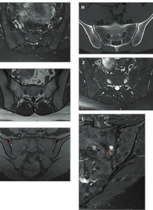
fIgure 1. Degenerative changes. 64-year-old woman, pain not fulfilling definition for inflammatory low back pain: A) coronal CT image, B) coronal oblique T1W and C) coronal oblique FS PDW MR images show subchondral sclerosis, more evident on the right iliac
side (arrows), osteophytosis (white asterisks in A and B) and subtle minor erosions/cortical irregularity (red asterisk in A and B). Note also the mild BME surrounding the area of subchondral sclerosis (red asterisk in C) -hyperaemia due to mechanical stress in a typical anterior-superior part of the SIJs. 34-year-old male, martial arts fighter: D) coronal CT image, E) coronal oblique T1W, F) axial oblique T1W and G) axial oblique FS T2W MR images depict marked anteroinferior osteophytosis (white asterisks in D, E and F), surrounded by subchondral sclerosis (arrows in D and E), more prominent on the left side, with very mild BME surrounding these changes (red asterisk in G). Notice how the structural changes are best depicted on CT as compared to MRI. 28-year-old male, soccer
player: H) coronal CT image, I) axial oblique T1W and J) axial oblique FS T2W MR images show mild subchondral sclerosis on an
anteriorsuperior location, with minor irregularity, more on the right SIJ (red asterisk in H and I) suggesting a mechanical cause, supported by the joint vacuum phenomenon (arrow in H). No frank erosions or fat metaplasia is seen. Notice the subtle BME adjacent to these degenerative/microtraumatic findings (white asterisks in J). 29-year-old female professional ballet dancer: K) coronal oblique FS T1W MR image shows subchondral sclerosis (arrows), osteophytosis (red asterisks) and cortical irregularity, with an erosion (white asterisk) better defined on the superioranterior aspect of the right SIJ. 50-year-old female: L) coronal oblique FS T2W MR image shows a subchondral cyst (geode) in the sacral side of the left SIJ(arrow), cortical irregularity and joint space narrowing.
I
J
K
l
antero-superior part of the SIJs7. Up to 27% of heathy
individuals with mechanical back pain show BME in the SIJs8. This can be even more extensive in
sports-ac-tive individuals. In fact, one should be aware that me-chanical/overload/microtraumatic changes that occur in sports-active individuals can induce changes in the SIJs, often BME and/or small erosions, an important confounding factor in imaging interpretation. A study of MRI of SIJs in athletes showed BME fulfilling ASAS criteria in 35% of hobby runners and 41% of ice-hock-ey players9. This non-inflammatory BME was more
common in the posterior lower ilium, followed by the anterior upper sacrum10. Similar results were found in
another study with military recruits, who fulfilled the ASAS MRI criteria in 23% and 36% before and 6 weeks after intensive physical training, respectively11. Fat
de-position is a non-specific finding, but may sometimes be seen in the degenerative setting and even in older healthy individuals12. Large erosions, subchondral cysts
and ankylosis are rare in osteoarthritis.
Learning Points - osteoarthritis:
• A young, sports-active individual may present with BME in the SIJs. Minor erosions, osteophytes and scle-rosis can also be seen. Look for age, history and look at the BME distribution (non-inflammatory BME more common in the posterior lower ilium>anterior upper sacrum).
• In the elderly with SIJs osteoarthritis, BME can also oc-cur (often in the anterosuperior part of the SIJs). Minor erosions can also be seen. Look for other signs sugges-tive of degenerasugges-tive disease (joint space narrowing, sub-chondral sclerosis, osteophytosis and joint vacuum phenomenon).
2.2. Degenerative changes of the lower lumbar spine are a very common finding in routine MRI of the SIJs. A study of 691 patients undergoing MRI of the SIJs for inflammatory back pain reported L5-S1 degenerative changes as the most common non-inflammatory find-ing (44%) and it consisted of disc degeneration (32%), facet joint arthrosis (8%) and disc herniation (4%)10. In
16% of this set of patients with spinal changes, adjacent BME was seen10. Not rarely, low back pain fulfilling the
definition of inflammatory back pain indicating axial SpA results from L5-S1 pathology10.
2.3. Transitional vertebrae, with or without adjacent BME, are found in up to 25% of population13. In these
patients, BME is more subtle and confined to the im-mediate surfaces involved in the transitional lum-bosacral transitional vertebra (that is, typically located
at the enlarged transverse process and the portion of the sacral bone adjacent to it)13. As such, the presence of a
BME-like pattern at the pseudoarticulation that does not reach the SIJs should not be confused with the usu-ally T2-brighter, subchondral/periarticular BME of the SIJs associated with axial SpA.
3. InfectIous sAcroIlIItIs
Infectious sacroiliitis is not uncommon but is more fre-quente in children and young individuals14. Clinical
history is critical, however, diagnosis is often delayed due to the typically insidious clinical presentation. Changes can be seen on MRI within up to three days after the onset of symptoms14. MRI appearance may be
similar to that of inflammatory sacroiliitis but is usually more dramatic (Figure 2). Extensive BME is typically present in the acute phase and often unila -teral (at least initially). Often, BME in infectious sacroiliitis shows sacral predominance or an even dis-tribution (not an iliac predominance, as in SpA)10. Joint
fluid (septic arthritis/synovitis), thick capsulitis, peri-and extra-articular edema peri-and fluid collections in the surrounding soft tissues may occur10,15. Regional
mus-cle edema is probably the most important predictor of infectious sacroiliitis10,15. Infection does not respect the
limits of the SIJs and infiltrates throughout the mus-cles (specially, the iliac muscle). In advanced stages, erosions (often large, >1cm), bony bridges, fatty re-placement and ankylosis may also be found16.
Also, indolent infections should be remembered, such as tuberculosis or fungi. Tuberculous septic arthri-tis is usually unilateral and an abscess containing cal-cifications anterior to the SIJs may develop. Erosions may be deep, from both iliac and sacral sides, and SIJs ankylosis may develop.
Learning point – infectious sacroiliitis:
• Features that can point out to infectious sacroiliitis (given the appropriate clinical setting) are that anato -mic boundaries are not respected (involvement can be unilateral or bilateral), usually with larger erosions, joint effusion, and more extensive BME and soft-tissue involvement, often with abscesses)
4. osteItIs condensAns IlII (ocI)
Osteitis Condensans Ilii (OCI) is reported to affect 0.9 to 2.5% of the population but may be underestimated17.
It is believed to be caused by mechani cal/stress-related remodelling across the SIJs. Usually bilateral and rela-tively symmetrical, it occurs more often in multiparous
females, and in the perinatal pe riod18-19. OCI may be
related to the increased tilt of the female pelvis. How-ever, men and nulliparous women are also affected19.
Although most frequently asym ptomatic, OCI may also be associated with axial low back pain20. Other cha
-racteristics are normal inflammatory markers and HLA B27 negativity18. The ima ging features (Figure 3) are a
triangular-shaped, anteriorly located well-circum-scribed subchondral sclerosis, mostly in the iliac side
(even though sacral sclerosis can also be seen, to a less-er extent), without less-erosions (even though small less- ero-sions can coexist due to degenerative changes) nor joint space narro wing21,17,22. According to Ma et al, the mean
value of the thickest part of sclerosis is about 13mm in OCI patients18. Concomitant BME surrounding
sclero-sis can be seen, especially in the postpartum period. Indeed, it may be more frequent than initially described – the same authors found BME beneath the iliac
sub-A b
c
d
e
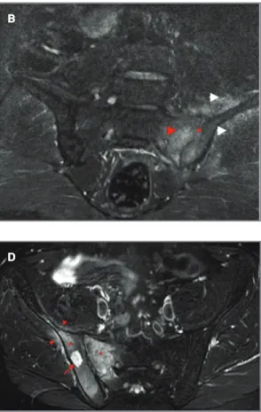
fIgure 2. Infectious sacroiliitis. 40-year-old male with
infectious sacroiliitis caused by Staphylococcus aureus following a skin injury: A) axial oblique FS T2W, and B) coronal oblique STIR
MR images show extensive BME (red arrowhead in A and B)
and enhancement (osteitis, red arrowhead in B) surrounding the left SIJ, left joint synovitis (asterisk in B) and
(inflammatory) edema involving the adjacent iliac and medial gluteus muscles (white arrowheads in B). 35-year-old male with
osteomyelitis (unknown agent): C) axial oblique T1W and D) axial
oblique STIR MR images show extensive BME in the right SIJ (asterisks in D) and surrounding soft tissue edema in the gluteus maximus and iliopsoas entheses (arrowheads in D). Note a bony collection in the posterior right ilium contacting the SIJ, with intermediate T1 and with fluid-signal on the STIR image (arrow in C and D), suggesting a Brodie abscess. 39-year
old male with sacroiliitis caused by Salmonella typhi: E) axial
oblique FS contrast-enhanced T1W MR image shows extensive unilateral osteitis surrounding the left SIJ (asterisks) with joint synovitis (red double arrow), anterior capsulitis (white arrow) and surrounding soft tissue edema/enthesitis (red arrowheads).
A b
c d
e
g
f
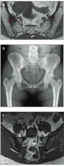
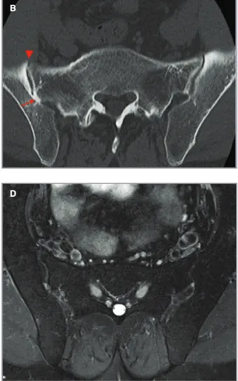
fIgure 3. Osteitis condensans ilii. 32-year-old male,
long-standing lumbar pain: A) axial CT image, B) axial oblique T1W
and C) axial oblique FS T2W MR images show bilateral anterior well-circumscribed triangular area of subchondral sclerosis (asterisks in A), more evident on the right side, involving both the iliac and (less) the sacral side, with
corresponding MRI T1 hypointensity (arrowheads in B). Do notice that sacral involvement can also be seen as well as minor erosions/loss of the cortical joint line (asterisks in B) and mild surrounding BME (arrow in C). 45-year-old female,
doubtful history for SpA: D) AP pelvic radiograph and E) coronal
CT image show unilateral triangular area of subchondral sclerosis, mainly on the iliac border of the right SIJ, with minimal involvement of the sacral side (arrowhead). F) coronal oblique T1W and G) coronal oblique PDW MR images of the same patient show corresponding well-defined area of hypointensity (arrowhead in F) and subtle BME surrounding the sclerotic area (arrowhead in G).
A b
chondral sclerosis in 48% of their patients, mostly cen-tered at the cartilaginous joint part, and 92% of these would also have BME on the sacral side18. This BME
was both deep (more than 10mm in 39%) and of high intensity (85% graded higher than mild)18. This
em-phasizes the need of differentiating OCI from sacroili-itis. Note also that both OCI and SpA may coexist, lead-ing to a difficult diagnostic challenge. Careful observation of the location and distribution pattern of BME may be helpful in differentiating OCI from early sacroiliitis.
Learning Points - OCI:
• Triangular-shaped, well-circumscribed, subchondral sclerosis (anteriorly located, iliac side > sacral), with-out gross erosions (minor irregularities and small ero-sions can exist) nor SIJs narrowing.
• BME surrounding sclerosis can be seen. Careful ob-servation of the location and distribution pattern of BME is helpful in the differential. If there is BME sur-rounding fat metaplasia, this may suggest SpA may co-exist with OCI.
• A practical hint that favors OCI is to look for char-acteristic imaging features and location in the anterior part of the SIJs, normal inflammatory markers and HLAB 27 negativity.
5. stress frActures (fAtIgue And InsuffIcIency)
In healthy, young, active adults, fatigue fractures (abnor mal stress on normal bone, Figure 4) occur
of-ten in the sacrum23. Even though clinically these sacral
fractures/stress changes may be confused with sacroili-itis, imaging findings are different, particularly since BME is generally located close to the sacral neurofora-mena23. In later stages, a hypointense irregular track
representing the fracture line can be observed, usually with vertical orientation, surrounded by BME and spar-ing the SIJs. They are not associated with SIJs struc-tural changes. In the elderly, insufficiency fractures (normal stress on abnormal bone, often secondary to osteoporosis or radiation therapy) may be seen. Besides the age and clinical setting, clues are the characteristic location (sacrum), fracture orientation (vertical orien -ted), and possible bilateral symmetric findings24-25. 6. dIffuse IdIopAthIc sKeletAl hyperostosIs (dIsh)
Diffuse Idiopathic Skeletal Hyperostosis (DISH) is a common condition, with an average prevalence of 10% in people >50 years of age10,26. Despite the extensive
structural changes, DISH is largely asymptomatic. It is a systemic disease characterized by continuous calcifi-cations and ossificalcifi-cations of soft tissues, primarily liga-ments and entheses27, which mainly involve the axial
skeleton, but also the peripheral joints. “Flowing os-teophytes” occur in the anterior longitudinal ligament and, to a lesser extent, involve the paravertebral con-nective tissue and the annulus fibrosus (Figure 5A)27.
The Resnick classification criteria for DISH are based on radiographs and require flowing osteophytes that bridge at least four contiguous thoracic vertebrae. Lite
-fIgure 4. Sacral stress fractures. 32-year-old female runner: A) coronal oblique FS T2W and B) coronal oblique T1W MR images
c
d
A b
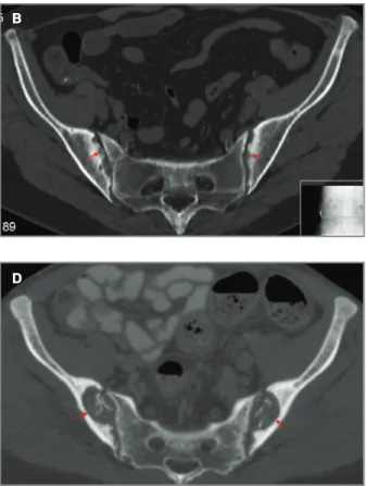
fIgure 5. DISH. 52-year-old male, DISH: A) sagittal CT image
of the lumbar spine shows flowing consecutive osteophytes (arrowheads), with disk space sparing; B) axial CT image of the SIJs shows posterior bone bridging (arrow) and anterior osteophytosis (arrowhead). Corresponding C) axial oblique T1W MR image reveals posterior bridging, more evident on the right (arrowheads); D) axial oblique FS T2W MR image shows absence of subchondral BME.
rature shows conflicting results regarding SIJs involve-ment and there is still a common misconception that SIJs are not involved in DISH28. In fact, DISH may also
affect the SIJ (Figures 5B-D) and lead to asymmetric in-traarticular partial fusion, osteophytes with or without bridging, ossification of the sacrotuberous and ili-olumbar ligaments27-29. The ligamentous portion of the
SIJs (upper portion if the SIJs) may show vacuum phe-nomena, space narrowing, sclerosis and partial or com-plete ankylosis. The lower two-thirds of the SIJs are usually spared. Ossification of the joint capsule in the anterior surface of the joint, resembling obliteration of the SIJs on a radiograph, may erroneously be inter-preted as SpA bone ankylosis.27Subchondral sclerosis,
erosions and subchondral cysts are typically absent27. Learning Points - DISH:
• SIJs are not spared in DISH! Look for overt, coarse bony/ossified bridges over the anterior and posterior SIJs articular margins and entheseal bridging. Intra-ar-ticular ankylosis, not previously included in the crite-ria for DISH, can also be seen.
• Pelvic locations of DISH are also suggestive (hyper-ostosis at ligamentous/tendinous attachments in the pelvis)
• Think in alternate diagnosis if there is presence of erosions and subchondral sclerosis.
7. hyperpArAthyroIdIsm
Secondary hyperparathyroidism is a response to low serum calcium levels, usually in the setting of chronic renal failure. It leads to subperiosteal, trabecular, in-tracortical, endosteal, subchondral, subligamentous or subtendinous bone resorption. Hyperparathyroidism often results in subchondral bone resorption around the SIJs, with irregularity, gross erosions and pseudowidening of the joint (Figure 6) that mimic sacroilii -tis. This usually occurs bilaterally, relatively symmetri-cally and is more pronounced on the iliac side. However, joint space narrowing or bone bridging/anky-losis do not develop30. Look for other signs of re
-sorption in the pelvis – re-sorption/erosions, and cysts at the hamstrings, adductor and at the gluteal entheses. Other imaging findings may also be a clue to the diagno sis - osteopenia, chondrocalcinosis in the pubic symphysis and brown tumors31,32. Although brown
tu-mors are more frequent in primary hyperparathy-roidism, secondary hyperparathyroidism is much more common, so most brown tumors seen today are asso-ciated with secondary hyperparathyroidism. They are
usually round, well-defined lesions extending to the bone cortex on radiographs. On MRI, they exhibit low signal intensity on T1W and T2W images with various foci of increased intensity (due to haemorrhage)33. The
most common site of involvement is the thoracic spine, while sacral involvement is very rare33.
Learning Points - Hyperparathyroidism:
• Subchondral resorption with irregularity, gross ero-sions and pseudowidening of the SIJs (more pro-nounced at the iliac side).
• Look for other signs of hyperparathyroidism: signs of resorption in the pelvis - subtendinous resorption at the hamstrings, adductor and at the gluteal origins, bony cysts in ischial and pubic bones; osteopenia, chondrocalcinosis and brown tumors.
• Look for a history of kidney disease (patients often tell they “cannot have contrast”)
8. synovItIs, Acne, pustulosIs, hyperostosIs syndrome (sApho) And chronIc recurrent multIfocAl osteomyelItIs (crmo)
Synovitis, Acne, Pustulosis, Hyperostosis syndrome (SAPHO) and chronic recurrent multifocal os-teomyelitis (CRMO) represents a spectrum of autoin-flammatory bone diseases (“non-bacterial osteitis”) in which non-infectious inflammatory chronic osteitis, in-volving both the cortex and the medullary canal (with endosteal and periosteal new bone formation) is the unifying feature, along with skin lesions. SAPHO is considered a rare disease. It may occur at any age but is more frequent <60 years of age34. CRMO is the
child-hood counterpart of SAPHO.
The site of disease involvement is age-related and most patients show polyostotic involvement34. In
adults, the sternoclavicular junction is the most com-mon location, followed by the spine and SIJs35. In
chil-dren, the metaphysis of long bones and clavicles are most commonly involved36. SAPHO/CRMO has va
-riable imaging appearances depending on the stage/age of the lesion and imaging modality (Figure 7) - it starts from BME in the acute stage, followed by osteolytic and later sclerotic and hyperostotic changes, erosions and ankylosis. Within the SIJs, the hyperostotic changes are more common in the iliac side. In a given patient there may be lesions in various stage, active (BME) or chro -nic (lytic, sclerotic). Usually, they tend to be unilater-al, but can be bilateral37. One should keep in mind that
de-veloping sacroiliitis, which is usually unilateral37.
Ulti-mately, the diagnosis may be based on the combina-tion of clinical and radiologic findings.
Learning Points - SAPHO/CRMO:
• Osteitis/BME (on either side of the SIJs) precedes erosive changes, sclerosis and hyperostosis in the SIJs (more marked on the iliac side). In practice, look for unilateral or bilateral asymmetric involvement of the SIJs, mainly involving the iliac side, with extensive os-teosclerosis.
• One should keep in mind that adult patients with SAPHO have a higher risk of developing sacroiliitis, which is usually unilateral.
9. gout
Gout is a metabolic disorder and the most common form of microcrystalline arthropathy38. Even though it
often affects the peripheral joints, gout may occasion-ally affect the axial skeleton, including the SIJs, and ax-ial gouty arthropathy may be, in fact, much more com-mon than initially thought39.
Based on radiographic findings alone, the incidence of SIJs involvement in chronic gout ranges between 7%
and 17%40. Unfortunately, imaging features of axial
gout are nonspecific. Deposition of urate crystals in the cartilage leads to irregular loss of joint space and su-perimposed degenerative changes. In this instance, it is difficult to distinguish gout from osteoarthritis.
In the chronic phase, intra- and para-articular ero-sions, often large, with a multilobulated base, and well-defined, thin sclerotic margins that may have over-hanging edges are found41. They are usually seen in
relation to tophi (juxta-articular, intra-articular, sub-chondral) because they represent intraosseous exten-sion of tophi42. Tophus gout has iso- to low- signal
in-tensity on T1W in relation to muscle, but it is quite variable on T2W images, due to differences in calcium concentration within a tophus. Calcifications within tophi are better visualized on computed tomography (CT) than MRI.
BME is usually mild in uncomplicated gout. The joint space is remarkably well preserved and there is no periarticular osteopenia until late in the disease39. The
lack of subchondral sclerosis also favours gouty sacroiliitis over SpA42.
Dual-energy CT can be used to detect urate deposits with an overall accuracy of 87–94%43.
d
A b
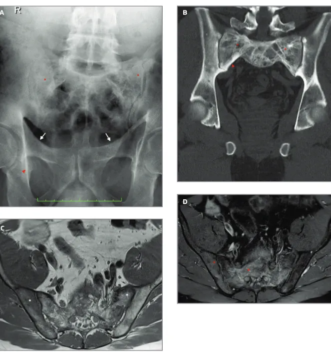
fIgure 6. Secondary hyperparathyroidism. 49-year-old
female, chronic renal disease: A) AP pelvic radiograph and B)
axial CT image show bilateral sclerosis of the SIJs (arrowheads in A) and subchondral bone reabsorption/erosions, more evident on the right (arrows in B) [Courtesy of Raquel Marques MD, Department of Rheumatology, Centro Hospitalar
Universitário Lisboa Norte, Lisbon]. 62-year-old female,
persistent lumbar pain and chronic renal disease: C) axial CT image
shows severe symmetrical subchondral bone reabsorption (arrows) of the SIJs, more on the iliac side.
Learning Points - gout:
• A tophus may form at the SIJs (juxta-articular, intra-articular and subchondral) and create large erosions with multilobulated base and well-defined, thin scle-rotic borders that may have overhanging edges. • The absence of subchondral sclerosis and relatively well-preserved joint space favor gouty sacroiliitis over SpA-sacroiliitis.
• To detect urate deposits and confirm the diagnosis, order dual-energy CT.
10. pAget’s dIseAse
Paget’s disease is a chronic bone disorder characterized by excessive bone remodelling, which includes diffe
-rent lytic, blastic and mixed phases44. The pelvis is
fre-quently involved. Common findings include cortical thickening and coarsening, enlargement of the bone af-fected by Paget’s when compared to the contralateral side, sclerosis of the iliac wing, thickening of the ilio -pectineal and ischiopubic lines, acetabular protrusion and enlargement of the pubic rami and ischium44,
which are often asymmetrical (Figure 8). Fusion of SIJs can uncommonly be seen in patients with Paget’s, either unilaterally or bilaterally45. Because Paget’s disease typi
-cally does not extend across healthy joints, it has been suggested that, when both SIJs are involved, inflam-matory sacroiliitis or other causes of previous joint damage must be present46,47. Similarly, concomitant c
A b
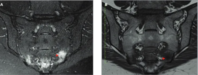
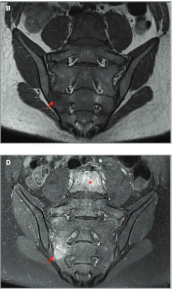
fIgure 7. SAPHO and CRMO. 40-year old female, SAPHO: A)
AP pelvic radiograph shows marked hyperostosis of the right SIJ (asterisks) with cortical erosions. 14-year-old boy, CRMO: B) coronal oblique T1W, C) coronal oblique FS T2W and D)
coronal oblique FS contrast-enhanced T1W MR images show BME in the right sacrum (arrow in C and D) with strong enhancement (arrow in D) in keeping with an acute stage. Concomitant extensive BME in L5 vertebra (asterisk in C and D) and two smaller areas of BME are seen on the upper S1/S2 segments. No sclerotic or lytic/erosive changes are yet seen.
ankylosis spondylitis and Paget’s disease is infrequent but has been reported in literature46-48.
Learning Points - Paget’s disease:
• Must have other Pagetic changes in the pelvis. • Fusion of the SIJs can be rarely seen in Paget’s disea se
in the course of coexisting sacroiliitis (not Paget’s itself).
11. mIscellAneous
11.1. Sarcoidosis: One of the rarest manifestations of osseous sarcoidosis is involvement of SIJs, usually in a unilateral fashion but bilateral cases have been des
-c
A b
fIgure 8. Paget’s disease. 58-year-old male: A) AP pelvic radiograph reveals trabecular coarsening of the sacrum (asterisks),
thickening of the right ischiopubic line (red arrowhead) and, to a lesser degree, of the iliopectineal line (white arrows). B) coronal CT image better depicts the course trabecular pattern (asterisks) and cortical thickening (arrowhead). C) axial oblique T1W and D) axial oblique FS T1W contrast-enhanced MR images confirm this course enlargement of the bony trabeculae with fat marrow in between (asterisks in C) and enhancement after contrast (asterisks in D) probably as a result of granulation tissue.
cribed, may be due to sarcoid osteitis or granulomatous joint infiltration49. Radiographic findings of sarcoidosis
are similar to those of inflammatory sacroiliitis. Multi-ple bony “punched out” lesions may coexist. MRI is very sensitive for bony changes in sarcoidosis, but not spe-cific50. MRI may aid in choosing a biopsy site51.
In the presence of known clinical sarcoidosis, the diagnosis of bone sarcoidosis should be considered if there is involvement of the SIJs, since these joints may be involved without a typical inflammatory back pain history50. It is rare but there are reports of sarcoidosis
coexisting with SpA52-54.
Learning Point – Sarcoidosis:
May mimic SpA in radiographs. In the presence of known clinical sarcoidosis, the diagnosis of bone sar-coidosis should be considered if there is concomitant involvement of the SIJs.
11.2. Behcet’s disease: Arthritis/arthralgia are the most common rheumatologic findings in Behcet’s. However, even though it has symptoms of SpA, most studies re-port no difference in the prevalence of sacroiliitis in Beh cet’s disease to that of the general popu lation. Ne -vertheless, joint involvement can be an early manifes-tation in Behcet’s disease55.
Learning Point - Behcet’s disease:
Look for the right clinical context.
11.3. Bone tumors/pseudotumors: usually a straight-forward imaging diagnosis. However, then can coexist in SpA patients and/or be a source of low back pain. The sacrum is a common site for metastases and myeloma56.
The most common primary sacral tumors are chordoma (25-40%), followed by giant cell tumor, aneurysmal bone cyst, chondrosarcoma, osteosarcoma and Ewing sarcoma56. Sacral meningeal cysts (Tarlov/perineural cysts)
are frequently found incidental pseudotumors and re -present abnormal dilatations of the meninges within the sacral canal or foramina, and may cause low back pain.
Learning Point - Tumors/pseudotumors:
Can be a source of pain or be incidental in SpA patients. They are usually a straightforward imaging diagnosis.
conclusIon
MRIs of the SIJs in patients with a clinical suspicion of
axial SpA sacroiliitis commonly demonstrate non-in-flammatory disease. We reviewed the ASAS criteria for a “positive” MRI (part I) and took a practical snapshot at the most common non-inflammatory causes of SIJs involvement that may mimic sacroiliitis in daily prac-tice (part II).
MRI interpretation will be boosted by adequate clini -cal information and/or by adequate patient referral from rheumatology centers. However, even with the engagement of both sides, rheumatologists and radio -logists, there will still be cases where neither clinical presentation or images are specific for SpA or other en-tity. In such instances, interdisciplinary discussions are needed to constantly increase our understanding of the disease and decide on the best approach for a given pa-tient.
correspondence to
Diana Afonso
Musculoskeletal Imaging Unit, Radiology Department, Hospital da Luz, Grupo Luz Saúde
Lisbon, Portugal
E-mail: p.diana.a@gmail.com
references
1. Zejden A, Jurik AG. Anatomy of the sacroiliac joints in children and adolescents by computed tomography. Pediatr Rheumatol Online J 2017;15(1):82.
2. Puhakka KB, Melsen F, Jurik AG, Boel LW, Vesterby A, Egund N. MR imaging of the normal sacroiliac joint with correlation to histology. Skeletal Radiol 2004;33(1):15–28
3. Demir M, Mavi A, Gümüsburun E, Bayram M, Gürsoy S, Nishio H. Anatomical variations with joint space measurements on CT. Kobe J Med Sci 2007;53(5):209–217.
4. Faflia CP, Prassopoulos PK, Daskalogiannaki ME, Gourtsoyian-nis NC. Variation in the appearance of the normal sacroiliac joint on pelvic CT. Clin Radiol 1998;53(10):742–746. 5. Li G, Yin J, Gao J, Cheng TS, Pavlos NJ, Zhang C, et al.
Sub-chondral boné in osteoarthritis: insight into risk factors and mi-crostructural changes. Arthritis Res Ther 2013;15(6):223 6. Lennard, T. A., Vivian, D. G., Walkowski, S. D. O. W., & Singla,
A. K. Pain Procedures in Clinical Practice E-Book. Elsevier Health Sciences, 2011.
7. Schueller-Weidekamm C, Mascarenhas VV, Sudol-Szopinska I et al. Imaging and interpretation of axial spondylarthritis: the ra-diologist's perspective—consensus of the Arthritis Subcom-mittee of the ESSR. Semin Musculoskelet Radiol 2014;18:265–79. doi:10.1055/s-0034-1375567.
8. Marzo-Ortega H, McGonagle D, O'Connor P et al. Baseline and 1-year magnetic resonance imaging of the sacroiliac joint and lumbar spine in very early inflammatory back pain. Relationship between symptoms, HLA-B27 and disease extent and persis-tence. Ann Rheum Dis 2009;68:1721–1727.
9. Rudwaleit M, VanderHeijde D, Landewé R et al. The develop-ment of Assessdevelop-ment of SpondyloArthritis international Society classification criteria for axial spondyloarthritis (part II): vali-dation and final selection. Ann Rheum Dis 2009; 68: 777–783.
10. Jans L, Van Praet L, Elewaut D, et al. MRI of the SI joints com-monly shows non-inflammatory disease in patients clinically suspected of sacroiliitis. Eur J Radiol 2014; 83(1):179-184 11. Varkas G, de Hooge M, Renson T, et al. Presence of BME and
structural lesions on MRI of the sacroiliac joints in young mili-tary recruits before and after 6 weeks of intensive physical train-ing. Arthritis Rheum;69(Suppl 10).
12. Haliloglu M, Kleiman MB, Siddiqui AR, Cohen MD. Os-teomyelitis and pyogenic infection of the sacroiliac joint. MRI findings and review. Pediatr Radiol 1994;24:333–335. 13. de Bruin F, Ter Horst S, Bloem JL, et al. Prevalence and clinical
significance of lumbosacral transitional vertebra (LSTV) in a young back pain population with suspected axial spondy-loarthritis: results of the SPondyloArthritis Caught Early (SPACE) cohort. Skeletal Radiol 2017;46(5):633–639. 14. Montandon C, Costa MAB, Carvalho TN, Montadon JMET,
San-tos KIS. Sacroiliitis: imaging evaluation, Radiolog Brasil 2007; vol. 40 (53-60).
15. Stürzenbecher A, Braun J, Paris S, Biedermann T, Hamm B, Bol-low M. MR imaging of septic sacroiliitis. Skeletal Radiol 2000;29(8): 439–446.
16. Rudwaleit M, Jurik AG, Hermann KG et al. Defining active sacroiliitis on magnetic resonance imaging (MRI) for classifica-tion of axial spondyloarthritis: a consensual approach by the ASAS/OMERACT MRI group. Ann Rheum Dis 2009;68: 1520–1527.
17. Mitra R. Osteitis condensans ilii. Rheumatol Int 2010;30 (3):293–296. doi: 10.1007/s00296-009-1100-7.
18. Ma L, Gao Z, Zhong Y, Meng Q. Osteitis condensans ilii may demonstrate bone marrow edema on sacroiliac joint magnetic resonance imaging. Int J Rheum Dis 2018;21(1):299-307. doi: 10.1111/1756-185X.13125.
19. Hutton CF. Osteitis condensans ilii. Br J Radiol 1953;26 (309): 490-3. doi:10.1259/0007-1285-26-309-490.
20. Lennard, T. A., Vivian, D. G., Walkowski, S. D. O. W., & Singla, A. K. Pain Procedures in Clinical Practice E-Book. Elsevier Health Sciences, 2011.
21. Klang E, Lidar M, Lidar Z, Aharoni D, Eshed I. Prevalence and awareness of sacroiliac joint alterations on lumbar spine CT in low back pain patients younger than 40 years. Acta Radiol 2017; 58(4):449-455.
22. Olivieri I, Ferri S, Barozzi L. Osteitis condensans ilii. Br J Rheumatol 1996;35:295–301. doi: 10.1093/rheumatolo-gy/35.3.295-a.
23. Heuft-Dorenbosch L, Weijers R, Landewe R, van der Linden S, van de Heijde D. Magnetic resonance imaging changes of sacroiliac joints in patients with recent-onset inflammatory back pain: inter-reader reliability and prevalence of abnormalities. Arthritis Res Ther 2006; 8: R11.
24. Girish G, Finlay K, Fessell D, Pai D, Dong Q, Jamadar D. Imag-ing review of skeletal tumors of the pelvis malignant tumors and tumor mimics. Scientific World Journal 2012;2012: 240281.
25. Cabarrus MC., Ambekar A., Lu Y, Link TM. MRI and CT of insufficiency fractures of the pelvis and the proximal femur. Ame -rican Journal of Roentgenol 2008;191(4), 995–1001. 26. Mader, R., Verlaan, J.-J., Eshed, I., Jacome, B.-A., Puttini, P. S.,
Atzeni, F., et al. Diffuse idiopathic skeletal hyperostosis (DISH): where we are now and where to go next. RMD Open 2017; 3(1), e000472–7. http://doi.org/10.1136/rmdopen-2017-000472. 27. Olivieri I, D’Angelo S, Palazzi C, Padula A, Mader R, Khan M.
Diffuse idiopathic skeletal hyperostosis: differentiation from ankylosing spondylitis. Curr Rheumatol Rep 2009;11:321–328. 28. Leibushor N, Slonimsky E, Aharoni D, Lidar M, Eshed I. CT Abnormalities in the Sacroiliac Joints of Patients With Diffuse Idiopathic Skeletal Hyperostosis. AJR Am J Roentgenol 2017;208(4):834-837.
29. Durback MA, Edelstein G, Schumacher HR Jr. Abnormalities of the sacroiliac joints in diffuse idiopathic skeletal hyperostosis: demonstration by computed tomography. J Rheumatol 1988;15(10):1506-1511.
30. Pialat JB, Di Marco L, Feydy A, Peyron C, Porta B, Himpens PH et al. Sacroiliac joints imaging in axial spondyloarthritis. Diag-nostic and Interventional Imaging 2016;97(7-8), 697-708, 31. Patel CN, Scarsbrook AF. Multimodality imaging in
hyper-parathyroidism. Postgrad Med J 2009;85(1009):597-605. doi: 10.1136/pgmj.2008.077842.
32. Chang CY, Rosenthal DI, Mitchell DM, Handa A, Kattapuram SV, Huang AJ. Imaging Findings of Metabolic Bone Disease. Ra-diographics 2016;36(6):1871-1887.
33. Karagoz A, Turkmen K, Yazici R, Arslan S, Baktik S, Karanis MI et al. An exceedingly rare localization of a brown tumor in a hemodialysis patient. Hemodial Int 2013;17(4):660-663. 34. Jurik A, Klicman R, Simoni P, Robinson P, Teh J. SAPHO and
CRMO: The Value of Imaging. Seminars in Musculoskeletal Ra-diol 2018; 22(02), 207–224.
35. Depasquale R, Kumar N, Lalam RK et al. SAPHO: What radiol-ogists should know. Clin Radiol 2012;67(03):195–206. 36. Khanna L, El-Khoury GY. SAPHO syndrome--a pictorial assay.
Iowa Orthop J. 2012;32:189-195.
37. Nguyen MT, Borchers A, Selmi C, Naguwa SM, Cheema G, Gershwin ME. The SAPHO syndrome. Semin Arthritis Rheum 2012;42 (03):254–265.
38. Monu JU, Pope TL Jr. Gout: a clinical and radiologic review. Ra-diol Clin North Am 2004 Jan;42(1):169-184.
39. Cardoso FN, Omoumi P, Wieers G, Maldague B, Malghem J, Lecouvet FE, Vande Berg BC. Spinal and sacroiliac gouty arthri-tis: report of a case and review of the literature. Acta Radiol Short Rep 2014; 17;3(8):2047981614549269.
40. Chen W, Wang Y, Li Y, Zhao Z, Feng L, Zhu J et al. Gout mim-icking spondyloarthritis: case report and literature review. Jour-nal of Pain Research 2017;8(10), 1511–1514.
41. Panwar J, Sandhya P, Kandagaddala M, Nair A, Jeyaseelan V, Danda. Utility of CT imaging in differentiating sacroiliitis asso-ciated with spondyloarthritis from gouty sacroiliitis: a retro-spective study. Clin Rheumatol 2018;37(3):779-788. doi: 10.1007/s10067-017-3865.
42. Teh J, McQueen F, Eshed I, Plagou A, Klauser A. Advanced Imaging in the Diagnosis of Gout and Other Crystal Arthropathies. Semin Musculoskelet Radiol 2018;22(2):225-236. doi: 10.1055/s-0038-1639484.
43. Glazebrook KN, Guimarães LS, Murthy NS, Black DF, Bongartz T, Manek NJ, Leng S, Fletcher JG, McCollough CH. Identifica-tion of intraarticular and periarticular uric acid crystals with dual-energy CT: initial evaluation. Radiolog 2011;261(2):516-24. doi: 10.1148/radiol.11102485.
44. Bezza A, Lechevalier D, Monreal M, Maghraoui A, Magnin J, Eulry F. L’atteinte sacro-iliaque au cours de la maladie de Paget. Presse Med 1999;28:1157–1159.
45. Seton M. Paget disease of bone: diagnosis and drug therapy. Cleve Clin J Med 2013;80(7):452–462. doi: 10.3949/ccjm. 80a.12142.
46. Banna M. Clinical radiology of the spine and the spinal cord. Gaithersburg, Md: Aspen,1985; 61-64.
47. Peel N, Barrington N, Austin C, Estell R. Paget’s disease in a pa-tient with ankylosing spondylitis- a diagnostic dilemma. Br J Rheumatol 1996;35:1011–1014. doi: 10.1093/rheumatolo-gy/35.10.1011.
48. Battafarano DF, West SG, Rak KM, Fortenbery EJ, Chantelois AE. Comparison of bone scan, com- puted tomography, and magnetic resonance im- aging in the diagnosis of active sacroili-itis. Semin Arthritis Rheum 1993;23:161–176.
49. Kötter I, Dürk H, Saal JG. Sacroiliitis in sarcoidosis: case reports and review of the literature. Clin Rheumatol 1995; 14(6):695--700.
50. Binicier O, Sari I, Sen G, Onen F, Akkoc N, Manisali M et al. Ax-ial sarcoidosis mimicking radiographic sacroiliitis. Rheumatol Int 2009;29(3):343–345. doi: 10.1007/s00296-008-0677-6. 51. Moore SL, Teirstein AE. Musculoskeletal sarcoidosis: spectrum
of appearances at MR imaging. Radiographics 2003;23(6):1389--1399.
52. Erb N, Cushley MJ, Kassimos DG, Shave RM, Kitas GD. An as-sessment of back pain and the prevalence of sacroiliitis in sar-coidosis. Chest 2005; 127(1):192-196.
53. Sezer I, Melikoglu MA, Cay HF, Kocabas H, Kacar C. A co-oc-currence of sarcoidosis and ankylosing spondylitis: a case re-port. Rheumatol Int 2008; 28(6):605-607.
54. Kremer P, Gallinet E, Banmansour A, Despaux J, Toussirot E, Wendling D. Sarcoidosis and spondyloarthropathy. Three case reports. Revue Rhum 1996;63(6):405–411
55. Cobankara V, Topaloglu M, Balkarlı A, Karabulut N, Akkoyun-lu N, KarabuAkkoyun-lut A. Determining the Prevalence of Sacroiliitis in Behcet's Disease by Magnetic Resonance Imaging. Annals of the Rheumatic Diseases 2014;73:699-700.
56. Diel J, Ortiz O, Losada RA, Price DB, Hayt MW, Katz DS. The sacrum: pathologic spectrum, multimodality imaging, and sub-specialty approach. Radiographics 2001; 21:83-104.