Esta licença está disponível em: https://creativecommons.org/licenses/by-nc/4.0/
Repositório Institucional da Universidade de Brasília
repositorio.unb.br
Este artigo está licenciado sob uma licença Creative Commons Atribuição-NãoComercial 4.0 Internacional.
Você tem direito de:
Compartilhar — copiar e redistribuir o material em qualquer suporte ou formato. Adaptar — remixar, transformar, e criar a partir do material.
De acordo com os termos seguintes:
Atribuição — Você deve dar o crédito apropriado, prover um link para a licença e indicar se mudanças foram feitas. Você deve fazê-lo em qualquer circunstância razoável, mas de maneira alguma que sugira ao licenciante a apoiar você ou o seu uso
Não Comercial — Você não pode usar o material para fins comerciais.
Sem restrições adicionais— Você não pode aplicar termos jurídicos ou medidas de caráter tecnológico que restrinjam legalmente outros de fazerem algo que a licença permita.
This article is licensed under a Creative Commons Attribution-NonCommercial 4.0 International License.
You are free to:
Share — copy and redistribute the material in any medium or format. Adapt — remix, transform, and build upon the material.
Under the following terms:
Attribution — You must give appropriate credit, provide a link to the license, and indicate if changes were made. You may do so in any reasonable manner, but not in any way that suggests the licensor endorses you or your use.
NonCommercial — You may not use the material for commercial purposes.
ABCD Arq Bras Cir Dig
Original Article
2010;23(4):243-246SHORT LENGHT OF THE ESOPHAGUS AND DISTAL REFLUX ARE
RISK FACTORS FOR PROXIMAL ESOPHAGEAL REFLUX?
Short length of the esophagus and distal reflux are risk factors for proximal esophageal reflux?
Humberto Oliveira SERRA1, Lenora GANDOLFI2, Riccardo PRATESI2
Trabalho realizado no 1Departamento de
Medicina II da Universidade Federal do
Maranhão, São Luis, MA e 2Universidade de
Brasília, Brasília, DF, Brasil.
HEADINGS – Gastroesophageal reflux.
Manometry. Esophageal pH monitorings.
ABSTRACT – Background - It is not clear whether patients suffering from distal esophageal reflux also present high risk to proximal esophageal reflux. Common sense suggests that reflux would more easily reach the pharynx in patients who have a smaller distance between the lower esophageal sphincter and the upper one and, thus, short esophagus. Aim - To Investigate if short esophageal length and presence of esophageal distal reflux are risk factors for proximal reflux among patients presenting respiratory symptoms. Methods – A hundred and seven patients were evaluated prospectively by interview, esophagoscopy, manometry and 24-hour esophageal pH monitoring. Student’s t test (two-sided), Spearman’s rank correlation, Chi-square and odds ratio were used in the statistical analysis. Significance level was set at 0.05.
Results - Respiratory symptoms that motivated the search for gastroesophageal reflux disease were cough 43 (40.2%); throat irritation 25 (23.4%), pharyngeal globe 23 (21.5%) and hoarseness 16 (14.9 %). By esophagoscopy, 22 (27.2%) presented some degree of esophagitis. A hiatal hernia was observed in 14 (17.5%) patients. According to manometry, 11 (10.8%) had lower esophageal sphincter hypotonia. The average esophageal body length was 24.3 (± 1.9) cm, ranging from 20 to 30 cm. The esophageal length was not associated with the presence of proximal esophageal reflux. As indicated by pH monitoring, 23 (21.5%) presented pathologic distal reflux and 12 (11.2%) had proximal reflux. Conclusion - The esophageal length was not associated with the presence of proximal esophageal reflux. Patients who had pathological distal esophageal reflux, independent of the esophageal length, were 4.6 times more likely to have proximal esophageal reflux.
RESUMO – Racional - Não está claro se pacientes que apresentam refluxo
gastroesofágico distal têm maior risco de apresentar também refluxo proximal. O senso comum sugere que um episódio de refluxo poderia chegar mais facilmente à faringe em pacientes que tivessem menor distância a percorrer entre o esfíncter inferior do esôfago e o superior. Objetivo - Investigar se o esôfago curto e a presença de refluxo esofágico distal são fatores de risco para refluxo proximal nos pacientes com sintomas respiratórios. Método – Cento e sete pacientes foram avaliados prospectivamente por meio de entrevista, esofagoscopia, manometria e pHmetria. Utilizaram-se o teste t de Student, o de correlação de Spearman, o do Qui-quadrado e odds-ratio. O nível de significância foi 0,05. Resultados - Os sintomas que motivaram a investigação da doença do refluxo gastroesofágico foram: tosse 43 (40,2%); pigarro 25 (23,4%), globo faríngeo 23 (21,5%) e rouquidão 16 (14,9%). No estudo endoscópico 22 apresentaram esofagite e 14 hérnia de hiato. Na avaliação manométrica 11 (10,8)% apresentaram hipotonia do esfíncter inferior. A média do comprimento do esôfago foi 24,3 (± 1,9) cm, variando de 20 a 30 cm. Na avaliação pHmétrica 23 (21,5%) apresentaram refluxo distal patológico e 12 (11,2%) refluxo proximal. Conclusões - O comprimento do esôfago não esteve associado com a
presença de refluxo proximal. Pacientes que apresentaram refluxo gatroesofágico distal, independente do comprimento do esôfago, tiveram risco aumentado de 4,6 vezes para apresentarem refluxo proximal.
Correspondence:
Humberto Oliveira Serra, e-mail: hoserra@gmail.com
Fonte de financiamento: não há Conflito de interesses: não há
Recebido para publicação: 22/06/2010 Aceito para publicação: 29/09/2010
DESCRITORES - Refluxo gastroesofágico,
Manometria, Monitoramento do pH esofágico.
INTRODUCTION
G
astroesophageal reflux disease (GERD)is considered one of the most prevalent digestive diseases in Western countries12.
It occurs as a result of exposure of esophageal or supra-esophageal mucosa to the intragastric content, containing harmful agents such as hydrochloric acid, pepsin, bile salts and pancreatic enzymes11.
GERD has long been recognized as a potential cause of many laryngeal and respiratory symptoms. Although cough, hoarseness, wheezing, and other airway symptoms may be caused by several etiologic factors, recent emphasis has been placed on the role of
gastroesophageal reflux as the culprit13.
Although several tests have been reported to
help determine whether reflux in a given patient is the
cause of respiratory symptoms, none is perfect. A “gold standard” for diagnosis has been 24-hour esophageal pH monitoring using a 2-channel pH-probe13. Because direct monitoring of laryngeal pH is impractical, the placement of a probe in the pharynx or in the upper esophageal sphincter has been employed to detect
extraesophageal acid reflux9.
Manometry is important to locate the lower esophageal sphincter (LES) and the upper esophageal sphincter (UES), to study the motor characteristics of the esophageal body and to determine its length.
The average length of the esophagus in adults varies from 20 to 35 cm3. In vivo measurement of
esophageal length can be accomplished through endoscopic visualization of the gastroesophageal junction15 and manometric analysis8.
It is not clear whether patients suffering from distal
esophageal reflux also presented high risk of proximal esophageal reflux. Common sense suggests that reflux
would more easily reach the pharynx of patients who have a smaller distance between the LES and UES and thus, short esophagus.
Thus, the aim of this study is to look for an association between esophageal length and presence of
proximal esophageal reflux, and to investigate whether those presenting distal reflux were at increased risk of presenting proximal reflux among patients presenting
respiratory symptoms.
METHODS
A hundred and seven patients of both genders were evaluated prospectively, with 81 (75.7%) being females and 26 (24.3%) males. The mean age was 43.1 (± 13.4) years, ranging from 20 to 77. All were referred by outpatient clinics of pneumology, otorhinolaryngology and gastroenterology who presented with pulmonary or laryngeal symptoms, whose symptoms were suspected
to be caused by gastroesophageal reflux, from January
2005 to June 2010. It was a convenience sample,
selected from a group of patients who presented the features of interest to the study.
Were included only patients who had respiratory symptoms, with suspected GERD, at least during two months, and nonsmokers. Were excluded all patients younger than 18 years.
Symptoms
An interview was performed by the same health professional with all patients before esophageal function testing, regarding the presence or absence of typical or
atypical gastroesophageal reflux symptoms (aphtha,
apnea, asthma, heartburn, bitter mouth, globus, dysphagia for liquids and solids, abdominal pain, chest pain, choking, belching, halitosis, acid tongue, nausea, odynophagia, throat irritation, bloating, regurgitation, hoarseness, hiccoughing, suffocation, coughing and vomiting).
Esophagoscopy
An upper gastrointestinal esophagoscopy was performed to detect erosive esophagitis (according
to Savary-Miller’s classification) and hiatal hernia,
diagnosed if >2 cm of gastric mucosa appeared above the diaphragm during endoscopy.
Manometry
A solid-state manometry catheter, inserted transnasally into the stomach with the patient in a sitting position, after six hours of fasting, was used to exclude motor disorders and to accurately identify the location of the upper and lower esophageal sphincter. The design combines three sensors, each separated by 5 cm. A station pull-through measurement of the LES pressure determined the characteristics of the sphincter. The LES pressure was averaged over a series of three respiratory cycles. The peristaltic pump of the esophageal body was assessed over a minimum of 10 episodes of deglutition with 5 mL aliquots of normal saline at 30-second intervals. Each swallow evaluated the speed, duration, amplitude, and propagation of the peristaltic wave through the esophagus. Esophageal
length was defined as the distance between the lower
border of the lower esophageal sphincter and the lower border of the upper esophageal sphincter. UES location, pressure, and relaxation were measured before completion of the procedure.
Ph monitoring
After the manometric study, 24-hour esophageal pH monitoring was performed using a 2-channel pH-probe with 18 cm spacing between sensors, connected to a continuous pH-recording device (Alacer Biomédica,
São Paulo, Brasil). Before the examination patients
were obtained for 24 hours. Patients were asked to maintain normal activity and diet, avoid acidic foods and alcoholic drinks. They were instructed to record episodes of symptoms, time and duration of meals, and time and duration of supine and upright position.
The patients kept a diary of their symptoms. The recordings and diary information were entered into a software program, which reported events (number
and duration of reflux episodes) and calculated acid
exposure times over the course of the study. Patients were considered to have pathological gastroesophageal
reflux when the fraction of total time with pH below 4
was higher than 3.0% of the total time of examination, i.e. more than 45 minutes, recorded by the distal sensor.
All tracings were individually reviewed, rather than relying only on the computer interpretation, to
determine episodes of proximal reflux (the pH in the
proximal sensor had to fall below 4, had to drop more than one point from its previous baseline, and had to be accompanied by a simultaneous drop in esophageal pH to below 4 in all distal sensors). One or more
episodes of proximal reflux recorded in proximal sensor
were regarded as abnormal.
Statistical analysis
Differences in mean esophageal length in patients
with and without proximal reflux were tested by the
Student’s t test (two-sided). Spearman’s rank correlation between esophageal length and LES pressures was estimated. Chi-square was used to compare prevalence
of proximal reflux according to presence of distal reflux. The odds-ratio for proximal reflux was calculated comparing those presenting distal reflux to those without distal reflux. Significance level was set at 0.05.
Ethics
This project was approved by the Ethics Committee of the University Hospital of the UFMA (Federal University
of Maranhão), Brazil, under number 33104-0451/2007.
RESULTS
Symptoms
Respiratory symptoms that motivated the search for GERD, reported in those interviews, were: cough 43 (40.2%); hoarseness 25 (23.4%), pharyngeal globe 23 (21.5%) and hoarseness 16 (14.9 %).
Esophagoscopy
Eighty-one patients underwent esophagoscopies with 59 (72.8%) having normal results, while 22 (27.2%) presented some degree of esophagitis. Hiatal hernia was observed in 14 (14.75) patients.
Manometry
Patients underwent manometry, and 11 (10.8%) had LES pressure below 10 mmHg (hypotonia). The
average length of the sphincter was 3.6 (± 0.8), ranging from 1 to 6 cm. The average body length of the esophagus was 24.3 (± 1.9) cm, ranging from 20 to 30 cm.
Table 1 shows that there was no statistical
significant difference between the mean esophageal length in patients with and without proximal reflux and
respiratory symptoms (p=0.15).
Ph monitoring
All patients underwent continuous esophageal pH monitoring for 24 hours. Twenty three (21.5%) had
pathologic distal reflux and 12 (11.2%) had proximal reflux.
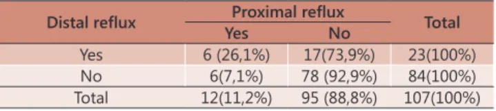
Table 2 shows that patients with distal reflux were 4.59 times more likely to present proximal reflux (p=0,01; OR=4.59 – 95% CI 1.32-15.97). A nonsignificant
correlation was found between esophageal length and LES pressure (Spearman r=0,12 and p=0,20).
DISCUSSION
The mechanisms involved in the genesis of
proximal esophageal reflux have not been fully
elucidated yet. Some causes have been proposed, such as the incompetence of the LES14, the transient lower
esophageal sphincter relaxation4,5,10 and the deficiency
of the esophageal body motility7.
In general they are considered as major changes when the pressure of the LES is lower than 6 mmHg, total length lower than 2 cm and intra-abdominal length of lower than 1 cm2. This study found that 10.8%
of patients had lower esophageal sphincter hypotonia (pressure <10 mmHg). One patient had total LES length
TABLE 1 – Mean esophageal length in patients presenting respiratory symptoms with and without proximal reflux
Proximal reflux N Mean (cm) 95% Confidence Interval
No 95 24,23 23,83 to 24,64 Yes 12 24,83 24,08 to 25,59
Student´s t test = 0.15
TABELA 2 – Prevalence of proximal reflux according to the presence of distal reflux among patients with respiratory symptoms
Distal reflux Proximal reflux Total
Yes No
Yes 6 (26,1%) 17(73,9%) 23(100%) No 6(7,1%) 78 (92,9%) 84(100%) Total 12(11,2%) 95 (88,8%) 107(100%)
lower than 2 cm. Spearman correlation showed an association between LES hypotonia and the presence
of pathological reflux in the distal canal (p=0.02).
Ineffective esophageal motility, the motor disorder more prevalent in GERD, has been found in patients with
respiratory symptoms and proven reflux6. In this study,
motor disorders were not associated neither with distal
esophageal reflux nor with proximal esophageal reflux.
Spearman correlation between variables that measured the esophageal body motor function (amplitude of the distal esophagus, peristaltic waves and waves that were not transmitted) with the presence of distal esophageal
reflux and proximal esophageal reflux did not show a statistically significant association.
The authors wished to determine if there was a relationship between esophageal length and distal
reflux with proximal esophageal reflux. In this study, Spearman correlation coefficient showed that proximal esophageal reflux was not significantly related to
esophageal length. Also, t test revealed that there
was no statistically significant difference between the
mean of the esophageal length in patients presenting respiratory symptoms with and those without proximal
reflux. However, presence of distal esophageal reflux
increased the risk of suffering from proximal esophageal
reflux (OR=4.6 p=0,017).
The esophagus begins in the neck at the cricoid cartilage and passes through the thorax within the posterior mediastinum and extends for a few centimeters past the diaphragm to its junction with the stomach3. The ideal method for measuring
esophageal length is controversial. It was measured esophageal length manometrically and its mean was 24.3 (± 1.9) cm, ranging from 20 to 30 cm, in accordance to what has been found by others3,13,15.
Awad, at al.1 performed a retrospective study
to test the relationship between esophageal length in normal control subjects, patients with esophageal disorders (achalasia, diffuse esophageal spasm, stricture, nutcracker esophagus), patients with GERD diagnosed by positive 24-hour pH monitoring, possible GERD but negative 24-hour pH monitoring. They found that patients with GERD (positive 24-hour pH monitoring) and patients with GERD-related stricture had a shorter than normal esophagus. In this study, the authors found that mean esophageal length did not differ between patients with and without
proximal esophageal reflux, probably because none of
these patients presented stricture GERD-related.
CONCLUSION
The esophageal length was not associated with presence of proximal esophageal reflux. P a t i e n t s
who presented pathological distal esophageal reflux,
independent of the esophageal length, were 4.6 times
more likely to have proximal esophageal reflux.
REFERENCES
1. Awad ZT, Watson P, Filipi CJ, Marsh RE, Tomonaga T, Shiino Y et al. Correlations between esophageal diseases and manometric length: a study of 617 patients. J Gastrointest Surg 1999; 3(5):483-488.
2. DeMeester TR, Peters JH, Bremner CG, Chandrasoma P. Biology
of gastroesophageal reflux disease: pathophysiology relating to
medical and surgical treatment. Annu Rev Med 1999; 50:469-506. 3. DeNardi FG, Riddell RH. The normal esophagus. Am J Surg Pathol
1991; 15(3):296-309.
4. Dent J, Dodds WJ, Friedman RH, Sekiguchi T, Hogan WJ, Arndorfer
RC et al. Mechanism of gastroesophageal reflux in recumbent
asymptomatic human subjects. J Clin Invest 1980; 65(2):256-267. 5. Dent J, Holloway RH, Toouli J, Dodds WJ. Mechanisms of lower
oesophageal sphincter incompetence in patients with symptomatic
gastrooesophageal reflux. Gut 1988; 29(8):1020-1028.
6. Fouad YM, Katz PO, Hatlebakk JG, Castell DO. Ineffective esophageal motility: the most common motility abnormality in patients with GERD-associated respiratory symptoms. Am J Gastroenterol 1999; 94(6):1464-1467.
7. Kasapidis P, Xynos E, Mantides A, Chrysos E, Demonakou M, Nikolopoulos N et al. Differences in manometry and 24-H ambulatory pH-metry between patients with and without
endoscopic or histological esophagitis in gastroesophageal reflux
disease. Am J Gastroenterol 1993; 88(11):1893-1899.
8. Li Q, Castell JA, Castell DO. Manometric determination of esophageal length. Am J Gastroenterol 1994; 89(5):722-725. 9. Machado MM, Cardoso PF, Ribeiro IO, Zamin J, I, Eilers RJ.
Esophageal manometry and 24-h esophageal pH-metry in a large sample of patients with respiratory symptoms. J Bras Pneumol 2008; 34(12):1040-1048.
10. Mittal RK, Holloway RH, Penagini R, Blackshaw LA, Dent J. Transient lower esophageal sphincter relaxation. Gastroenterology 1995; 109(2):601-610.
11. Moraes-Filho JP, Chinzon D, Eisig JN, Hashimoto CL, Zaterka S.
Prevalence of heartburn and gastroesophageal reflux disease in
the urban Brazilian population. Arq Gastroenterol 2005; 42(2):122-127.
12. Nasi A, Moraes-Filho JP, Cecconello I. [Gastroesophageal reflux disease: an overview.]. Arq Gastroenterol 2006; 43(4):334-341.
13. Oelschlager BK, Eubanks TR, Maronian N, Hillel A, Oleynikov D, Pope CE et al. Laryngoscopy and pharyngeal pH are complementary in the diagnosis of gastroesophageal-laryngeal
reflux. J Gastrointest Surg 2002; 6(2):189-194.
14. Sloan S, Rademaker AW, Kahrilas PJ. Determinants of gastroesophageal junction incompetence: hiatal hernia, lower esophageal sphincter, or both? Ann Intern Med 1992; 117(12):977-982.