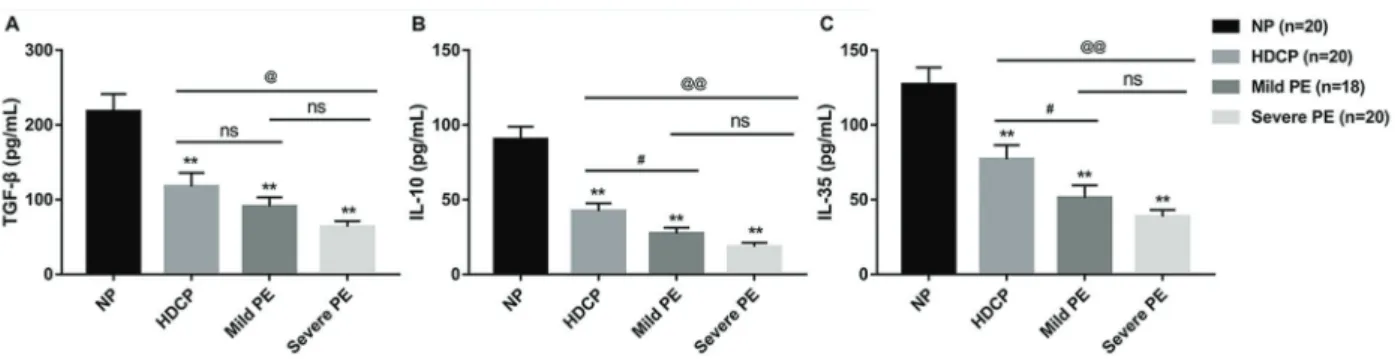
PD-1PD-L1 regulates Treg differentiation in pregnancy-induced hypertension
Texto
Imagem
Documentos relacionados
Indeed, protein deprivation increases the density of PV-IR neurons in the granule cell layer and in the hilus but decreases the number of CR-IR cells in hilus and
Desta forma, podemos concluir que realizar a filtragem na s´ erie temporal antes de model´ a-la incorre em ganho preditivo e que ´ e poss´ıvel utilizar a energia e´ olica como
a better understanding of the role of Tax mRNA in the gene expression of cellular markers we measured Tax, Foxp3, CTLA- 4, GITR, TGF-β, and IL-10 mRNA in Treg cells of 50 patients
Assim, uma vez constatada a presença de elementos que conferem credibilidade às declarações, estas deverão passar por um verdadeiro juízo de corroboração, à procura de uma
Legenda: Menos de 1 kg/ano.km 2 Entre 1 e 10 kg/ano.km 2 Entre 10 e 50 kg/ano.km 2 Entre 50 e 100 kg/ano.km 2 Entre 100 e 250 kg/ano.km 2 Castro Daire São Pedro do Sul Ovar
Os principais resultados obtidos a partir da realização do trabalho foram: compreender a incidência de emoções positivas e negativas, ou seja, emoções ambivalentes, simultaneamente
ACCORD
(2005), a rugosidade de acetábulo diminui com o início do ensaio, possivelmente pela redução das marcas de fabricação. As avaliações em 0,75 Mc foram as primeiras medidas
