Cohort of Patients Referred for Brugada Syndrome Investigation in
an Electrophysiology Service - 19-Year Registry
Stefan Warpechowski Neto, Tiago Luiz Luz Leiria, Laura Lessa Gaudie Ley, Antonio Lessa Gaudie Ley, Luiza Zwan
Dutra, Leonardo Martins Pires, Marcelo Lapa Kruse, Gustavo Glotz de Lima
Instituto de Cardiologia - Fundação Universitária de Cardiologia (IC/FUC), Porto Alegre, RS - Brazil
Mailing Address: Tiago Luiz Luz Leiria •
Av. Princesa Isabel, 370. Postal Code 90620-000, Santana, Porto Alegre, RS – Brazil
E-mail: pesquisa.leiria@gmail.com, editoracao-pc@cardiologia.org.br Manuscript received October 10, 2017, revised manuscript February 22, 2018, accepted February 22, 2018
DOI: 10.5935/abc.20180094
Abstract
Background: Brugada syndrome (SBr) is an arrhythmic condition characterized by ST-T segment abnormalities in the right precordial leads associated with a high risk of ventricular arrhythmias and sudden death. Local data regarding the clinical characteristics of patients with a typical electrocardiographic (ECG) pattern undergoing electrophysiological study are scarce. Objective: To evaluate patients with an ECG pattern suggestive of SBr referred for electrophysiological evaluation in a specialized center.
Methods: Cohort study of patients referred for electrophysiological study because of an ECG pattern compatible with SBr between January 1998 and March 2017.
Results: Of the 5506 procedures, 35 (0.64%) were for SBr investigation, 25 of which (71.42%) were performed in men. The mean age was 43.89 ± 13.1 years. The ECG patterns were as follows: type I, 22 (62.85%); type II, 12 (34.30%); and type III, 1 (2.85%). Twenty-three patients (65.7%) were asymptomatic, 6 (17.14%) had palpitations, 5 (14.3%) had syncope, and 3 (8.6%) had a family history of sudden death. Electrophysiological study induced ventricular tachyarrhythmias in 16 cases (45.7%), the mean ventricular refractory period being 228 ± 36 ms. Ajmaline / procainamide was used in 11 cases (31.4%), changing the ECG pattern to type I in 7 (63.6%). Sixteen cases (45.7%) received an implantable cardioverter defibrillator (ICD). In a mean 5-year follow-up, 1 of the 16 patients (6.25%) with ICD had appropriate therapy for ventricular fibrillation. There was no death. Other arrhythmias occurred in 4 (11.4%) cases.
Conclusions: Most patients are men, and a type I ECG pattern is the main indication for electrophysiological study. Class IA drugs have a high ECG conversion rate. The ICD event rate was 6%. (Arq Bras Cardiol. 2018; 111(1):13-18)
Keywords: Brugada Syndrome; Ventricular Tachycardia; Sudden Death.
Introduction
Brugada syndrome (BrS) is a genetic arrhythmogenic disorder characterized by typical electrocardiographic changes of the ST-T segment in the right precordial leads (V1-V3), associated with an increased risk for sudden death due to ventricular arrhythmias, mainly polymorphic ventricular tachycardia, in the absence of structural heart disease.1
The BrS was first described in 1992, relates to the loss of function in the sodium ion channels of ventricular cardiomyocytes and results from the decrease in that channel amount and failure of expression, its voltage change, time-dependent action and accelerated or prolonged inactivation recovery,2 leading to a reduction
in the sodium ion inflow and in the physiological duration
of the action potential. Despite its autosomal dominant inheritance, BrS is currently known to be sporadic in two-thirds of its cases (65%),3 due to mutations leading
to the failure of the SCN5A gene function that encodes sodium channels – initially re-written in 19984 – or to other
350 pathogenic mutations in several sodium, potassium or calcium channel genes, currently representing percentages of genetic changes lower than 35%.
Because of its multifactorial etiology that involves the contribution of genetic, environmental and hormonal factors, the clinical manifestation varies, affecting mainly men (proportion of 8-9:1),5 with clinical onset, on average,
at the age of 40 years, and major outcome of sudden death triggered by sleep, vagotonia or fever. Brugada syndrome accounts for 20% of the sudden cardiac deaths with structurally normal hearts6 and 4-12% of all sudden
cardiac deaths.7
Methods
This is a cohort study of patients referred for electrophysiological study at the ICFUC electrophysiology laboratory between January 1998 and March 2017. Of the 5506 studies performed in that period, 35 (0.67%) corresponded to assessment of patients with electrocardiographic pattern compatible with BrS (Brugada pattern), who were followed up from that study on.
The inclusion criteria were: absence of structural heart disease, absence of personal history of aborted sudden death, electrocardiogram (ECG) compatible with type I, II or III Brugada pattern, and electrophysiological study under a preestablished protocol of ventricular stimulation with three baseline cycles (600, 500 and 400 ms) and introduction of up to three extra stimuli. Diagnostic challenge with infusion of class IA antiarrhythmic drugs according to the Vaughan Williams classification (ajmaline at the dose of 1 mg/kg for 10 minutes or procainamide 10 mg/kg for 10 minutes) was performed in type II electrocardiographic presentations, in accordance with the most used drugs in European and American studies.8
From the electrophysiological study on, the patients were followed up through medical appointments at regular six-month intervals, medical record review and/or telephone contact.
Statistical analysis
Our data bank was stored in Microsoft Excel sheets and analyzed by use of the Statistical Package for Social Sciences (SPSS) software, version 20.0 (Armonk, NY, USA: IBM Corp). The continuous variables were expressed as mean (± standard deviation) and compared by use of independent samples t test. The continuous variables of non-gaussian distribution were expressed as median [interquartile range (IQR)] and compared by using Mann-Whitney U test. The categorical variables were expressed as percentages and compared by use of chi-square test. The comparisons between groups were performed by using z test, with post-hoc Bonferroni analysis to identify the statistical difference. Kaplan-Meyer event-free survival analysis was performed, with percentage survival and standard error. Differences between the frequency of events over time according to the variables identified were compared by use of log-rank test. A p value < 0.05 was considered statistically significant.
Follow-up outcomes
By use of electronic medical record review or telephone call, the occurrence of the following events was investigated: death, syncope, hospitalization due to arrhythmia, and recurrent palpitations requiring medical care. In patients receiving an implantable cardioverter defibrillator (ICD), the occurrence of shock was investigated, and, when present, the appropriateness (shock due to ventricular arrhythmia) or inappropriateness (shock due to supraventricular tachycardia, increased T-wave sensitivity or electromagnetic interference) of the event was assessed.
Results
Of the 35 patients included in the cohort, 22 (62.85%) showed a type I electrocardiographic pattern, 12 (34.30%)
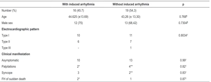
Regarding sex, 25 patients (71.42%) were of the male sex. The mean age was 43.89 ± 13.1 years, and most patients (65.71%) were asymptomatic at the time of inclusion. Regarding the symptoms, 6 patients (17.14%) had palpitations, 5 (14.28%) reported syncope, and 3 (8.57%) reported sudden death of a first-degree relative. Sixteen patients (45.7%) had induced ventricular tachyarrhythmias on stimulation – mean refractory ventricular period of 228 ± 36 ms. Eleven patients (31.4%) with type II ECG pattern received ajmaline or procainamide, and 7 of them (63.6%) changed to type I ECG pattern. Table 1 summarizes the clinical, electrocardiographic and electrophysiologic characteristics of the patients included in this study. No difference was observed between the groups with and without induced arrhythmia (Table 2).
Sixteen patients (45.7%) received an ICD. Of those patients, only 2 had no arrhythmia triggered (reason for implantation: history of sudden death and syncope). Two patients with ventricular arrhythmia (1 with nonsustained ventricular tachycardia and another with ventricular fibrillation) refused to receive the ICD despite the clinical indication. In a mean follow-up of 5 years, 1 of the 16 patients (6.25%) who received the ICD had appropriate therapy for ventricular fibrillation, and 1 (6.25%) attended no consultation after implantation (Figure 1). No death was reported during follow-up. Four patients (11.4%) had other arrhythmic events, such as episodes of nonsustained supraventricular tachyarrhythmias and frequent premature ventricular complexes. Figure 2 shows the discrimination of events in patients with ICD.
Discussion
The long-term event rate of patients diagnosed with BrS or electrocardiographic pattern of BrS is little known, because of the relative short time since that syndrome initial description in 1992,1 in addition to the limited follow-up duration of current
studies, most of which no longer than 3 years.
Table 1 – Clinical, electrocardiographic and electrophysiological study characteristics
Clinical presentations N = 35
Men 25 (71.42%)
Age 43.89 ± 13.1 years
Asymptomatic 23 (65.7%)
Syncope 5 (14.3%)
Palpitation 6 (17.14%)
Electrocardiographic presentations
Type I 22 (62.85%)
Type II 12 (34.30%)
Type III 1 (2.85%)
Electrophysiological study
Ventricular tachyarrhythmia 16 (45.7%)
Refractory period 228 ± 36 ms
HV interval 49 ± 8.6 ms
Figure 1 – Event-free survival curve of the patients with implantable cardioverter defibrillator (ICD).
100
90
80
70
60
50
40
30
20
10
0
Likelihood of appropriate-shock-free
survival ICD (%)
Patients with ICD
0 500 1000 1500 2000 2500 3000 3500
Days
18 10 6 4 4 3 3 0
Number at risk
Table 2 – Characteristics regarding arrhythmia induction during the electrophysiological study
With induced arrhythmia Without induced arrhythmia p
Number (%) 16 (45.7) 19 (54,3)
Age 44.625 (±13.69) 43,26 (± 13,30) 0.768¶
Male sex 12 (75) 13 (68,42) 0.7304¶
Electrocardiographic pattern
Type I 10 11 0.8034†
Type II 6 7
Type III - 1
Clinical manifestation
Asymptomatic 10 13 0.99†
Palpitations 2* 4** 0.82†
Syncope 3 2** 0.83†
FH of sudden death 2* 1 0.87†
* In the group of patients with induced arrhythmia, one had palpitations and sudden death in the family. ** In the group of patients without induced arrhythmia, one had palpitations and syncope. FH: family history. ¶: Student t test; †: Chi-square / Fisher exact test.
The worldwide prevalence of BrS is heterogeneous, because of its nonpermanent electrocardiographic tracings, disparate genetic changes or undiagnosed patients. In addition, potential arrhythmic events and sudden death can occur,
Figure 2 – Discrimination of events in patients with implantable cardioverter defibrillator (ICD).
ICD – Nonsustained supraventricular
tachycardia
ICD – Frequent premature ventricular complexes
ICD – Ventricular fibrillation
ICD – No arrhythmic event detected
No implantable device
2 2
1
11
19
The male predominance found in this study, already reported in the initial description of the disorder as 75%,1
is in accordance with data from the global literature, whose percentages range according to the geographic location: 84.3% in a large Japanese cohort,5 70% in a Spanish cohort,9
and 57.9% in a Belgian study.10 The proportion is maintained
in geographically close populations, such as an Argentinian cohort of similar size to ours (43 patients), whose male percentage reached 85%.11 Likewise, the mean age of
43.89 years coincides with the findings of several populations studied, even those with larger samples,12-14 clearly and
repeatedly showing the impairment of young individuals with high productive capacity, emphasizing the importance of the correct identification of those at higher risk based on a common epidemiological profile.
Although the history of ventricular arrhythmias of the fibrillation or tachycardia type is a predictor of mortality in patients with BrS, and the arrhythmia recurrence rates are around 7.7% per year,14 most of our patients were asymptomatic
at the time of the electrophysiological study. If, on the one hand, asymptomatic patients without additional risk factors are currently classified as of low risk,14,15 on the other it is
difficult to predict the potential risk based solely upon the ECG assessment, requiring a multifactorial approach in the search for other complications, such as family history of sudden death, personal history of syncope or induced arrhythmia, because the electrocardiographic pattern in isolation seems insufficient to define high risk for events.16
The incorporation of the advances in cardiology in the search for risk predictors has diverging results in a scenario where the identification of susceptibility is the key point, and, because therapy showed no significant changes in past years, it remains without any effective pharmacological alternative, being limited
to have a significant, although indirect, contribution to the patients’ quality of life because of their daily social or professional repercussions,17 adding arguments to the already challenging
process of identifying its real beneficiaries.
In 2003, the assessment of 547 patients with the BrS pattern and no previous history of sudden death, with a mean 24-month follow-up, a positive electrophysiological study was associated with arrhythmic outcomes on a multivariate analysis, with a 6-fold higher risk in 2 years versus a 2.5-fold for the second better predictor, the previous history of syncope.13 In a cohort14 of 1029 patients (72% of men, mean
age of 45 years, and 64% asymptomatic - a population profile similar to ours), the electrophysiological study was performed in 638 individuals and had a 41% positivity, but was not a risk predictor on multivariate analysis, leaving only personal history and electrocardiographic pattern correlated with events.
Two years later, a prospective multicenter study,15 assessing
specifically the accuracy of arrhythmia induced by stimulation and the identification of new risk predictors, evidenced that induced arrhythmia was not an event predictor in a 36-month follow-up (and only 34% of the patients with induced arrhythmia experienced a new induction when repeating the protocol), in addition to the same findings regarding type I ECG and personal history of syncope, and the additional positive finding for ventricular refractory period shorter than 200 ms and QRS fragmentation. Of the 14 events, only 1 showed no spontaneous type I pattern, with a number needed to treat (NNT) of 25.2.
follow-up, with higher risks for patients induced with one or two extra stimuli.18 The overall analysis of data indicates that
the electrophysiological study is useful, mainly in patients at intermediate risk, to whom the clinical characteristics cannot provide a dichotomous classification of high or low risk.
In 2017, Sieira et al.19 proposed a model of risk classification
based on a cohort of 400 patients from a single Belgian center, with mean age and percentage of asymptomatic individuals similar to those of our cohort, in which the clinical factors associated with outcomes were categorized into a score model including the following variables: type I electrocardiographic pattern, history of sudden death of a first-degree relative younger than 35 years, arrhythmia induced on electrophysiological study, syncope, sinus node disease and history of sudden death. In the model proposed, a score equal to or greater than 2 represents high risk for outcome, with positive predictive value of 90%, maintained at 81% when having external validity.
In the present study, the rate of the implantable device events was lower than that reported in the literature, including national studies with patients with BrS,20 and the
one patient with appropriate therapy received it in the first year of follow-up. Nevertheless, the mean 5-year follow-up showed a temporal gain as compared to many similar studies, allowing for the analysis of events in a larger time window – knowing that the risks are continuous throughout life – with the potential advantage of overcoming occasional inaccurate clinical data, manly family history, because the information is patient-dependent and previous data might not be well characterized in the generation immediately before the proband.
Although controversial, the use of electrophysiological study for stratification has shown to be a useful tool to identify high-risk patients, representing a clear signal that the ventricle is more excitable, and, thus, prone to arrhythmic events.21
Limitations
This study has limitations such as the fact that the cohort is not constituted by patients identified by ECG, but by those, who, according to their attending doctors would benefit from an electrophysiological study for risk stratification, a fact that limited the sample size and can be a bias by selecting patients that raise more concern about future events. Another fact is that, of the 35 patients, 5 did not undergo follow-up at the same institution where the electrophysiological study was performed. In such cases, data were limited to information collected via telephone, with checking up on neither the electronic medical records nor the devices. Moreover, we
performed no genetic study of the population assessed, because it is not routinely available in the healthcare system in addition to its costs.
Conclusion
Brugada syndrome is a potentially fatal arrhythmic condition, and reports on it increased substantially in past years. In this cohort, similarly to the world literature, most patients are of the male sex and had spontaneous type I electrocardiographic pattern. Class IA antiarrhythmic drugs of the Vaughan Williams classification have high rates of electrocardiographic conversion when used for diagnostic challenge. The rate of arrhythmic event was 6.25%, and mortality was lower than that in the literature. The electrophysiological study for risk assessment, although controversial, is currently a useful tool for patient’s stratification, mainly when the clinical characteristics are poor and do not allow for estimating accurately the risks of future events.
Author contributions
Conception and design of the research, Acquisition of data, Analysis and interpretation of the data, Statistical analysis, Obtaining financing, Writing of the manuscript and Critical revision of the manuscript for intellectual content: Warpechowski Neto S, Lima GG, Ley LLG, Ley ALG, Dutra LZ, Pires LM, Kruse ML, Leiria TLL.
Potential Conflict of Interest
No potential conflict of interest relevant to this article was reported.
Sources of Funding
There were no external funding sources for this study.
Study Association
This study is not associated with any thesis or dissertation work.
Ethics approval and consent to participate
1. Brugada P, Brugada J. Right bundle branch block, persistent ST segment elevation and sudden cardiac death: a distinct clinical and electrocardiographic syndrome: a multicenter report. J Am Coll Cardiol. 1992;20(6):1391-6.
2. Tse G, Liu T, Li KH, Laxton V, Chan YW, Keung W, et al. Electrophysiological mechanisms of Brugada syndrome: insights from pre-clinical and clinical studies. Front Physiol. 2016 Oct 18;7:467.
3. Campuzano O, Brugada R, Iglesias A. Genetics of Brugada syndrome. Curr Opin Cardiol. 2010;25(3):210-5.
4. Chen Q, Kirsch GE, Zhang D, Brugada R, Brugada J, Brugada P, et al. Genetic basis and molecular mechanism for idiopathic ventricular fibrillation. Nature. 1998;392(6673):293-6.
5. Matsuo K, Akahoshi M, Nakashima E, Suyama A, Seto S, Hayano M, et al. The prevalence, incidence and prognostic value of the Brugada-type electrocardiogram: a population-based study of four decades. J Am Coll Cardiol. 2001;38(3):765-70.
6. Juang JM, Huang SK. Brugada syndrome—an underrecognized electrical disease in patients with sudden cardiac death. Cardiology. 2004;101(4):157-69.
7. Vohra J, Rajagopalan S; CSANZ Genetics Council Writing Group. Update on the diagnosis and management of Brugada Syndrome. Heart Lung Circ. 2015;24(12):1141-8.
8. Polovina MM, Vukicevic M, Banko B, Lip GH, Potpara TS. Brugada syndrome: a general cardiologist’s perspective. Eur J Intern Med. 2017 Oct;44:19-27.
9. Benito B, Sarkozy A, Mont L, Henkens S, Berruezo A, Tamborero D, et al. Gender diferences in clinical manifestations of Brugada syndrome. J Am Coll Cardiol. 2008;52(19):1567-73.
10. Sieira J, Conte G, Ciconte G, de Asmundis C, Chierchia GB, Baltogiannis G, et al. Clinical characterisation and long-term prognosis of women with Brugada syndrome. Heart. 2016;102(6):452-8.
11. Abud AM, Carlessi A, Goyeneche R, Strada B, Arceluz M, Fernández A, et al. Retrospective analysis of patients with Brugada syndrome and implantable cardioverter defibrillator. Rev Argent Cardiol. 2014;82(1):21-5.
12. Priori SG, Napolitano C, Gasparini M, Pappone C, Della Bella P, Giordano U, et al. Natural history of Brugada syndrome: insights for risk stratification and management. Circulation. 2002;105(11):1342-7.
13. Brugada J, Brugada R, Brugada P. Determinants of sudden cardiac death in individuals with the electrocardiographic pattern of Brugada syndrome and no previous cardiac arrest. Circulation. 2003;108(25):3092-6.
14. Probst V, Veltmann C, Eckardt L, Meregalli PG, Gaita F, Tan HL, et al. Long-term prognosis of patients diagnosed with Brugada syndrome: results from the FINGER Brugada Syndrome Registry. Circulation. 2010;121(5):635–43.
15. Priori SG, Gasparini M, Napolitano C, Della Bela P, Ottonelli AG, Sassone B, et al. Risk Stratification in Brugada Syndrome: Results of the PRELUDE (Programmed ELectrical stimUlation preDictive valuE) Registry. J Am Coll Cardiol. 2012;59(1):37-45.
16. Delise P, Allocca G, Marras E, Giustetto C, Gaita F, Sciarra L, et al. Risk stratification in individuals with the Brugada type 1 ECG pattern without previous cardiac arrest: usefulness of a combined clinical and electrophysiologic approach. Eur Heart J. 2011;32(2):169-76.
17. Probst V, Plassard-Kerdoncuf D, Mansourati J, Mabo P, Sache F, Fruchet C, et al. The psychological impact of implantable cardioverter defibrillator implantation on Brugada syndrome patients. Europace. 2011;13(7):1034-9.
18. Sroubek J, Probst V, Mazzanti A, Delise P, Hevia JC, Ohkubo K, et al. Programmed ventricular stimulation for risk stratification in the Brugada Syndrome: a pooled analysis. Circulation. 2016; 133(7):622-30.
19. Sieira J, Conte G, Ciconte G, Chierchia GB, Casado-Arroyo R, Baltogiannis G, et al. A score model to predict risk of events in patients with Brugada Syndrome. Eur Heart J. 2017;38(22):1756-1763.
20. da Fonseca SM, Belo LG, Carvalho H, Araújo N, Munhoz C, Siqueira L, et al. Clinical follow-up of patients with implantable cardioverter-defibrillator. Arq Bras Cardiol. 2007;88(1):8-16.
21. Brugada R, Campuzano O, Sarquella-Brugada G, Brugada J, Brugada P. Brugada syndrome. Methodist Debakey Cardiovasc J. 2014;10(1):25-8.