CHALLENGING CLINICAL CASES
Laparoscopic approach to pheochromocytoma in pregnancy:
case report
_______________________________________________
Felipe de Almeida e Paula
1,2,3, Ravisio Israel dos Santos Junior
1,2,3, Odivaldo Antonio Ferruzzi
2, Rafael Osti
de Melo
3, Mariana Takaku
1,21 Hospital Regional do Câncer de Presidente Prudente, Presidente Prudente, SP, Brasil; 2 Santa Casa
de Misericórdia de Presidente Prudente, Presidente Prudente, SP, Brasil; 3 Faculdade de Medicina de Presidente Prudente, Universidade do Oeste Paulista, Presidente Prudente, SP, Brasil
ABSTRACT
ARTICLE
INFO
______________________________________________________________ ______________________
A 32-year-old 22-week pregnant hypertensive woman with sporadic episodes of headaches, sweating, and facial flushing was diagnosed with pheochromocyto-ma through biochemical and ipheochromocyto-maging tests. Perioperative pheochromocyto-management included a multidisciplinary approach, symptom stabilization with ɑ blockade followed by ß blockade, and tumor resection by laparoscopic adrenalectomy at 24 weeks gesta-tion. The diagnosis was confirmed by histopathological examination and immuno-histochemistry tests. The decision for surgical removal of the tumor was based on maternal symptoms, tumor size, gestational age, the possibility of doing a laparos-copy, and the expertise of the surgical team.
INTRODUCTION
Pheochromocytoma (PCC) is a neuroendo-crine tumor derived from chromaffin cells and lo-cated within the adrenal medulla in approximately 90% of cases (1). Although extraordinarily rare during pregnancy with a reported incidence less than 0.2 per 10,000 pregnancies, PCC should be considered when severe hypertension occurs before 20 weeks gestation or when hypertension is asso-ciated with disease symptoms such as headaches, palpitations, and diaphoresis (2). PCC in gestation, when untreated, raises maternal and fetal mortality
to up to 50% (3, 4). However, early diagnosis and appropriate treatment reduce maternal mortality to less than 5% and fetal mortality to below 15% (3, 5). The diagnosis can be established by detecting elevated levels of catecholamines and their metab-olites in plasma or urine. Ultrasound and magnetic resonance imaging (MRI) are the preferred imaging modalities for tumor localization during pregnancy (6). Surgery is the definitive treatment for PCC after management of hypertension with α blockade, but
the timing of tumor removal depends on the gesta-tional age, the severity of maternal symptoms, and the risks associated with pregnancy termination (4).
Vol. 44 (3): 629-633, May - June, 2018 doi: 10.1590/S1677-5538.IBJU.2017.0540
Keywords:
Pheochromocytoma; Pregnancy; Laparoscopy
Int Braz J Urol. 2018; 44: 629-33
_____________________
Submitted for publication: October 05, 2017
_____________________
Accepted after revision: November 22, 2017
_____________________
CASE PRESENTATION
A 32-year-old Caucasian woman at 22 weeks of gestation with singleton live fetus pre-sented 5 weeks prior with sporadic episodes of headaches, facial flushing, and sweating. The obstetric ultrasound showed a healthy fetus. Her first pregnancy four years earlier was compli-cated by severe preeclampsia at 34 weeks gesta-tion for which she was induced and delivered by emergency cesarean section. Since then, she was treated with methyldopa 500 mg/day, but her blood pressure was not monitored and she had no other complaints or complications. She was not on any regular medications, was not a smoker or an alcohol user, and there was no history of illicit drug use nor family history of multiple endocrine neoplasia (MEN). Due to palpitations, nine months earlier she underwent a cardiac stress test, which detected sustained
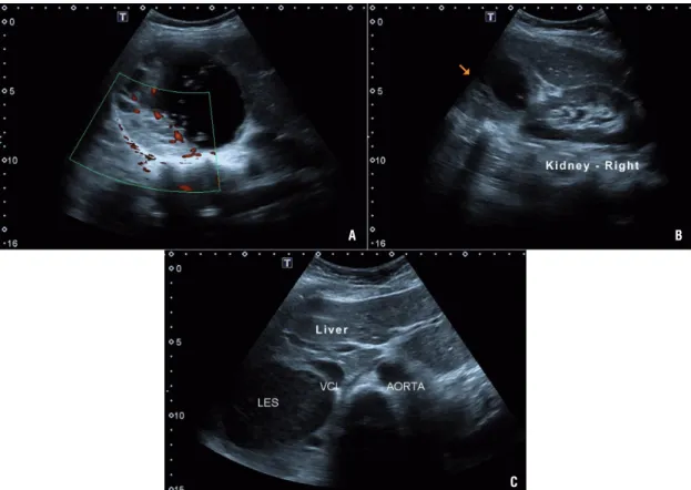
supraventricular paroxysmal tachycardia, fol-lowed by an electrophysiology study that failed to reveal any disorder or dysfunction. The echo-cardiogram was also normal. Symptoms of head-aches, facial flushing, and sweating started at 17 weeks gestation. Routine laboratory tests re-vealed a fasting glucose of 92 mg/dL and serum creatinine of 0.5 mg/dL. Complete blood count, sodium, potassium, calcium, phosphorus, AST, ALT, total cholesterol and fractions, TSH, free T4, urinary sediment levels, serum aldosterone, calcitonin, parathyroid hormone (PTH), and cor-tisol were all normal. However, metanephrine and normetanephrine on 24 hours urine collec-tion were 6873.2 µg/24 h (reference value <280 µg/24 hours) and 6299.2 µg/24 hours (reference value <732 µg/24 hours), respectively. Ultraso-nography of the abdomen revealed a 10.1 × 9.5 cm mass in the right adrenal gland consistent with PCC (Figure-1). Blood pressure was
con-A B
C
IBJU| LAPAROSCOPIC APPROACH TO PHEOCHROMOCYTOMA
trolled with prazosin 2 mg/day for 10 days fol-lowed by propranolol 20 mg/day for four days, when a consultation by a multidisciplinary team, including surgeons, anesthesiologist, obstetrician, and endocrinologist, was held to determine the most appropriate management. The surgical ap-proach selected was laparoscopic transperitoneal right adrenalectomy due to the considerable ex-perience of the surgical team with the technique, the patient’s significant symptoms, and the ges-tational age (second trimester, 24 weeks gestation at the time of surgery). The patient was placed in a modified flank position, 45 degrees back from vertical. Three ports were used in triangulation, and an additional fourth port for the liver retrac-tion. The patient received combined epidural and general anesthesia with an epidural catheter placed for postoperative analgesia and was continuously monitored with a cardioscope, pulse oximetry, in-vasive arterial pressure monitoring from the radial artery, and capnography. Serial arterial blood gas-es and glucose were monitored to prevent maternal acidosis and hypoglycemia. Central venous access was accomplished via her right jugular vein for administration of liquids and vasoactive agents. General anesthesia was maintained with continu-ous remifentanil and propofol infusion. Special consideration was taken with pneumoperitoneum pressure, which was maintained below 12 mmHg for nearly the entire duration of surgery and was only raised to 15 mmHg for a few minutes during vessel dissection to prevent a reduction in utero-placental blood flow. During dissection of the ad-renal vessels, a sodium nitroprusside infusion (0.8 µg/kg/min for approximately 7 min.) and inter-mittent boluses of esmolol 10 mg IV (total 20 mg) were used to control mean arterial pressure (MAP reduced from 136 mmHg to 83 mmHg) and heart rate (lowered from 133 bpm to 86 bpm). An ultra-sonic dissector - Harmonic scalpelTM (Ethicon Endo
Surgery INC - Johnson & Johnson, NJ, USA) was used for adrenal gland dissection. Blood loss was less than 50 mL. The patient remained normoten-sive for the remainder of the surgery and postop-eratively. The operative time was 120 min and no complications were recorded. The patient was ex-tubated immediately after surgery with good pain control, and the epidural catheter was removed on
the same day of surgery. Obstetric ultrasound per-formed soon after anesthesia recovery revealed no abnormalities. Antihypertensive therapy was not required postoperatively or during the remainder of pregnancy. The patient was discharged on post-operative day four with no symptoms and normal blood pressure.
Histopathological examination and immu-nohistochemistry tests (positive chromogranin A and synaptophysin staining) confirmed the diag-nosis of PCC.
Five weeks after surgery, while remain-ing normotensive without antihypertensive drug therapy, the patient was admitted due to prema-ture rupprema-ture of membranes, and one day later suffered placental abruption with severe bleed-ing. She underwent emergency cesarean section but the newborn, who was born alive, died pre-maturely within 48 hours.
DISCUSSION
PCC in pregnancy can cause fatal hyperten-sive crisis that can be triggered by vaginal delivery, general anesthesia, the physical effects of the en-larging uterus, uterine contractions, or fetal move-ments (2), and may impose a serious risk to the fetus because extreme vasoconstriction in the uteropla-cental circulation may result in intrauterine hypoxia and premature placental abruption (5). Undiagnosed and/or untreated PCC carry a risk of mortality for both mother and fetus as high as 58% (4, 6).
Detecting elevated levels of catecholamines and their metabolites in plasma and urine establish-es the diagnosis of PCC. Once a diagnosis has been confirmed biochemically, ultrasonography and MRI are the preferred modalities of tumor localization for the safety of the fetus (2, 4, 5).
The success of treatment will depend on the appropriate preoperative medical management, which should always be started with ɑ-adrenergic blockade (phenoxybenzamine or prazosin) for ad-equate control of blood pressure (9). Pre-surgical preparation with this class of drugs is one of the main reasons why surgical mortality has decreased over the last 30 years to <3% (5). Even though there is no consensus regarding the optimal duration of medical pretreatment, it should be given for 10‒14 days or until a stable hemodynamic condition is achieved. In case of tachyarrhythmias, β-adrenergic blockade should only be started after some days of appropriate α-adrenergic blockade (2, 4, 7). A collab-orative multidisciplinary team, including surgeons, anesthesiologist, obstetrician, and endocrinologist, is decisive for case management (10). Surgical re-section of the tumor is the definitive treatment for PCC, which can be achieved either by open, laparo-scopic, or robotic approaches (1, 6, 8). Laparoscop-ic adrenalectomy is the surgLaparoscop-ical approach of first choice if tumor size is <7 cm (4, 10, 11); this is a safe procedure with a complication rate <8% (5, 9). Al-though the posterior retroperitoneoscopic approach seems to be a good alternative for adrenalectomy than laparoscopic transperitoneal approach, avoid-ing the need to enter the peritoneal cavity, there is no evidence that supports the superiority of one over the other, both showing similar low morbidity and mortality (11). The choice of surgical approach must be based mainly on the expertise of the surgi-cal team and the greater maternal and fetal safety. In this case, laparoscopic transperitoneal approach was chosen, considering the greater experience with this technique by the surgical team, as well as the unfeasible prone position of pregnant patient to perform a posterior retroperitoneoscopic approach.
Timing of tumor excision in pregnant wom-en will depwom-end on the gestational age at which di-agnosis is made, fetal development, and the severity of maternal symptoms. Surgery should be avoided in the first trimester — as organogenesis is
incom-plete and miscarriage is highly likely — as well as in the third trimester — because by then the enlarging uterus precludes adequate access and visualization of the abdomen (12). Thus, the second trimester is recommended as the ideal time for surgical inter-vention. Surgical removal is recommended before 24 weeks of gestation (3, 5, 7, 8). However, after 24 weeks of gestation, the patient can be treated with the appropriate ɑ-adrenergic blockade until the fe-tus is viable, when the tumor can be removed after an elective cesarean section (4, 7, 8). In the current case, following a multidisciplinary consultation and with the patient’s consent, and having been treated with prazosin for 10 days, she underwent laparo-scopic adrenalectomy at 24 weeks gestation.
IBJU| LAPAROSCOPIC APPROACH TO PHEOCHROMOCYTOMA
Importantly, perioperative management in-cludes treatment of both mother and fetus (4). In the current case, management was effective, with-out intra or postoperative complications, resulting in complete resolution of the disease. Despite the perinatal fetal demise, maternal safety was pre-served and delaying surgical resection could have increased the risk of catastrophic complications in late pregnancy.
CONCLUSIONS
PCC is a rare but important cause of hy-pertension in pregnancy. It should be included and investigated as a differential diagnosis in cases of
labile hypertension in pregnant women with or without associated symptoms, because inadequate treatment considerably increases maternal and fetal mortality. Management of PCC during pregnancy should be discussed on a case by case basis by a multidisciplinary team. Surgical resection is recom-mended during the second trimester of pregnancy. Considering its benefits and the expertise of the surgical team, laparoscopic surgery is the favored technique.
CONFLICT OF INTEREST
None declared.
REFERENCES
1. Castilho LN, Simoes FA, Santos AM, Rodrigues TM, Santos Junior CA. Pheochromocytoma: A Long-Term Follow-Up of 24 Patients Undergoing Laparoscopic Adrenalectomy. Int Braz J Urol. 2009;35:24-35.
2. Dugas G, Fuller J, Singh S, Watson J. Pheochromocytoma and pregnancy: a case report and review of anesthetic management. Can J Anesth. 2004; 51: 134-8.
3. Santos DRP, Barbisan CC, Marcellini C, Santos RMVR. Feocromocitoma e gravidez: Relato de caso e revisão atualizada. J Bras Nefrol. 2015;37:496-500.
4. Oliva R, Angelos P, Kaplan E, Bakris G. Pheochromocytoma in Pregnancy. A Case Series and Review. Hypertension. 2010; 55:600-6.
5. Lenders JWM. Pheochromocytoma and pregnancy: a deceptive connection. Eur J Endocrinol. 2012;166:143-50. 6. Memon MA, Aziz W, Abbas F. Surgical management of
pheochromocytoma in a 13-week pregnant woman. BMJ Case Rep. 2014. pii: bcr2013202838
7. Kiroplastis K, Kambaroudis A, Andronikou A, Reklou A, Kokkonis D, Petras P, et al. Dealing with Pheochromocytoma during the First Trimester of Pregnancy. Case Rep Obstet Gynecol. 2015 ;2015:439127.
8. Podolsky ER, Feo L, Brooks AD, Castellanos A. Robotic Resection of Pheochromocytoma in the Second Trimester of Pregnancy. JSLS. 2010;14:303-8.
9. George J, Tan LYL: Pheochromocytoma in Pregnancy. A Case Report and Review of Literature. Obstet Med. 2010;3:83-5. 10. Conzo G, Pasquali D, Gambardella C, Pietra CD, Esposito
D, Napolitano S et al. Long-term outcomes of laparoscopic adrenalectomy for Cushing disease. Int J Surg. 2014;12:107-11.
11. Conzo G, Tartaglia E, Gambardella C, Esposito D, Sciascia V, Mauriello C, et al. Minimally invasive approach for adrenal lesions: Systematic review of laparoscopic versus retroperitoneoscopic adrenalectomy and assessment of risk factors for complications. Int J Surg. 2016;28:118-23. 12. Kitayama K, Kashiwagi S, Amano R, Noda S, Ohira G,
Yamazoe S et al. A Case of Bilateral Pheochromocytoma During Pregnancy. BMC Surg. 2015;15:55.