online | memorias.ioc.fiocruz.br
Recombinant hepatitis C virus-envelope protein 2
interactions
with low-density lipoprotein/CD81 receptors
Ana Carolina Urbaczek1/+, Valdecir Farias Ximenes2, Ana Afonso3,4,5, Wesley Cardoso Generoso6,
Camila Tita Nogueira1, Aline Tansini1, Luciana Teresa Dias Cappelini5, Wilson Malagó Júnior6,
Flávio Henrique da Silva6, Luiz Marcos da Fonseca1, Paulo Inácio da Costa1
1Departamento de Análises Clínicas, Faculdade de Ciências Farmacêuticas de Araraquara 2Departamento de Química, Faculdade de Ciências, Universidade Estadual Paulista, Bauru, SP, Brasil 3Departamento de Parasitologia Médica, Unidade de Parasitologia Médica e Microbiologia,
Instituto de Higiene e Medicina Tropical, Universidade Nova de Lisboa, Lisboa, Portugal 4Departamento de Morfologia e Patologia 6Departamento de Genética e Evolução, Universidade Federal de São Carlos, São Carlos, SP, Brasil
5Departamento de Química e Física Molecular, Instituto de Química de São Carlos, Universidade de São Paulo, São Carlos, SP, Brasil
Hepatitis C virus (HCV) envelope protein 2 (E2) is involved in viral binding to host cells. The aim of this work was to produce recombinant E2B and E2Y HCV proteins in Escherichia coli and Pichia pastoris, respectively, and to study their interactions with low-density lipoprotein receptor (LDLr) and CD81 in human umbilical vein endothelial cells (HUVEC) and the ECV304 bladder carcinoma cell line. To investigate the effects of human LDL and differences in protein structure (glycosylated or not) on binding efficiency, the recombinant proteins were either associated or not associated with lipoproteins before being assayed. The immunoreactivity of the recombinant proteins was analysed using pooled serum samples that were either positive or negative for hepatitis C. The cells were immunophenotyped by LDLr and CD81 using flow cytometry. Binding and binding inhibition assays were performed in the presence of LDL, foetal bovine serum (FCS) and specific antibodies. The results revealed that binding was reduced in the ab-sence of FCS, but that the addition of human LDL rescued and increased binding capacity. In HUVEC cells, the use of antibodies to block LDLr led to a significant reduction in the binding of E2B and E2Y. CD81 antibodies did not affect E2B and E2Y binding. In ECV304 cells, blocking LDLr and CD81 produced similar effects, but they were not as marked as those that were observed in HUVEC cells. In conclusion, recombinant HCV E2 is dependent on LDL for its ability to bind to LDLr in HUVEC and ECV304 cells. These findings are relevant because E2 acts to anchor HCV to host cells; therefore, high blood levels of LDL could enhance viral infectivity in chronic hepatitis C patients.
Key words: HCV - E2 - LDLr - CD81
doi: 10.1590/0074-02760140441
Financial support: FAPESP (2008/58957-0) + Corresponding author: [email protected] Received 23 November 2014
Accepted 20 March 2015
Hepatitis C virus (HCV) is a small, enveloped RNA virus. A single open reading frame encodes a precursor polyprotein that is cleaved via the signalling mechanisms of host and viral proteases, generating 10 different struc-tural [core (C), envelope 1 (E1) and envelope 2 (E2)] and nonstructural 2-5 proteins, in addition to the protein P7 (Bartenschlager & Lohmann 2000, Penin et al. 2004).
The E2 protein is a 70-kDa viral envelope glyco-protein that includes an N-terminal ectodomain and a C-terminal transmembrane domain. After synthesis, the E2 protein undergoes a process of post-translational modification and presents nine to 11 potential glycosyla-tion sites, which differ depending on virus genotype and subtype, but are nevertheless well conserved and are therefore good antigenic targets for developing antiviral molecules (Liu et al. 2001). E1 and E2 proteins perform essential functions during different stages of the HCV
replication cycle and are necessary for initiation of the in-fection process; their functions include receptor binding, fusion with the host cell membrane and invasion (Barten-schlager & Lohmann 2000, Dubuisson et al. 2008).
con-ducted by Albecka et al. (2012) investigated the role of LDLr in the HCV life cycle by comparing virus entry to a process of lipoprotein uptake and showed that HCV par-ticles can interact with LDLr. However, this interaction does not necessarily lead to a productive infection, but rather indicates a role for LDLr as a lipid-providing recep-tor, which modulates viral RNA replication.
Vascular changes in the cirrhotic livers of patients with chronic hepatitis C have attracted increasing inter-est because little is known about the relationship of HCV with endothelial cells. As this pathology is associated with major complications and prognostic implications, the necessity of acquiring a more detailed characterisa-tion of the interaccharacterisa-tions between HCV and cells during infection is required. Previous studies using hepatoma cell lines, lymphocyte cell lines and nonhuman cell lines have already demonstrated that the E2 protein can bind to these cells. In the current study, we tested E2 binding to human umbilical vein endothelial cells (HUVEC) and ECV304 cells, both of which are endothelial cell mod-els. Therefore, we aimed to study the binding interac-tions between recombinant E2 proteins and LDLr and CD81, two well known binding partners of E2, in two different endothelial cell lines: HUVEC, which have a high concentration of LDLr but no CD81 receptors, and ECV304 cells, which are a derivative of the human uri-nary bladder carcinoma T24 cell line and possess a high concentration of LDLr and a low concentration of CD81. To investigate how human LDL and differences in pro-tein structure (glycosylated or not) affected binding ef-ficiency, we examined recombinant proteins that were either associated or not associated with lipoproteins.
MATERIALS AND METHODS
Strains, cell lines and media - The DH5α Escherichia coli strain (Invitrogen, USA) was used for general prop-agation of plasmids and the E. coli Rosetta (DE3) strain was used to express the E2 protein. The KM71H(Muts)
Pi-chia pastoris strain (Invitrogen) was used as an expres-sion host. HUVEC (ATCC® CRL-2873™) and ECV304
cells (ATCC® CRL-1998TM) were used to analyse viral
binding to host receptors. All cell lines were grown ac-cording to Urbaczek et al. (2014).
Cloning, expression and purification of E2 protein
- HCV cDNA was obtained from viral RNA that was ex-tracted with a QIAamp Viral RNA Mini Kit (QIAGEN, USA) using sera that was collected and pooled from in-dividuals with HCV genotype 1a. E. coli and P. pastoris
were used to express E2 proteins. The E2B protein was expressed in E. coli and the E2Y protein was expressed in P. pastoris. Cloning, expression and purification were performed according to Urbaczek et al. (2014).
Immunoreactivity of recombinant proteins - Recom-binant E2 proteins were transferred to polyvinylidene difluoride (PVDF) membranes. Pooled sera from HCV positive patients was diluted 1:100 and added to the membrane for 1 h. As a negative control, pooled sera that tested negative for HCV and select other infectious dis-eases (Chagas, syphilis, hepatitis B, human immunode-ficiency virus-1/2 and human T-lymphotropic virus) was
used at a dilution of 1:10. The reaction was visualised using biotinylated human immunoglobulin G (IgG) di-luted 1:1000, avidin-peroxidase and chromogenic sub-strate 3,3’,5,5’-tetramethylbenzidine/hydrogen peroxide.
Immunophenotyping - HUVEC and ECV304 [culti-vated with and without foetal bovine serum (FCS)] cells were suspended at a concentration of 1 x 10⁶ cells/mL
and immunophenotyped for CD81 (TAPA-1) and LDLr cell surface receptors following the addition of anti-CD81 mouse monoclonal antibody (MAb), IgG1-fluo-rescein isothiocyanate (FITC) conjugate and anti-LDLr mouse MAb IgG1-phycoerythrin (PE)-conjugated; cells were incubated with the indicated immunoglobulins for 30 min at 4ºC. The cells were analysed by flow cytom-etry (FACS Canto BD Biosciences and FACSDiva soft-ware v.6.1.3). In total, 30,000 cells were analysed. All experiments were repeated on at least three different oc-casions and their results were combined. The results are expressed as average percentage fluorescence.
Binding assay - HUVEC and ECV304 cells were grown in media with and without the addition of FCS and were then washed twice in phosphate buffered saline, pH 7.2. Following this, the cultures were suspended at a con-centration of 1 x 10⁶ cells/mL and incubated at 37°C for 45
min with 5 µg/mL untreated E2 protein (E2B or E2Y) or 5 µg/mL E2 protein pre-incubated with 10 µg/mL human LDL. For these assays, human LDL was obtained from pooled sera taken from individuals who were HCV nega-tive and who exhibited high concentrations of LDL (val-ues above the normal reference). The cells were incubated with an anti-His mouse MAb IgG1-Allophycocyanin (APC) conjugate for 30 min at 4ºC and were then analysed by flow cytometry. A total of 30,000 cells were analysed for each experiment and all experiments were repeated a minimum of three times. The proportions of cells bound to the proteins were measured and pooled and the mean was calculated. A total of 10 experiments were performed to calculate the means and standard deviation (SD).
Binding inhibition - To inhibit the binding of E2 re-combinant proteins to HUVEC and ECV304 cell surface receptors, cell cultures were grown to 1 x 10⁶ cells/mL
(grown in medium with and without FCS) and LDLr and CD81 receptors on the cell surface were blocked through the addition of anti-LDLr and anti-CD81. The blocked cells were then incubated at 37ºC for 45 min with 5 µg/mL E2 protein (E2B or E2Y) or 5 µg/mL E2 protein pre-incu-bated with 10 µg/mL human LDL (obtained from pooled sera taken from HCV negative patients that exhibited high concentrations of LDL) at 37ºC for 1 h. After washing, an IgG1-APC anti-His mouse MAb conjugate was added and the cells were incubated at 4ºC for 30 min. The cells were analysed by flow cytometry. A total of 30,000 cells were analysed for each experiment and all experiments were repeated a minimum of three times. A total of 10 experi-ments were performed to calculate the means and SD.
RESULTS
Sequence amplification and correspondence to E2 recombinant proteins - A sequence was amplified that corresponded to an E2 protein without a transmembrane domain (GenBank accession AF009606); when including restriction sites, this resulted in an 852-bp-long sequence. A recombinant pET-42a vector was sequenced and analy-sed using BLAST by alignment with the HCV subtype-1a genome sequence (GenBank accession M67463). The recombinant E2 proteins corresponded to the native form of E2 but without the transmembrane domain (amino acid residues from 384-661), which increased their solubility.
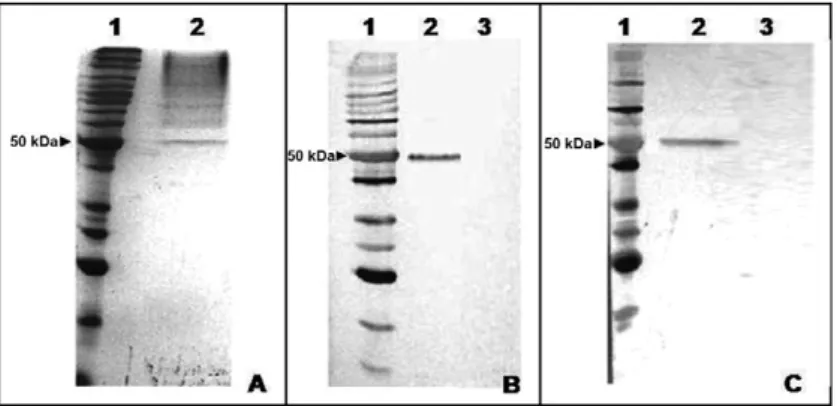
Expression analysis and immunoreactivity of E2 recombinant proteins - Protein expression levels were confirmed by their corresponding electrophoretic bands. The protein that was produced in E. coli and fused to a glutathione-S-transferase protein (GST) and 6x His tag was found to be 63.5 kDa (E2B) (Fig. 1). The protein that was produced in P. pastoris and fused to a 6x His-tag was found to be 50 kDa (E2Y) (Fig. 2) because of N-glycosylation. Molecular weight differences were re-lated to the different fusion proteins that were employed and to the varying post-translational modifications that were imparted by different organisms. These results are
Fig. 1A-C: protein size markers. Expression and reactivity of the envelope 2 (E2) protein produced in Escherichia coli (E2B). A: sodium dodecyl sulfate polyacrylamide gel electrophoresis stained with Coomassie blue [2 in A: supernatant of the induced bacterial culture; 3 in A: recombi-nant E2 protein fused to glutathione-S-transferase protein (GST) and 6x His tag (63.5 kDa) after purification in the GST column]; B: polyvi-nylidene difluoride (PVDF)membrane transferred with the E2 recombinant protein [2 in B: recombinant E2 protein localised by anti-His tag primary antibody and alkaline phosphatase anti-immunoglobulin G (IgG)secondary antibody; 3 in B: negative control and alkaline phosphatase anti-IgGsecondary antibody]; C: PVDF membrane transferred with the E2 recombinant protein for test of reactivity with human serum [2 in C: reactivity of the E2 recombinant protein with a pool of hepatitis C virus (HCV) (+) human serum (diluted 1:100); 3 in C: no reactivity of protein with a pool of HCV (-) human serum (diluted 1:10)].
in agreement with previous publications that evaluated the same organisms and gene sequences (Yurkova et al. 2004, Martinez-Donato et al. 2006).
The antigenicity of each recombinant protein was tested using serum samples taken from chronic hepatitis C carriers. We found that human anti-IgG demonstrated a specific recognition of E2 proteins [Figs 1 (2 in C), 2 (2 in C)]. As a control, a PVDF membrane that had been incubated with E2 proteins was tested against a pool of HCV negative serum; no interactions were found [Figs 1 (3 in C), 2 (3 in C)]. No immunoreactivity was observed with respect to 6x His-tag and GST fusion proteins in HCV-negative and positive serum pools.
Cellular immunophenotyping - LDLr and CD81 (TA-PA-1) surface receptors were characterised in HUVEC and ECV304 cells by flow cytometry and representative histograms that were obtained using FITC-conjugated anti-CD81 and PE-conjugated anti-LDLr are shown in Fig. 3. In HUVEC, 93% were positive for LDLr, whereas no significant fluorescence was found with respect to CD81 (approximately 1%). In ECV304 cells, 15% were positive for CD81 and 98% were positive for LDLr. The presence or absence of FCS in culture medium did not significantly alter the expression of receptors.
Binding of E2 recombinant proteins to LDLr and CD81 receptors - Assessing the binding of the
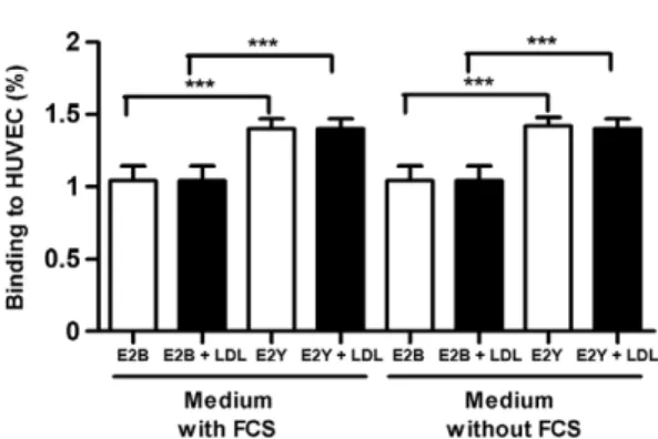
recombi-nant E2 proteins to HUVEC and ECV304 cells revealed that E2 proteins could bind to receptors without having to form a heterodimer with E1. In HUVEC, binding ca-pacity was found to be totally dependent on the presence of FCS, as the percentage of binding was reduced for both E2 proteins when FCS was removed from culture media (Fig. 4). However, the addition of human LDL to the media both rescued and increased the binding capac-ity of both E2 for all combinations studied and regard-less of the presence or absence of FCS (Fig. 4). It is no-table that in HUVEC, these recombinant proteins were only found to bind to LDLr because this cell line did not present significant amounts of CD81 receptors under the experimental conditions that were used in this work.
With respect to ECV304 cells, the presence of FCS was important for the binding efficiency of both recom-binant E2 proteins. However, these results differed from those obtained with HUVECs, which, in the absence of LDL, were totally dependent on the presence of FCS in culture media. Similar to the results obtained using HU-VEC, in ECV304 cells, the addition of human LDL led to a significant increase in binding in all of the experi-mental conditions that were tested (Fig. 5).
Furthermore, we also observed more significant binding (p < 0.01) of E2Y vs. E2B in both cell types in the presence of LDL or FCS.
Inhibiting recombinant E2 proteins from binding to cell receptors - In HUVEC, blocking LDLr led to a sig-nificant reduction in E2B and E29 binding both in the presence and absence of FCS and/or LDL in the media (Fig. 6). Blocking the CD81 receptor produced a mark-edly different result, as both the binding and the effects produced by FCS and/or LDL were similar to the results that were obtained in experiments that did not include the CD81 inhibitory antibody (Fig. 7). In another set of ex-periments, both LDLr and CD81 receptors were blocked and only a small amount of fluorescence was detected (Fig. 8), suggesting that these proteins might be binding to other receptors that are also able to bind E2 proteins, such as SR-BI, CLDN1, OCLN and TfR1. We also can-not rule out the possibility that LDL might modify E2 (e.g., conformation) and alter its interactions with CD81 or other cellular receptors in the absence of LDLr. Nev-ertheless, these results suggest that E2 proteins primar-ily bound to LDLr and not to another cell receptor. In ECV304 cells, blocking LDLr also led to a decrease in the binding of both recombinant proteins under all of the experimental conditions that were tested. However, this effect was not as intense as that which was observed in HUVEC. In Fig. 9, it can be observed that blocking LDLr could not completely eliminate binding. This may indicate that both CD81 and other E2 receptors that are present in this cell line, such as SR-BI, CLDN1, OCLN and TfR1, represent alternative binding sites, which may be more relevant in the context of E2Y than E2B. When blocking CD81, a small but significant decrease in bind-ing capacity was also observed under all of the experi-mental conditions that were tested in ECV304 cells (Fig. 10). These results confirm that CD81 is also relevant to the binding of E2 recombinant proteins to ECV304 cells and they differ from what was observed in HUVEC, which were practically insensitive to blockage of this receptor. Finally, the simultaneous blockage of both receptors (Fig. 11) reduced the binding of E2 proteins to ECV304 cells, similarly to what was observed in HUVECs.
Fig. 4: binding of recombinant E2B and E2Y to human umbilical vein endothelial cells (HUVEC) - effect of the addition of low-density li-poprotein (LDL) and foetal bovine serum (FCS). The results are mean and standard deviation. **: p < 0.01; ***: p < 0.001 (ANOVA and Tukey’s multiple comparison test).
Fig. 5: binding of recombinant E2B and E2Y to ECV304 - effect of the addition of low-density lipoprotein (LDL) and foetal bovine serum (FCS). The results are mean and standard deviation. ** p < 0.01; ***: p < 0.001 (ANOVA and Tukey’s multiple comparison test).
Fig. 6: binding of recombinant E2B and E2Y to human umbilical vein endothelial cells (HUVEC) with blocked low-density lipoprotein re-ceptor (LDLr) - effect of the addition of LDL and foetal bovine serum (FCS). The results are mean and standard deviation. ***: p < 0.001 (ANOVA and Tukey’s multiple comparison test).
DISCUSSION
Many details of HCV receptors in host cells are not completely understood; however, there is evidence that E2 is the main protein responsible for the initial events that occur during the coupling of the virus to host cells (Zibert et al. 1995, Farci et al. 1996). Moreover, LDL and CD81 receptors are believed to mediate this process (Scar-selli et al. 2002, Voisset & Dubuisson 2004). The absence of significant fluorescence that was noted with respect to CD81 is an indication that this receptor is not constitu-tively expressed in HUVEC under physiological condi-tions. Consistent with this, the expression of CD81 has previously been found to be reduced in endothelial cells in the absence of inflammation (Rohlena et al. 2009). In ECV304 cells, fluorescence was low but significant, which is an indication that the CD81 receptor was expressed. This result can be explained by the fact that ECV304 is a hybrid cell line that was created from endothelial cells that were contaminated with epithelial cells from bladder carcinoma (T24/83) (Brown et al. 2000). We believe that this might explain the very low but constitutive expres-sion of CD81 in this cell line. However, we cannot rule out that the low percentage of ECV304 cells expressing CD81 may be related to a particular cell cycle phase.
The binding capacity of the recombinant E2 proteins to surface receptors in ECV304 cells was dependent on the presence of FCS and/or LDL in the culture media. These results suggest that E2 proteins are binding to CD81, as this receptor was present in a lower percentage of cells than was LDLr. Moreover, an absence of LDL in the cul-ture media impeded the binding of E2 to LDLr, which was found to be present in a higher percentage of cells.
The presence of human LDL was also critical to the binding of both proteins to HUVEC when FCS was ab-sent from the medium. This is an indication that the virus primarily bound to LDLr. Coupled with the absence of CD81 in HUVEC, this finding suggests that, in both of the cell types that were used in this study, the interaction of E2 with LDLr seems to be at least partially mediated by LDL. Consistent with our findings, experiments that Fig. 8: binding of recombinant E2B and E2Y to human umbilical vein
endothelial cells (HUVEC) with blocked low-density lipoprotein re-ceptor (LDLr) and CD81 - effect of the addition of LDL and foetal bovine serum (FCS). The results are mean and standard deviation. ***: p < 0.001 (ANOVA and Tukey’s multiple comparison test).
Fig. 9: binding of recombinant E2B and E2Y to ECV304 with blocked low-density lipoprotein receptor (LDLr) - effect of the addition of LDL and foetal bovine serum (FCS). The results are mean and standard devi-ation. ***: p < 0.001 (ANOVA and Tukey’s multiple comparison test).
Fig. 10: binding of recombinant E2B and E2Y to ECV304 with blocked CD81 - effect of the addition of low-density lipoprotein (LDL) and foetal bovine serum (FCS). The results are mean and standard deviation. **: p < 0.01; ***: p < 0.001 (ANOVA and Tukey’s multiple comparison test).
were performed to assess the binding properties of E2 proteins that were expressed in Chinese Hamster Ovar-ian cells revealed that binding was increased in a human acute lymphoblastic leukaemia cell line (MOLT-4) when using media that contained both LDL and FCS (Wün-schmann et al. 2006). It is notable that several studies have used HCV particles that were produced in vitro (Barto-sch et al. 2003, Zhong et al. 2005), which are structurally different from each other and from particles produced in vivo(André et al. 2002). In this study, we tested the re-combinant proteins in two different organisms and found effects that were similar to previously reported findings.
When HUVEC were blocked against CD81 and cul-tured in media that lacked FCS, neither of the recombi-nant proteins was able to significantly bind to the cells until after the addition of LDL. Additionally, when cells were incubated with anti-LDLr, the binding of E2 pro-teins was not significant, suggesting that the binding of E2 proteins to cells occurs completely via LDLr. Alto-gether, these results suggest that LDLr is the primary target of both proteins under conditions in which they are associated with LDL. In agreement with these find-ings, CD81-deficient cells have previously been demon-strated to internalise HCV after it binds to LDL and uses LDLr (Agnello et al. 1999). Additionally, LDLr-deficient cells are less susceptive to HCV infection (Hishiki et al. 2010). Indeed, LDLr has long been considered HCV’s route of entry into host cells (Wünschmann et al. 2000, Seipp et al. 1997). LDLr has also been shown to be rel-evant for the cell entry of various members of the Flavi-virus genus (Wünschmann et al. 2000).
Another important finding was the verification that E2Y always exhibited a greater binding capacity than E2B. The primary difference between these two proteins is the glycosylation of E2Y. These results are in agreement with previous reports in which glycosylation was found to be indispensable for the global folding and conforma-tion of this viral envelope protein (Goffard & Dubuisson 2003, Voisset & Dubuisson 2004, Bian et al. 2009, Lin et al. 2009). Moreover, glycosylation is also important for the ability of E2Y to bind to LDL and to other receptors of host cells, thereby enabling the coupling and entry of HCV into cells (Dubuisson & Rice 1996, Goffard et al. 2005, Tello et al. 2010). A previous report by Yurkova et al. (2004) found that glycosylation was not important for the binding of E2 to CD81. This finding was not corrobo-rated by our experiments, as we found that E2Y more efficiently bound to both CD81 and LDLr than did E2B.
In vivo assays have shown the presence of HCV RNA-containing particles in low-density fractions of plasma, a finding that has been associated with high infectivity. However, the nature of circulating HCV particles and their association with immunoglobulins or lipoproteins, as well as the details of their cell entry mechanisms, have all been subject to conflicting reports (André et al. 2002). Nevertheless, the importance of HCV’s associa-tion with LDL during viral entry through LDLr has been observed both in vivo and in vitro.
In conclusion, we have observed that the recombinant E2 HCV protein is dependent on LDL for its ability to bind to LDLr in HUVEC and ECV304 cell lines. The
as-sociation between E2 and LDL during the coupling of E2 to host cells has been demonstrated elsewhere (Thomssen et al. 1992, Nahmias et al. 2006); hence, the major contri-bution of this work is the demonstration that endothelial cells are also a potential target for this association. This may be related to the initial inflammation that is caused by HCV in endothelial cells of the hepatic portal vein, which may contribute to HCV-mediated cirrhosis.
This finding is very relevant because E2 serves as an anchor protein during the binding of HCV to host cells and high blood levels of LDL might enhance viral infec-tivity in chronic hepatitis C patients. In fact, LDL has been shown to promote an increase in HCV infectivity in several previous studies (Lavillette et al. 2005, Meu-nier et al. 2005, Nahmias et al. 2006, Siagris et al. 2006, Hishiki et al. 2010). The glycosylation of E2 is of critical importance for enabling the virus to anchor to LDLr. Ad-ditionally, although many studies have indicated that the E1E2 heterodimer is necessary for viral association with host cell receptors, we demonstrated here that the E2 protein is able to independently bind to these receptors. Finally, the putative mechanism of cellular infection in-volving E2, LDL, CD81 and LDLr could offer potential new targets for the development of novel antiviral thera-pies. Moreover, controlling serum LDL levels might also be helpful for the treatment of chronic hepatitis C.
ACKNOWLEDGEMENTS
To Prof Iracilda Zeppone Carlos (School of Pharmaceu-tical Sciences/UNESP), Prof Dulcinéia Saes Parra Abdalla (School of Pharmaceutical Sciences/USP), Prof Marcelo Dias Baruffi (School of Pharmaceutical Sciences of Ribeirão Preto/USP), Marisa Polezi Placeres, Felipe Fortino Verdan da Silva, Lívia Carolina de Abreu Ribeiro, Thalita Athiê Néo and Thaís Ferreira Isabel (School of Pharmaceutical Sciences of Araraquara/UNESP.
REFERENCES
Agnello V, Abel G, Elfahal M, Knight GB, Zhang QX 1999. Hepatitis C virus and other Flaviviridae viruses enter cells via low density lipoprotein receptor. Proc Natl Acad Sci USA 96: 12766-12771.
Albecka A, Belouzard S, de Beeck AO, Descamps V, Goueslain L, Bertrand-Michel J, Tercé F, Duverlie G, Rouillé Y, Dubuisson J 2012. Role of low-density lipoprotein receptor in the hepatitis C virus life cycle. Hepatology 55: 998-1007.
André P, Komurian-Pradel F, Deforges S, Perret M, Berland CJL, Sodoyer M, Pol S, Bréchot C, Paranhos-Baccalà G, Lotteau V 2002. Characterization of low and very-low-density hepatitis C virus RNA-containing particles. J Virol 76: 6919-6928.
Bartenschlager R, Lohmann V 2000. Replication of hepatitis C virus. J Gen Virol 81: 1631-1648.
Bartosch B, Vitelli A, Granier C, Goujon C, Dubuisson J, Pascale S, Scarselli E, Cortese R, Nicosia A, Cosset FL 2003. Cell entry of hepatitis C virus requires a set of co-receptors that include the CD81 tetraspanin and the SR-B1 scavenger receptor. J Biol Chem 278: 41624-41630.
Bian T, Zhou Y, Bi S, Tan W, Wang Y 2009. HCV envelope protein function is dependent on the peptides preceding the glycopro-teins. Biochem Biophys Res Commun 378: 118-122.
Critical evaluation of ECV304 as a human endothelial cell model defined by genetic analysis and functional responses: a comparison with the human bladder cancer derived epithelial cell line T24/83. Lab Invest 80: 37-45.
Dubuisson J, Helle F, Cocquerel L 2008. Early steps of the hepatitis C virus life cycle. Cell Microbiol 10: 821-827.
Dubuisson J, Rice CM 1996. Hepatitis C virus glycoprotein folding: disulfide bond formation and association with calnexin. J Virol 70: 778-786.
Evans MJ, von Hahn T, Tscherne DM, Syder AJ, Panis M, Wölk B, Hatziioannou T, McKeating JA, Bieniasz PD, Rice CM 2007. Claudin-1 is a hepatitis C virus co-receptor required for a late step in entry. Nature 446: 801-805.
Farci P, Shimoda A, Wong D, Cabezon T, De Gioannis D, Strazzera A, Shimizu Y, Shapiro M, Alter HJ, Purcell RH 1996. Prevention of hepatitis C virus infection in chimpanzees by hyperimmune serum against the hypervariable region 1 of the envelope 2 pro-tein. Proc Natl Acad Sci USA 93: 15394-15399.
Germi R, Crance JM, Garin D, Guimet J, Lortat-Jacob H, Ruigrok RW, Zarski JP, Drouet E 2002. Cellular glycosaminoglycans and low density lipoprotein receptor are involved in hepatitis C virus adsorption. J Med Virol 68: 206-215.
Goffard A, Callens N, Bartosch B, Wychowski C, Cosset FL, Montpel-lier C, Dubuisson J 2005. Role of N-linked glycans in the functions of hepatitis C virus envelope glycoproteins. J Virol 79: 8400-8409.
Goffard A, Dubuisson J 2003. Glycosylation of hepatitis C virus en-velope proteins. Biochimie 85: 295-301.
Hishiki T, Shimizu Y, Tobita R, Sugiyama K, Ogawa K, Funami K, Ohsaki Y, Fujimoto T, Takaku H, Wakita T, Baumert TF, Mi-yanari Y, Shimotohno K 2010. Infectivity of hepatitis C virus is influenced by association with apolipoprotein E isoforms. J Virol 84: 12048-12057.
Lavillette D, Morice Y, Germanidis G, Donot P, Soulier A, Pagkalos E, Sakellariou G, Intrator L, Bartosch B, Pawlotsky JM, Cosset FL 2005. Human serum facilitates hepatitis C virus infection and neutralizing responses inversely correlate with viral repli-cation kinetics at the acute phase of hepatitis C virus infection. J Virol 79: 6023-6034.
Lin X, Zhang Y, Bi S, Lu J, Zhao H, Tan W, Li D, Wang Y 2009. Hep-atitis C virus envelope glycoproteins complementation patterns and the role of the ecto and transmembrane domains. Biochem Biophys Res Commun 385: 257-262.
Liu J, Zhu L, Zhang X, Lu M, Kong Y, Wang Y, Li G 2001. Expres-sion, purification, immunological characterization and applica-tion of Escherichia coli-derived hepatitis C virus E2 proteins. Biotechnol Appl Biochem 34: 109-119.
Lozach PY, Lortat-Jacob H, de Lavalette AL, Staropoli I, Foung S, Amara A, Houles C, Fieschi F, Schwartz O, Virelizier JL, Aren-zana-Seisdedos F, Altmeyer R 2003. DC-SIGN and L-SIGN are high affinity binding receptors for hepatitis C virus glycoprotein E2. J Biol Chem 278: 20358-20366.
Lupberger J, Zeisel MB, Xiao F, Thumann C, Fofana I, Zona L, Da-vis C, Mee CJ, Turek M, Gorke S, Royer C, Fischer B, Zahid MN, Lavillette D, Fresquet J, Cosset FL, Rothenberg SM, Pi-etschmann T, Patel AH, Pessaux P, Doffoël M, Raffelsberger W, Poch O, McKeating JA, Brino L, Baumert TF 2011. EGFR and EphA2 are host factors for hepatitis C virus entry and possible targets for antiviral therapy. Nat Med 17: 589-595.
Martin DN, Uprichard SL 2013. Identification of transferrin receptor 1 as a hepatitis C virus entry factor. Proc Natl Acad Sci USA 110: 10777-10782.
Martinez-Donato G, Acosta-Rivero N, Morales-Grillo J, Musacchio A, Vina A, Alvarez C, Figueroa N, Guerra I, Garcia J, Varas L, Muzio V, Dueñas-Carrera S 2006. Expression and processing of hepatitis C virus structural proteins in Pichia pastoris yeast. Bio-chem Biophys Res Commun 342: 625-631.
Meunier JC, Engle RE, Faulk K, Zhao M, Bartosch B, Alter H, Emer-son SU, Cosset FL, Purcell RH, Bukh J 2005. Evidence for cross-genotype neutralization of hepatitis C virus pseudo-particles and enhancement of infectivity by apolipoprotein C1. Proc Natl Acad Sci USA 102: 4560-4565.
Nahmias Y, Casali M, Barbe L, Berthiaume F, Yarmush ML 2006. Liver endothelial cells promote LDL-R expression and the uptake of HCV-like particles in primary rat and human hepatocytes. He-patology 43: 257-265.
Penin F, Dubuisson J, Rey FA, Moradpour D, Pawlotsky JM 2004. Structural biology of hepatitis C virus. Hepatology 39: 5-19.
Ploss A, Evans MJ, Gaysinskaya VA, Panis M, You H, de Jong YP, Rice CM 2009. Human occludin is a hepatitis C virus entry factor required for infection of mouse cells. Nature 457: 882-886.
Rohlena J, Volger OL, van Buul JD, Hekking LH, van Gils JM, Bonta PI, Fontijn RD, Post JA, Hordijk PL, Horrevoets AJ 2009. En-dothelial CD81 is a marker of early human atherosclerotic plaques and facilitates monocyte adhesion. Cardiovasc Res 81: 187-196.
Sainz B, Barretto N, Martin DN, Hiraga N, Imamura M, Hussain S, Marsh KA, Yu X, Chayama K, Alrefai WA, Uprichard SL 2012. Identification of the Niemann-Pick C1-like 1 cholesterol absorption receptor as a new hepatitis C virus entry factor. Nat Med 18: 281-285.
Scarselli E, Ansuini H, Cerino R, Roccasecca RM, Acali S, Filocamo G, Traboni C, Nicosia A, Cortese R, Vitelli A 2002. The human scavenger receptor class B type I is a novel candidate receptor for the hepatitis C virus. EMBO J 21: 5017-5025.
Seipp S, Mueller HM, Pfaff E, Stremmel W, Theilmann L, Goeser T 1997. Establishment of persistent hepatitis C virus infection and replication in vitro. J Gen Virol 78: 2467-2476.
Siagris D, Christofidou M, Theocharis GJ, Pagoni N, Papadimitriou C, Lekkou A, Thomopoulos K, Starakis I, Tsamandas AC, Labro-poulou-Karatza C 2006. Serum lipid pattern in chronic hepatitis C: histological and virological correlations. J Viral Hepat 13: 56-61.
Tello D, Rodríguez-Rodríguez M, Yélamos B, Gómez-Gutiérrez J, Ortega S, Pacheco B, Peterson DL, Gavilanes F 2010. Expression and structural properties of a chimeric protein based on the ect-odomains of E1 and E2 hepatitis C virus envelope glycoproteins. Protein Expr Purif 71: 123-131.
Thomssen R, Bonk S, Propfe C, Heermann KH, Köchel HG, Uy A 1992. Association of hepatitis C virus in human sera with beta-lipoprotein. Med Microbiol Immunol 181: 293-300.
Urbaczek AC, Ribeiro LCA, Ximenes VF, Afonso A, Nogueira CT, Generoso WC, Alberice JV, Rudnicki M, Ferrer R, da Fonseca LM, da Costa PI 2014. Inflammatory response of endothelial cells to hepatitis C virus recombinant envelope glycoprotein 2 protein exposure. Mem Inst Oswaldo Cruz 109: 748-756.
Voisset C, Dubuisson J 2004. Functional hepatitis C virus envelope glycoproteins. Biol Cell 96: 413-420.
Wünschmann S, Medh JD, Klinzmann D, Schmidt WN, Stapleton JT 2000. Characterization of hepatitis C virus (HCV) and HCV E2 interactions with CD81 and the low-density lipoprotein receptor. J Virol 74: 10055-10062.
en-velope glycoprotein E2 and serum lipoproteins (LPs) results in enhanced cellular binding of both HCV E2 and LPs. J Infect Dis 194: 1058-1067.
Yurkova MS, Patel AH, Fedorov AN 2004. Characterisation of bacte-rially expressed structural protein E2 of hepatitis C virus. Protein Expr Purif 37: 119-125.
Zhang J, Randall G, Higginbottom A, Monk P, Rice CM, McKeat-ing JA 2004. CD81 is required for hepatitis C virus glycoprotein-mediated viral infection. J Virol 78: 1448-1455.
Zhong J, Gastaminza P, Cheng G, Kapadia S, Kato T, Burton DR, Wie-land SF, Uprichard SL, Wakita T, Chisari FV 2005. Robust hepatitis C virus infection in vitro. Proc Natl Acad Sci USA 102: 9294-9299.
Zibert A, Schreier E, Roggendorf M 1995. Antibodies in human sera specific to hypervariable region 1 of hepatitis C virus can block viral attachment. Virology 208: 653-661.