Adenotonsillar Hypertrophy in Pre-School
Children with Sickle Cell Disease and Diagnostic
Accuracy of the Sleep Disturbance Scale for
Children
Carlos Rodolfo Tavares de Góis
1Jeferson Sampaio D’Ávila
1Rosana Cipolotti
1Amanda da Silva Lira
2Ana Letícia Leite Silva
11Department of Medicine, Universidade Federal de Sergipe (UFS),
Aracaju, SE, Brazil
2Department of Statistics and Actuarial Sciences, Universidade
Federal de Sergipe (UFS), São Cristóvão, SE, Brazil
Int Arch Otorhinolaryngol 2018;22:55–59.
Address for correspondence Carlos Rodolfo Tavares de Góis, PhD, Rua Dr. Celso Oliva, 250, Torre B, Apto. 802, Bairro 13 de Julho, Aracaju, SE, Brazil CEP: 49020-090 (e-mail: carlosrtgois@yahoo.com.br).
Introduction
Sickle cell disease (SCD) is a systemic nosological disease characterized by progressive damage in several organs; it is
considered one of the most serious monogenic diseases. It received this denomination because of the sickle-shaped erythrocytes of the patients. Sickle cell anemia (SCA) is the most common form of the disease, and specifically refers to Keywords
►
hypertrophy
►
pharyngeal tonsil
►
palatine tonsil
►
sickle cell disease
Abstract
Introduction
Adenotonsillar hypertrophy is more common in children with sickle cell
disease, and can lead to sleep-disordered breathing.
Objectives
To determine the frequency of adenotonsillar hypertrophy in pre-school
children with sickle cell disease and assess the diagnostic accuracy of the
sleep-disordered breathing subscale in the Sleep Disturbance Scale for Children.
Method
Observational study with a group of 48 children with sickle cell disease and a
control group of 35 children without the disease. The children underwent
orophar-ingoscopy and video nasal endoscopy. The parents and/or guardians answered the
questions of the subscale.
Results
Adenotonsillar hypertrophy was observed in 25% of the children in the study
group, and in 20% of the children in the control group, with no statistical difference
between the groups. The subscale score ranged from 3 to 11 in both groups. There was
a statistical signi
fi
cance in the study group. The average was 4.79 (standard deviation
[SD]
2.50), with 4.19 (SD
1.72) among the children without adenotonsillar
hypertrophy, and 6.5 (SD
3.40) among the children with adenotonsillar
hypertro-phy. There was also a statistical signi
fi
cance in the control group. The average was 5.23
(SD
2.81), with 4.44 (SD
2.2) among the children without adenotonsillar
hyper-trophy, and 7.87 (SD
2.89) among the children with adenotonsillar hypertrophy.
Conclusion
Adenotonsillar hypertrophy was not associated with sickle cell disease in
pre-school children. The subscale of sleep-disordered breathing in the Sleep
Distur-bance Scale for Children was a useful tool for the diagnostic suspicion of adenotonsillar
hypertrophy in children in this age group.
received
November 12, 2016 accepted
March 18, 2017 published online May 2, 2017
DOIhttps://doi.org/ 10.1055/s-0037-1602702. ISSN 1809-9777.
Copyright © 2018 by Thieme Revinter Publicações Ltda, Rio de Janeiro, Brazil
the homozygosity of theβS-globin allele, which leads to the production of a particular type of hemoglobin, that is, hemoglobin S (HbS). There is also sickle cell (SC) hemoglo-binopathy, which results from the relationship of the β
S-globin allele and theβC-globin allele withβ-thalassemia resulting from the combination of theβS-globin allele with
theβ-thalassemia allele, causing the HbS/β-thalassemia.1 The clinical manifestation of SCD basically occurs due to two processes: a) vas occlusion, with ischemia and reperfu-sion injuries; and b) hemolytic anemia.2Bacterial infections are the leading cause of morbidity and mortality, and they result from hyposplenism, disorders in the activation of the complement system, micronutrient deficiency, and tissue ischemia.3
Adenotonsillar hypertrophy (ATH) is the term commonly used to describe the abnormal growth of the pharyngeal tonsil (adenoid vegetations) and palatine tonsils. Although this growth can arise from a hyperplastic process of the lymphoid cells of these tissues, this differentiation is clini-cally irrelevant.4
The causes of ATH are not yet fully known; however, recurring chronic or acute inflammation seem to be involved, since children subject to these processes usually exhibit an abnormal growth of cervical and pharyngeal lymphoid tissues.5
Adenotonsillar hypertrophy seems to be more frequent and to have a tendency to be extended in children with SCD, which may result in recurrent pharyngitis. There are three hypotheses suggested for the association between ATH and SCD: compensation for autosplenectomy; consequence of recurrent infections of the upper airways due to failed opsonization of pathogenic bacteria; and the function of pharyngeal and palatine tonsils as hematopoietic centers due to hemolysis. Adenotonsillar hypertrophy can lead to sleep-disordered breathing (SDB), which varies from snoring to obstructive sleep apnea syndrome, with resulting hypox-emia, hypercapnia, and acidosis, raising the risk of HbS polymerization and, consequently, vaso-occlusive phenom-ena and other complications, such as transient ischemic attacks and cerebrovascular accident.6,7
The Sleep Disturbance Scale for Children (SDSC) is a questionnaire validated for the Portuguese language that has been increasingly used in studies on SDB in children and adolescents.8
The goals of the present study were to assess the associa-tion between SCD and ATH, and the diagnostic accuracy of the SDCS for ATH in pre-school children with SCD.
Method
The study group was composed of children aged between 2 and 6 years, who attended the specialized outpatient clinic of the regional university public service. They were recruited by the researcher during routine appointments. The control group consisted of healthy children of the same age, who attended two-day care centers. One of these centers belonged to the municipal network, and the other to the private network, and both were located in the suburbs of the
capital of the state. In order to recruit the children of the control group, we sent invitation letters to their parents and/ or legal guardians from the two day care centers.
The criteria for inclusion in the study group were: a) having a diagnosis of SCD confirmed by the quantitative analysis of the hemoglobin, performed through hemoglobin electrophoresis or high-performance liquid chromatography; and b) being clinically stable. The criteria for inclusion in the control group were: a) no diagnosis of SCD; and b) no acute infectious process. The exclusion criteria for the two groups were: a) exhibiting craniofacial malformations; b) debilitating diseases; c) recent craniofacial trauma; d) adenoidectomy surgery and/or previous tonsillectomy; and e) undergoing systemic therapy with corticosteroids during the study.
The parents and/or guardians of the children who were willing to participate in the study signed an informed con-sent form. Subsequently, they answered three questions related to SDB in the Portuguese version of the SDSC. The subscale was composed of three items: a) the child does not breathe well during sleep; b) the child stops breathing for a few moments during sleep; and c) the child snores. The score of each item varied from 1 (never) to 5 (always), considering the frequency during the previous 6 weeks.8Subsequently, the children underwent oropharingoscopy with frontal light-ing and tongue depressor, and video nasal endoscopy uslight-ing a 3.2-mmflexible endoscope (Machida Endoscope Co., Tokyo, Japan) coupled to a LED light source (Lumen), and a digital microcamera (Pro-HD II - Pillertech Equip. Ltda, Curitiba, Brazil) to record the images for analysis.
The palatine tonsils were classified according to Brodsky’s criteria,9namely: grade 0¼palatine tonsils located inside the tonsillar fossae; grade 1¼tonsils located beyond the tonsillar fossae, occupying less than 25% of the airspace in the oropharynx; grade 2¼tonsils occupying more than 25% and less than 50% of the oropharyngeal space; grade 3¼tonsils occupying more than 50% and less than 75% of the orophar-yngeal space; grade 4¼tonsils occupying more than 75% of the oropharyngeal space. Palatine tonsils with grades 3 or 4 were considered obstructive. The pharyngeal tonsil (ade-noid) was considered obstructive when there was choanal occlusion of at least 70%.10The children with adenoid and/or obstructive palatine tonsils were diagnosed with ATH.11
The data were analyzed using the Statistical Package for the Social Sciences (SPSS) software, version 16 (SPSS Inc., Chicago Il, US). After determining the normality of the sample distribution, the data were compared using the chi-squared test for categorical variables, and the Mann-Whitney test for continuous variables. Values ofp<0.05 were considered significant.
The project was approved by the Research Ethics Com-mittee of the institution, under CAAE Protocol No. 23646413.6.0000.5546.
Results
was composed of 35 children (27 boys and 8 girls). There was no statistical difference between the groups (p¼0.369). The average age of the study group was 4.04 years (standard deviation [SD]:1.458), and the average age of the control group was 4.25 years (SD:1.120), with no statistically significant difference (p¼0.5764).
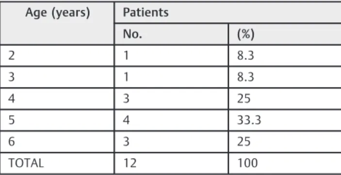
We observed that ATH occurred in 12 children of the study group (25%), 7 boys and 5 girls. In the control group, 7 children (20%) exhibited ATH (6 boys and 1 girl). There was no significant difference between the frequencies in the two groups (p¼0.246). As it can be seen in►Table 1, there was no association between ATH and age in the study group (p¼0.831); however, that association occurred in the control group (p¼0.043), in which we observed a higher frequency of ATH in children under 5 years of age (►Table 2). The score obtained in the three items of the SDB subscale in the SDSC ranged from 3 to 11in both groups. In the study group, the average was 4.79 (SD2.50), with 4.19 (SD1.72) among the children without ATH, and 6.5 (SD x2009;3.40) among the children with ATH. There was a statistical significance in the study group (p¼0.0025). In the control group, the average was 5.23 (SD2.81), with 4.44 (SD2.2) among the children with-out ATH, and 7.87 (SD2.89) among the children with ATH. There also was a statistical significance in this group (p¼0.0026).
Discussion
The results demonstrated that the groups were comparable, since both of them exhibited statistically similar sex and age distribution. Even though there was a significantly greater proportion of boys in the control group, this fact would probably not interfere in the frequency of ATH because, as already demonstrated in several studies, this disease has shown similar prevalence between sexes.5,10 We also ob-served equal SCD distribution between the sexes, which is in line with thefindings of national and international medical studies.12,13
The age group most affected by ATH ranges from 2 to 6 years and, after this age, there is an involution process of the lymphoid tissue in the pharynx.5,10,14,15For this reason, we decided to study children in this age group. One study found a prevalence peak between 3 and 5 years.5 In the present study, we only found an association between age and ATH in the control group, and it was more frequent among 5-year-old children.
Regarding the frequency of ATH, the two groups also showed a similarity. In the literature consulted, the preva-lence of ATH ranged from 11 to 37.6%,16,17which was in line with thefindings of our study. Other studies have estimated
the prevalence of SDB, whose main cause in children is ATH,4,14,18 characterized by the occurrence of snoring as an isolated signal (primary snoring), or associated with an increased resistance of the upper airways, with obstructive hypoventilation until the complete collapse, leading to ob-structive sleep apnea syndrome.18,19While primary snoring affects 9 to 29% of children, SDB with hypoventilation and apnea occurs in 1 to 4% of children.18,20Among the other causes of SDB, obesity, craniofacial malformations, neurolo-gical diseases, and gastroesophageal reflux disease stand out.19,21,22
One limitation of the present study was that we did not assess body mass index, craniofacial measures, and the Mallampati classification, which is based on the proportion between the tongue and the pharynx, and is routinely used in sleep medicine. However, as the focus of the study was the association between ATH and SCD, and considering the difficulty of a detailed assessment in children in this age group, we only observed the degree of nasal obstruction caused by the pharyngeal and palatine tonsils. It is possible that the use of topical and systemic corticosteroids may transiently affect the degree of nasal obstruction and, even, the tonsillar dimensions,23,24although this hypothesis has been refuted by some authors.25
Following the methodology of other studies,11,12we chose to consider only the use of systemic corticosteroids during the otorhinolaryngologic examination as an exclusion criterion, because there was great difficulty in obtaining reliable infor-mation about the recent use of medications from the parents and/or guardians of the children. In addition, there is scarce literature on the duration of the effects caused by various corticosteroids on the pharyngeal and palatine tonsils.
Although some authors have reported a higher prevalence of ATH among children with SCD,6,11,26the analysis of the Table 2 Age-related distribution of patients with
adenotonsillar hypertrophy in the control group
Age (years) Patients
No. (%)
2 0 0
3 1 14.3
4 1 14.3
5 4 57.1
6 1 14.3
TOTAL 7 100
Table 1 Age-related distribution of patients with adenotonsillar hypertrophy in the study group
Age (years) Patients
No. (%)
2 1 8.3
3 1 8.3
4 3 25
5 4 33.3
6 3 25
results of the present study did not detect a significant difference between the children with SCD and those of the control group. It is worth mentioning that the children with SCD were recruited at the time of their pediatric appoint-ments. They were accompanied by the parents in the health care service, and there was no refusal to participate in the study.
On the other hand, the children of the control group were recruited by means of invitation letters, which explained what ATH was, the symptoms, the effects, and the method that we would use for the diagnosis. In this way, we may have created a selection bias, since the parents of the children with SDB symptoms may have had greater interest in the assess-ment, allowing the physical examination and the video nasal endoscopy. However, given the wide acceptance on the part of the parents, possibly due to the difficult access to specia-lized examinations, as well as the fact that we found the frequency of ATH to be within the expected range according to the literature,16,17we believe that this was not an im-portant limitation of our study.
In fact, we observed a clear divergence with respect to the ATH frequency in children with SCD, when compared with thefindings of previous studies. A study showed palatine tonsil hypertrophy in 55% of 53 patients with SCD, whose ages ranged from 1.9 to 16.5 years, although the measure-ment scale for the tonsils was not the same as the scale used in our study.26
A national study found that ATH had a frequency of 55.3% in patients with SCD aged between 2 and 19 years. Although the authors used Brodsky’s classification for the assessment
of the palatine tonsils in a way similar to ours, they con-sidered that the pharyngeal tonsils were hypertrophic when they occluded more than 50% of the choanae, which certainly contributed to increase the frequency of ATH in comparison to our sample.11Even though other authors have also worked using this percentage to consider adenoidal obstruction,27 we used a minimal choanal occlusion of 70% to consider the pharyngeal tonsil obstructive.
The data from the literature indicate an absence of asso-ciation between the dimensions of the palatine and phar-yngeal tonsils and the severity of the SDB10,14; however, with the daily practice of otorhinolaryngology physicians can perceive that children with pharyngeal tonsil exhibiting an occlusion degree lower than 70% exhibit signs and symptoms compatible with SDB. On the other hand, regarding the palatine tonsils, we agreed that the occupation of more than 50% of the oropharynx (Brodsky’s grades 3 and 4) should be considered obstructive, because it is unlikely to
find palatine tonsils with those dimensions without the presence of adenoid vegetations closing at least half of the choanae. This fact was confirmed in our sample, as well as in a previously published study.11In this case, it is believed that the joint obstructive action of the palatine and pharyngeal tonsils would lead to nasal obstruction symptoms.
Studies have demonstrated that children with SCD and adenoid vegetations, even with obstruction below 70%, ex-hibit a higher apnea-hypopnea index than children without SCD.6,11,18 However, it is possible that this occurs due to
other reasons relating to SCD, because obstructive sleep apnea syndrome has a multifactorial etiology.19,21,22
One study used magnetic resonance imaging to compare the dimensions of the lymphatic organs of the head and neck in patients with and without SCD. The authors observed that all of these organs, including the pharyngeal and palatine tonsils, had significantly increase in the patients with SCD.6 On the other hand, the present study aimed to detect an association between SCD and obstructive ATH, and not simply the relative increase in lymphoid tissue in patients with SCD.
The SDSC, which assesses a variety of sleep-related beha-vior patterns in children, has six subscales for diagnosing parasomnias, difficulty in initiating and maintaining sleep, SDB, excessive daytime sleepiness, hyperhidrosis, and non-restorative sleep. The six subscales have a total of 26 items, with scores ranging from 1 (never) to 5 (always).8,28In the present work, we initially tried to work using the SDSC in its entirety; however, there was a great difficulty on the part of the parents and/or guardians of the children to properly understand and answer the questions, probably due to their low level of formal education.
In addition, the sample would hardly be sufficient to generate reliable results in the assessment of some sleep disorders. As the priority of the study was to assess ATH in children with SCD and in controls, we decided to use only the items related to SDB (13 to 15), and the total score ranged from 3 to 15 points.
As expected, when we analyzed the SDSC score, we did not
find a higher score in children with ATH in the two groups;
however, we only observed a statistical significance in the group of children with SCD. It is possible that the number of children in the control group has hindered the observation of the statistical evidence of this association. There are no studies comparing the score of SDB in the SDSC in pre-school children with or without ATH, and only two studies have used the SDSC in pre-school children. In thefirst one, which assessed a sample of 904 children aged from 3 to 6 years, the average score was 4.4 (SD1.6).29The second study involved 601 children in the same age group, and the score ranged from 3 to 14, with an average of 4.79 (SD1.91).30These results were very close to those of our study. In the study by Bruni, Ottaviano and Guidetti, with patients aged from 6 to 16 years, the average score was 3.77 (SD1.45),28which probably reflected the lower prevalence of ATH in this age group.
Conclusion
In the present study, ATH was not associated with SCD in pre-school children. The subscale of SDB in the SDSC proved to be a useful tool for the diagnostic suspicion of ATH in pre-school children.
References
2 Franceschi L. Pathophisiology of sickle cell disease and new drugs for the treatment. Medit J Hemat Infect Dis. 2009. Open Journal System
3 Serjeant GR, Serjeant BE. Sickle cell disease. Oxford UK: Oxford University Press; 2001
4 Potsic WP. Assessment and treatment of adenotonsillar hyper-trophy in children. Am J Otolaryngol 1992;13(05):259–264
5 Yaseen ET, Khammas AH, Anbaky FA. Adenoid enlargement assessment by plain X- ray and nasoendoscopy. Iraqui J Comm Med. 2012;1:88–91
6 Strauss T, Sin S, Marcus CL, et al. Upper airway lymphoid tissue size in children with sickle cell disease. Chest 2012;142(01): 94–100
7 Warrier R, Chauhan A, Athale U. Tonsillectomy and adenoidect-omy for obstructive sleep apnea in sickle cell anemia. Indian J Pediatr 2010;77(06):669–672
8 Ferreira VR, Carvalho LBC, Ruotolo F, de Morais JF, Prado LBF, Prado GF. Sleep disturbance scale for children: translation, cultural adaptation, and validation. Sleep Med 2009;10(04): 457–463
9 Brodsky L. Modern assessment of tonsils and adenoids. Pediatr Clin North Am 1989;36(06):1551–1569
10 Valera FCP, Avelino MAG, Pettermann MB, et al. OSAS in children: correlation between endoscopic and polysomnographicfindings. Otolaryngol Head Neck Surg 2005;132(02):268–272
11 Salles C, Ramos RTT, Daltro C, Nascimento VM, Matos MA. Association between adenotonsillar hypertrophy, tonsillitis and painful crises in sickle cell disease. J Pediatr (Rio J) 2009;85(03): 249–253
12 Felix AA, Souza HM, Ribeiro SBF. Aspectos epidemiológicos e sociais da anemia falciforme. Rev Bras Hematol Hemoter 2010;32(3): 203–208
13 Platt OS, Brambilla DJ, Rosse WF, et al. Mortality in sickle cell disease. Life expectancy and risk factors for early death. N Engl J Med 1994;330(23):1639–1644
14 Tagaya M, Nakata S, Yasuma F, et al. Relationship between adenoid size and severity of obstructive sleep apnea in preschool children. Int J Pediatr Otorhinolaryngol 2012;76(12):1827–1830
15 Pac A, Karadag A, Kurtaran H, Aktas D. Comparison of cardiac function and valvular damage in children with and without adenotonsillar hypertrophy. Int J Pediatr Otorhinolaryngol 2005;69(04):527–532
16 Kara CO, Ergin H, Koçak G, Kiliç I, Yurdakul M. Prevalence of tonsillar hypertrophy and associated oropharyngeal symptoms in
primary school children in Denizli, Turkey. Int J Pediatr Otorhi-nolaryngol 2002;66(02):175–179
17 Abreu RR, Rocha RL, Lamounier JA, Guerra AF. Etiology, clinical manifestations and concurrentfindings in mouth-breathing
chil-dren. J Pediatr (Rio J) 2008;84(06):529–535
18 Salles C, Ramos RTT, Matos MA. Apneia obstrutiva do sono em portadores de anemia falciforme. Rev Bras Hematol Hemoter 2010;32(01):70–75
19 Aubertin G. Le syndrome d’apnées obstructives du sommeil chez
l’enfant. Rev Pneumol Clin 2013;69(04):229–236
20 Piteo AM, Lushington K, Roberts RM, et al. Prevalence of snoring and associated factors in infancy. Sleep Med 2011;12(08): 787–792
21 Miranda GN. Trastornos respiratorios del sueño en la edad pediátrica. Rev Med Clin Las Condes 2013;24:403–411
22 Salles C, Bispo M, Trindade-Ramos RT. Association between morphometric variables and nocturnal desaturation in sickle-cell anemia. J Pediatr (Rio J) 2014;90(04):420–425
23 Cho D-Y, Sinha SR, Gardner JM, et al. Effect of intratonsillar injection of steroids on the palatine tonsils of rabbits. Laryngo-scope 2014;124(12):2811–2817
24 Demain JG, Goetz DW. Pediatric adenoidal hypertrophy and nasal airway obstruction: reduction with aqueous nasal beclometha-sone. Pediatrics 1995;95(03):355–364
25 Al-Ghamdi SA, Manoukian JJ, Morielli A, Oudjhane K, Ducharme FM, Brouillette RT. Do systemic corticosteroids effectively treat obstructive sleep apnea secondary to adenotonsillar hypertro-phy? Laryngoscope 1997;107(10):1382–1387
26 Samuels MP, Stebbens VA, Davies SC, Picton-Jones E, Southall DP. Sleep related upper airway obstruction and hypoxaemia in sickle cell disease. Arch Dis Child 1992;67(07):925–929
27 D’Ávila JS, Naves AB, Chagas L, et al. Adenoidectomia: novos
princípios. Estudo interdisciplinar. Rev Bras Otorrinolaringol 1999;65:511–516
28 Bruni O, Ottaviano S, Guidetti V, et al. The Sleep Disturbance Scale for Children (SDSC). Construction and validation of an instrument to evaluate sleep disturbances in childhood and adolescence. J Sleep Res 1996;5(04):251–261
29 Simola P, Niskakangas M, Liukkonen K, et al. Sleep problems and daytime tiredness in Finnish preschool-aged children-a commu-nity survey. Child Care Health Dev 2010;36(06):805–811