1
Update
Determination of Hemodynamic Parameters
Using Doppler Two-dimensional Echocardiography:
A Searching Tool for Therapeutic Optimization in
Patients with Congestive Heart Failure on an
Outpatient Care Follow-up
Dora V. Palombini, Luis E. Rohde, Leticia Crestana, Lívia Goldraich, Marta Pereira Lima,
Candice Campos, Nadine Clausell
Porto Alegre, RS - Brazil
Hospital de Clínicas de Porto Alegre and Programa de Pós-Graduação em Ciências Cardiovasculares: Cardiologia
Mailing address: Nadine Clausell - Rua Honorio Silveira Dias, 873/901 Cep 90550-150 - Porto Alegre, RS, Brazil
E-mail: [email protected], [email protected] Received for publication: 03/30/2004
Accepted for publication: 07/15/2004 English version by Stela Maris Costalonga
One of the major problems in the management of patients with severe heart failure is frequent hospital readmissions due to lack of compensation, which, in addition to causing a great burden to the public health system, is one of the major causes of loss in quality of life of those patients. On average, 30 to 50% of the patients are readmitted 3 to 6 months after hospital discharge 1. A recent
meta-analysis indicated that the frequency of hospital readmissions may be reduced through a constant follow-up by a multidisciplinary team of physicians and nurses in a specialized heart failure clinic2,3.
Therefore, a careful follow-up with individualized treatment directed to the specific and current situation of each patient can have medium- and long-term benefits in the outpatient care of chronic patients with moderate to severe congestive heart failure. In this article, we report the potential applications of methods, such as echocardiography, for the optimized management of patients with congestive heart failure and present the initial results of the use of such an instrument for guiding the therapy.
Peculiarities of the development and manifestations of con-gestion and low cardiac output in congestive heart failure - In patients with congestive heart failure, an important cause of re-hospitalization or visits to the emergency unit, or both, is related to congestive states 4,5. On the other hand, the clinical diagnosis
of congestive states may be particularly difficult in chronic patients, due to several pathophysiological adaptations that make the clinical manifestations of congestion subtle in those patients, frequently leading to underuse of diuretics and vasodilating agents 6, which,
in turn, contributes to increase the risk of decompensation. The identification of elevated ventricular filling pressures in congestive heart failure does not follow the same pattern of the semiotic clinical findings in the acute settings considered in the-rapeutic decision making processes. One of the major signs and symptoms of congestion is an elevation in jugular venous pressure
and orthopnea. The clinical assessment of jugular venous pressure requires a careful technique, because it seems to be the most important clinical sign for assessing the real congestive status of patients with congestive heart failure 7.
In congestive heart failure, the concordance between the es-timates of right- and left-sided pressures is known to be 80%, because Drazner et al 8 have shown that, through a right atrial
pressure (RAP) > or < 10 mmHg, the left atrial pressure (LAP) may be estimated as > or < 22 mmHg, respectively 8,9. Other
factors, such as abdominal compression with positive hepatojugular reflux, a “square root”-type pressure response to the Valsalva ma-neuver, and increased intensity of the second cardiac sound may also indicate an elevation in the right-sided chamber cavities, which, in turn, may estimate high left-sided pressures 9,10.
Due to the compensatory lymphatic drainage chronically present in patients with congestive heart failure, pulmonary rales are la-cking in more than 80% of cases, even in the presence of elevated filling pressures. The symptoms of dyspnea and fatigue are due to the fact that although the residual interstitial fluid does not hinder oxygenation it may restrict inspiration and reduce pulmonary compliance 6.
Lower limb edema occurs in only 25% of patients with con-gestive heart failure below the age of 70 years, while in older patients it may rather be caused by local factors than by an eleva-tion in the central venous pressure itself. These data allow us to suggest that peripheral edema and elevated jugular venous pressure are more specific than sensitive for detecting elevated left-sided filling pressures.
The third cardiac sound is present in most patients with con-gestive heart failure, although in some patients it may never be identified. It is worth emphasizing that the degree of concordance about the presence of the third cardiac sound, even within expe-rienced physicians, is moderate or low. This fact may be related to the loss of auscultatory acuity in a time of more sophisticated and accessible diagnostic methods 11-13. Some authors have
sug-gested that the third cardiac sound is not a reliable parameter for assessing the congestive status of patients with congestive heart failure 6. Nevertheless, the presence of the third cardiac sound
2
Determination of Hemodynamic Parameters Using Doppler Two-dimensional Echocardiography: A Searching Tool for Therapeutic Optimization in Patients with Congestive Heart Failure on an Outpatient Care Follow-up
failure are independently associated with a higher incidence of hospitalization due to heart failure (RR=1.32, P<0.01), death, or hospitalizations due to heart failure (RR=1.30, P<0.005) death due to heart failure (RR=1.37, P<0.05) 4.
The reason why the elevation in jugular venous pressure or the presence of the third cardiac sound is associated with an elevated risk in congestive heart failure progression is not clear. The elevated jugular venous pressure reflects an increase in right atrial pressure, which, by itself, correlates with elevated left pres-sures in patients with congestive heart failure. The elevated left pressures, in turn, are associated with a poor prognosis perhaps because of the apoptosis triggered by the myocardial parietal ten-sion and by the activation of the sympathetic nervous system 14,15.
The third cardiac sound, which relates more to low left ventricular compliance and the elevated filling pressures and velocities, has also been associated with a poor prognosis due to a disorder in diastole in patients with systolic dysfunction 4.
One of the few useful clinical bedside instruments for patients with congestive heart failure is pulse pressure (systolic pressure -diastolic pressure/systolic pressure), which, when < 25%, esti-mates a cardiac index of 2.2 L/min/m2-6. The ratio between the
pulse pressure obtained with normal breathing and the pulse pres-sure during the Valsalva maneuver may also be used to estimate left atrial pressure with a 93% accuracy 16.
In addition to those characteristics, in patients with congestive heart failure and enlarged ventricular diameters, the degree of mitral regurgitation is usually significant, significantly contributing to the appearance of signs and symptoms of congestion and low cardiac output. The mechanism responsible for mitral regurgitation in congestive heart failure is the lack of appropriate coaptation of the mitral leaflets due to dilation of the mitral valve annulus and impairment of the geometry of the subvalvular apparatus 17. With
compensation of the congestive setting and the reduction in left ventricular and atrial volumes, mitral regurgitation and the effective regurgitant orifice decline significantly. Thus, the systolic volume “steal” greater than 50%, which occurs in symptomatic patients at rest, is redistributed and the effective left ventricular systolic volume increases 18. Because the dominant symptoms are
secon-dary to congestion, the consequent relief in symptoms at rest is mainly due to the reduction in the elevated filling pressures. Other factors positively affected by the reduction in filling pressures are as follows: the improvement in coronary percussion; the reduction in neurohormonal activation with a reduction in norepinephrine release and parietal stress 14,19,20; the improvement in functional
capacity 21; a reduction in the circulating cytokines, in the natriuretic
peptides, in liver congestion, and, later, in the progressive malnu-trition of chronic patients 22.
Contrary to that which occurs in patients after acute myocardial infarction, whose stiff myocardium, not yet dilated, benefits from high filling pressures, patients with chronically dilated ventricles have maximally elongated sarcomeres, in which a greater pressure or volume overload will not elongate them any more, but increase the wall tension and valvular incompetence. Therefore, the decrease in the filling pressures to normal or low levels with diuretics and vasodilators reflects in systolic volume increase. This increase is due not to an improvement in the ejection fraction, which elevates just a little, but to a great reduction in the systolic volume lost during mitral regurgitation 23.
Hemodynamic optimization in heart failure - The basic ob-jectives of the clinical therapy of heart failure guided by hemody-namic optimization are those defined by Stevenson et al 24, which
are as follows: left atrial pressure of 15 mmHg or lower; right atrial pressure ≤ 8 mmHg; systemic vascular resistance around 1200 dynes/sec/cm5; and systolic blood pressure ≥ 80 mmHg.
Classically this strategy requires an intensive care environment with the use of a catheter in the pulmonary artery. By redesigning therapy for 24 to 72 hours according to hemodynamics, essentially using a vasodilator (sodium nitroprusside) and intravenous loop diuretic (furosemide), the hemodynamic profile may be optimized and the patient may be maintained clinically stable for the following months, with a reasonable improvement in symptoms, which allows the patient’s withdrawal, although temporarily, from the heart transplantation list 25-28. Although the strategy of
hemody-namic optimization using a catheter in the pulmonary artery has repeatedly resulted in an improvement in functional and clinical outcomes, these are observational data, which have not been tested in a clinical trial. Therefore, the multicentric randomized study called ESCAPE is being carried out, comparing the invasive strategy and clinical management in congestive heart failure 29.
Such studies should clarify whether that strategy in fact alters the outcomes, creating survivors, or whether it simply identifies those with greater chances to survive, regardless of the therapeutic strategy used.
In addition to providing an adequate hemodynamic profile with that approach, a recent study 30 has shown that in patients with
severe hemodynamic impairment, interventions with diuretics and vasodilators, with no inotropic agents, directed to normalize the conditions of overload and systemic resistance reduce the markers of neurohumoral activation, such as atrial natriuretic peptide (ANP), brain natriuretic peptide (BNP), and the vasoconstrictors endothelin and norepinephrine. Thus, intensification of the con-gestive heart failure therapy with diuretics and angiotensin-con-verting enzyme inhibitors (ACEI) has been shown to reduce BNP serum levels, and, in addition, patients treated with a therapy guided by BNP values had a 35% reduction in cardiovascular events (P=0.02) as compared with those undergoing clinical follow-up only 31. On the other hand, recent data indicate that
54% of patients with baseline BNP levels of 480 pg/mL or greater will have a new congestive episode within 6 months 32. These
data indicate that the guidance of the treatment of decompensated congestive heart failure, aiming at reducing the filling pressures, either through numeric hemodynamic data, or through a reduction in BNP levels (a marker whose increase indicates distension of the sarcomeres), has clinical benefits to the patients.
echocardio-3
Determination of Hemodynamic Parameters Using Doppler Two-dimensional Echocardiography: A Searching Tool for Therapeutic Optimization in Patients with Congestive Heart Failure on an Outpatient Care Follow-up
graphy with a bioimpedance device, may be useful for the follow-up and individualization of the congestive heart failure therapy33,9.
A recent study 34 was carried out using a hemodynamic monitor
of right ventricular pressure (Medtronic Incorporation) implanted as a pacemaker to guide daily clinical management of 32 patients with congestive heart failure, which resulted in a 57% reduction in hospitalizations (P<0.01).
Echocardiographic hemodynamic assessment - Echocardio-graphy may be an attractive alternative as a tool of hemodynamic assessment, because, in most patients with heart failure, it may provide hemodynamic data with an excellent correlation with those concomitantly collected by using right catheterization 35,36.
Right-sided pressures, for example, may be obtained 97% of the time when assessing the diastolic (89%) and systolic (73.5%) pressures in the pulmonary artery 37.
In congestive heart failure, right-filling pressures may be esti-mated by using echocardiographic parameters, such as the infe-rior vena cava diameter associated with its collapse index during inspiration. The tendency of the inferior vena cava towards total or partial collapse occurs because, during inspiration, the negative intrathoracic pressure increases, and, as long as the inferior vena cava and the right cavities are not overloaded, blood flow should increase towards the right atrium, causing a partial or total collapse of that vessel. When, due to technical reasons, the assessment of the inferior vena cava is not possible and no evidence of elevation in the pressures of the right cavities exists, the simple use of an arbitrary mean value of 10 mmHg is the general consensus 38.
Pulmonary artery pressure may be obtained by measuring the velocities of regurgitation between the right cavities, basically tricuspid and pulmonary regurgitation. With the complete asses-sment of all echocardiographic “windows”, the tricuspid transval-vular gradient and pulmonary regurgitation may be recorded in 86 and 89% of the tests, respectively. It has been shown that, by adding the 2 possibilities, a pressure value was detected in the pulmonary artery in 97% of a series of 200 patients 37,39. In patients
who do not have a sufficient degree of pulmonary regurgitation to estimate the gradient, tricuspid regurgitation is used at the mo-ment of pulmonary valve opening to estimate the diastolic pressure in the pulmonary artery. This simple technique showed a correlation with cardiac catheterization (r=0.92), except for the patients with severe tricuspid regurgitation and those undergoing mecha-nical ventilation 40,41.
Another important hemodynamic parameter, cardiac output, can also be calculated by use of echocardiographic data and shows a correlation coefficient of r=0.97 with the data obtained through right catheterization 35. For such, one should multiply heart rate
by the time of the velocity integral (TVI) and the area of left ventricular outflow tract, corrected to body surface, for obtaining the cardiac index values.
Pulmonary capillary pressure is a well-established parameter for assessing heart function and left ventricular filling. In patients with congestive heart failure, an elevated pulmonary capillary pressure is associated with a poor prognosis, frequent decompen-sations of symptoms and low exercise tolerance 42-45. The reduction
in those values after adequate treatment improves the patients’ quality of life 46. The use of data referring to those values may
help in the routine therapeutic management of patients with con-gestive heart failure.
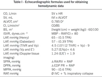
Pulmonary capillary or left atrial pressure may be estimated by assessing blood flow velocity in the pulmonary artery, if the patient has no pulmonary disease, or by assessing the transmitral and pulmonary venous flow in the left side. Transmitral flow analysis also provides information on left atrial pressure and heart failure prognosis. However, it depends on multiple conditions, such as heart rate, ventricular relaxation and suction, left atrial and ven-tricular compliance, and mitral valve conditions. To avoid the interference of these factors, other parameters, such as pulmonary venous flow, the response of transmitral flow to different volume loads, color M mode, and Doppler tissue imaging of the mitral ring, may be used. Left atrial pressure cannot be estimated by using isolated rules, but after assessing several parameters and respecting the limitations of each one. For practical purposes, pulmonary capillary pressure and left atrial pressure are considered equivalent, because the pulmonary capillaries, the pulmonary veins, and the left atrium communicate freely, forming a large chamber. Simultaneous measurements of right catheterization and left atrium through transeptal catheterization have already confirmed the similarity of those pressures 47-49. The several calculations used to
obtain hemodynamic data through echocardiography are shown in table I.
Other methods - Doppler tissue imaging is a relatively new technique that records the systolic and diastolic myocardial velo-cities at the level of the mitral valve annulus, preferentially medial or lateral. Filters should be used to eliminate the high frequencies, and the Nyquist limit should be maintained between –15 and 20 cm/s. Contrary to the transmitral flow (E wave), the velocity re-corded at the mitral valve annulus by using Doppler tissue imaging (Ea) does not undergo the influence of left atrial pressure and reflects isolated left ventricular relaxation. Thus, it is an excellent
Table I - Echocardiographic formulas used for obtaining hemodynamic data
CO, L/min SV x HR
SV, mL tVI x ALVOT
ALVOT, cm2 0.785 D2
CI, L/min/m2 CO/BS
BS, m2 [height (cm) + weight (kg)] - 60/100
ISVR, dynes.cm –5 MBP – RAP/CI × 80
LAP, mmHg (Henry) 65 – (0.5 TPA)
LAP, mmHg (Dabestani) 57 – (0.39 TPA)
LAP, mmHg (TIVR and Vp) 4.5 (103/ [2 TIVR] + Vp) - 9
LAP, mmHg (Vp and E’) 5.27 [E/Vp]+ 4.6
LAP, mmHg (Doppler tissue 1.24 (E/E’) + 1.9 imaging)
SPPA, mmHg ∆ RA/RV + RAP
DPPA, mmHg ∆ EDP PR + RAP
MPPA, mmHg 90 – (0.6 TPA)
RAP, mmHg Ø IVC + % inspiratory collapse
4
Determination of Hemodynamic Parameters Using Doppler Two-dimensional Echocardiography: A Searching Tool for Therapeutic Optimization in Patients with Congestive Heart Failure on an Outpatient Care Follow-up
index for assessing the relaxation deficit, even in pseudo-normal cases, and, when coupled to nontissue transmitral valve flow, it may estimate left atrial pressure. An E/Ea ratio > 10 may foretell a LAP > 12 mmHg with 91% sensitivity and 81% specificity, or an E/Ea ratio > 10 may foretell a LAP > 15 mmHg with 97% sensitivity and 78% specificity. Ommen et al 50 have shown a
correlation level between the E/Ea medial ratio and the mean left ventricular diastolic pressure of r=0.64, greater in patients with ejection fraction lower than 50%, as compared with those without ventricular dysfunction. These authors have also shown a positive predictive value of 64% for a left ventricular end-diastolic pressure > 12 mmHg, if E/Ea is> 15 mmHg, and a negative predictive value of 97% for low left atrial pressures, if the E/Ea ratio is < 8. In the presence of infarction in the laterobasal wall, atrial fibrillation, and severe mitral regurgitation, significant technical limitations exist and may render those measurements unfeasible 51,52.
In addition to the already cited methods, several other formu-las may be used to estimate left atrial pressure, using the following echocardiographic parameters: time of isovolumetric relaxation; velocity of propagation of the transmitral valve flow obtained by using color M mode (TMVF) 53-57; velocity of deceleration of the
early transmitral valve flow 58-60; systolic fraction of the pulmonary
venous flow; time of deceleration of the diastolic pulmonary flow61;
systolic fraction of the integral of velocity of the anterograde pul-monary venousflow 62; and ratio between the time of atrial
con-traction of the retrograde pulmonary venous flow and the antero-grade transmitral valve flow 63. The choice of the most appropriate
technique may vary according to the individual characteristics of the patients.
Therefore, the noninvasive hemodynamic assessment with in-formation provided by echocardiography may be obtained with a good accuracy by using more than one parameter and knowing the advantages and limitations of each method. Technically, it requires time and training of the echocardiographer, and its per-formance depends on the need of the clinical cardiologist in patients’ management.
Clinical application of echocardiography in the search for optimum hemodynamic parameters - Our group is carrying out a randomized clinical trial comparing the echocardiography-guided outpatient care, which aims at tailoring the hemodynamic profile (a reduction in the filling pressures and peripheral resistance), with the conventional clinical management, which aims at im-provement of symptoms. Our study comprised 99 patients diagnosed with congestive heart failure of any etiology, in functional class II-IV, with an ejection fraction ≤ 40%, who were hospitalized or visited the emergency unit due to decompensated heart failure in the prior 3 months. Our preliminary study comprised 70 patients, 31% of whom had congestive heart failure of ischemic etiology, a mean age of 60±15 years, an ejection fraction of 27±7%, and 60% were males. The preliminary data have shown that, in the group with the echocardiography-guided treatment, a reduction occurred in the right atrial pressure from 10.1±5 to 7.8±4 mmHg (P=0.004), in the maximum systolic pressure of the pulmonary artery from 47±12 to 39±12 mmHg (P=0.003), and in the systemic vascular resistance index from 3821±1265 to 3390± 1142 dynes/sec/cm5 (P=0.048). No significant difference was
ob-served in those parameters in the conventional clinical management group. These data and the conduction of this study so far have
indicated that the use of echocardiography-guided treatment based on hemodynamic data is feasible and more effective than the treatment based on traditional clinical management for obtaining a favorable profile of reduction in the filling pressures and peripheral resistance in patients with heart failure being followed up on an outpatient care basis.
Role of the hemodynamic profile for prognosis of chronic heart failure and predictive factors - The importance of the volemic status in the prognosis of patients with congestive heart failure may also be observed in data indicating that survival may range from 80% in 2 years in patients without congestion to less than 50% in 6 months in patients with refractory symptoms at rest 64.
The maintenance of a clinical status free from congestion, aiming at low or normal filling pressure levels within 4 to 6 weeks after hospital discharge in patients with functional class IV was asso-ciated with survival improvement in the following 2 years 65.
Hemodynamic monitoring may also be useful to stratify risk, because patients with normal initial filling pressures had a one-year survival of 95%, while in those with elevated initial pressures, a better prognosis was attributed to the therapy for reducing the pressures rather than to the level of initial congestion 66.
In addition to the congestive status of patients with congestive heart failure, the following important prognostic data may be obtai-ned by use of echocardiography: degree of preservation of right ventricular function67,68; left ventricular end-diastolic diameter69; left
atrial pressure; restrictive pattern in transmitral diastolic flow70-72;
and degrees of mitral and tricuspid regurgitation73,74. The BNP
le-vels, which are elevated in congestive heart failure perhaps indicating the degree of congestion, also correlate with sudden death75, NYHA
functional class76, and future events77.
Filling pressures, therefore, are useful in the management of patients with congestive heart failure, because they may foretell outcomes even after adjustments in therapy 66 and may be inferred
not only through BNP levels, but also through echocardiographic parameters, such as those already cited.
Final considerations - The treatment of systolic heart dysfunc-tion has advanced considerably in the past 20 years, mainly in the pathophysiological field due to the discoveries of the role of the neurohormonal factors78, which led to the use of drugs that proved
to reduce mortality, such as ACEI79-81, beta-blockers82,83, and
spiro-nolactone84. The participation of multidisciplinary teams has been
important for specifically reducing hospital readmissions85,86.
Despite these advances, mortality and hospital readmission due to symptoms of decompensation are still high 87,88, and this is
partially due to the limitation in the clinical setting to indicate adequacy of the treatment before hospital discharge, especially in defining the congestive/volemic status of those patients. Con-ventional tests, such as echocardiography, have not been routinely used in their plain potential to provide additional information to clinical examination in regard to the real hemodynamic conditions of the patient. On the other hand, the frequent use of right cardiac catheterization in managing patients with decompensated heart failure is not feasible, not only due to the risks and high costs of the procedure, but also because it requires the use of intensive care unit beds 89-92. Therefore, alternative methods should be
5
Determination of Hemodynamic Parameters Using Doppler Two-dimensional Echocardiography: A Searching Tool for Therapeutic Optimization in Patients with Congestive Heart Failure on an Outpatient Care Follow-up
long-term outpatient care treatment of those patients. The current ample availability of echocardiography in cardiology practice may allow better control of patients with congestive heart failure on an outpatient care basis, if the potential of that method for
deter-References
1. Roglieri JL, Futterman R, McDonough KL et al. Disease management intervention to improve outcomes in congestive heart failure. Am J Mang Care 1997;3:1831-9. 2. McAlister FA, Lawson FM, Teo KK et al. A systematic review of randomized trials of disease management programs in heart failure. Am J Med 2001;110:378-84. 3. Hunt AS, Baker DW, Chin MH, Cinquegrani MP et al. ADD/AHA Guidelines for the evaluation and management of chronic heart failure in the adult. J Am Coll Cardiol 2001;38:2101-13.
4. Drazner MH, Rame JE, Stevenson, LW, Dries DL. Prognostic importance of eleva-ted jugular venous pressure and a third heart sound in patients with heart failure. The New Engl J Med 2001; 345, 574-81.
5. Shah NB, Der E, Ruggerio C, Heindenreich PA et al. Prevention of hospitalizations for heart failure with an interactive home monitoring program. Am Heart J 1998; 135:373-8.
6. Stevenson LW, Perloff J. The limited reliability of physical signs for estimating he-modynamics in chronic heart failure. JAMA 1989;261:884-88.
7. McGee SR. Physical examination of venous pressure: a critical review. Am Heart J 1998;136:10-18.
8. Drazner MH, Hamilton MA, Fonarow G et al. Relationship between right and left -sided filling pressures in 1000 patients with advanced heart failure. J Heart Lung Transplant 1999;18:1126-32.
9. Badgett RG, Lucey CR, Mulrow CD. Can the clinical examination diagnose left-si-ded heart failure in adults? JAMA 1997;277:1712-9.
10. Butman SM, Ewy GA, Standen JR et al. Bedside cardiovascular examination in patients with severe chronic heart failure: importance of rest or inducible jugular venous distension. J Am Coll Cardiol 1993;22:968-74.
11. Zema MJ, Restivo B, Sos T et al. Left ventricular dysfunction: bedside Valsalva maneuver. Br Hear J 1980;44:560-9.
12. Westman EC, Matchar DB, Samsa GP, Mulrow CD et al. Accuracy and reliability of apical S3 gallop detection. J Gen Intern Med 1995;10:455-7.
13. Lok GE, Morgan CD, Ranganathan N. The accuracy and interobserver agreement in detecting the “gallop sounds” by cardiac auscultation. Chest 1998; 114: 1283-8. 14. Azevedo ER, Newton GE, Floras JS et al. Reducing cardiac filling pressure lowers
norepinephrine spillover in patients with chronic heart failure. Circulation 2000; 101:2052-9.
15. Kaye DM, Lambert GW, Lefkovits J, Morris M, Jennings G, Esler MD. Neurochemi-cal evidence of cardiac sympathetic activation and increased central nervous sys-tem norepinephrine turnover in severe congestive heart failure. J Am Coll Cardiol 1994; 23:570-8.
16. Weilenmann D, Hans Rickli, Follath Ferenc et al. Noinvasive evaluation of pulmo-nary capillary wedge pressure by BP response to the Valsalva maneuver. Chest 2002; 122, 140-5.
17. Rosario LB, Stevenson LW, Solomon SD, Lee RT, Reimold SC. The mechanism of decrease in dynamic mitral regurgitation during heart failure treatment: importance of reduction in the regurgitant orifice size. J Am Coll Cardiol 1998; 32:1819-24. 18. Rosario LB, Stevenson LW, Chelimsky-Fallick C et al. Sustained hemodynamic
effi-cacy of therapy tailored to reduce filling pressures in survivors with advanced heart failure. Circulation 1997; 96:1165-72.
19. Johson W, Omland T, Collins CM et al. Neurohormonal activation rapidly decreases after intravenous vasodilator and diuretic therapy for class IV heart failure [abs-tract]. Circulation 1998; 98:I-780.
20. Lucas C, Johnson W, Hamilton MA et al. Freedom from congestion predicts good survival despite previous class IV symptoms of heart failure. Am Heart J 2000; 140:840-7.
21. Chomsky DB, Lang CC, Rayos G et al. Treatment of subclinical fluid retention in patients with symptomatic heart failure: effect of exercise performance. J Heart Lung Transplant 1997;16:846-53.
22. Carr JG, Stevenson LW, Walden JÁ et al. Prevalence and hemodynamic correlates of malnutrition in severe congestive heart failure secondary to ischemic or idi-opathic dilated cardiomyopathy. Am J Cardiol 1989;63:709-13.
23. Stevenson LW, Bellil D, Grover-Mckay et al. Effects of afterload reduction on left ven-tricular volume and mitral regurgitation in severe congestive heart failure secondary to ischemic or idiopathic dilated cardiomyopathy. Am J Cardiol 1987; 60: 654-8. 24. Stevenson LW, Tillisch JH. Maintenance of cardiac output with normal filling
pres-sures in patients with dilated heart failure. Circulation 1986; 74, 1303-8. 25. Pierpont GL, Cohn J, Franciosa JA. Combined oral hydralazine-nitrate therapy in
left ventricular failure: hemodynamic equivalency to sodium nitroprusside. Chest 1978;73:8-13.
26. Rohde LE, Furian T, Campos C et al. Implications of the hemodynamic
optimiza-tion approach guided by right heart catheterizaoptimiza-tion in patients with severe heart failure. Arq Bras Cardiol 2002;78:261-6.
27. Stevenson L. Tailored therapy to hemodynamic goals for advanced heart failure. European J of Heart Failure 1999; 1: 251-7.
28. Steimle AE, Stevenson LW, Chelimsky-Fallick C et al. Sustained hemodynamic effi-cacy of therapy tailored to reduce filling pressures in survivors with advanced heart failure. Circulation 1997;96, 1165-71.
29. Shah MR, O’Connor CM, Sopko G et al. Evaluation study of congestive heart fail-ure and pulmonary artery catheterization effectiveness (ESCAPE): design and ra-tionale. Am Heart J 2001;141:528-35.
30. Johnson W, Ormland T, Hall C et al. Neurohormonal activation rapidly decreases after intravenous therapy with diuretics and vasodilators for class IV heart failure. J Am Coll Cardiol 2002; 39:1623-9.
31. Troughton RW, Frampton CM, Yandle TG, Espiner EA et al. Treatment of heart fai-lure guided by plasma aminoterminal brain natriuretic (N-BNP) concentrations. Lancet 2000; 355:1126-30.
32. Maisel, AS, Krishnaswamy P, Nowak RM et al. Rapid measurement of B-type na-triuretic peptide in the emergency diagnosis of heart failure. N Engl J Med 2002; 347:161-7.
33. Cheng V, Kazanagra R, Garcia A et al. A rapid bedside test for B-type peptide pre-dicts treatment 2001;37:386-91.
34. Adamson PB, Magalski A, Braunschweig F et al. Ongoing right ventricular hemo-dynamics in heart failure. J Am Coll Cardiol 2003;41:565-71.
35. Stein JH, Neumann A, Preston LM, Costanzo MR et al. Echocardiography for he-modynamic assessment of patients with advanced heart failure and potential heart transplant recipients. J Am Coll Cardiol 1997; 30:1765-72.
36. Dini FL, Bezante GP, Faggiano P, Odaglia F et al. Is a totally non-invasive assess-ment of the hemodynamic profile possible in patients with chronic heart failure? Ital Heart J 2000;1:1395-403.
37. Borgeson DD, Seward JB, Miller FA, Oh JK, Tajik J. Frequency of Doppler measur-able pulmonary artery pressures. J Am Soc Echocardiogr 1996;9:832-7. 38. Moreno FL, Hagan AD, Holmen JR, Pryor TA, Strickland RD, Castle CH. Evaluation
of size and dynamics of the inferior vena cava as an index of right-sided cardiac function. Am J Cardiol 1984;53:579-85.
39. Abramson SV, Burke JB, Pauletto FJ, Kelly JK. Use of multiple views in the echo-cardiographic assessment of pulmonary artery systolic pressure. J Am Soc Echo-cardiogr 1995;8:55-60.
40. Stephen B, Dalal P, Berger M, Schweitaer P, Hecht S. Noninvasive estimation of pulmonary artery diastolic pressure in patients with tricuspid regurgitation by Doppler echocardiography. Chest 1999; 116: 73-77.
41. Steven JL. Noninvasive estimation of right –sided pressures from spectral Doppler recordings of tricuspid and plutonic regurgitation velocities. Editorial: chest 1999; 116: 1-3.
42. Werner GS, Schaefer C, Dirks R et al. Prognostic value of Doppler echocardiographic assessment of left ventricular filling in idiopathic dilated cardiomyopathy. Am J Cardiol 1994;73:792-8.
43. Franciosa JÁ, Backer BJ, Seth L. Pulmonary versus systemic hemodynamics in de-termining exercise capacity of patients with chronic left ventricular failure. Am Heart J 1985;110:807-13.
44. Vanoverscherlde JLJ, Raphael DA, Robert AR, Cosyns R. Left ventricular filling in dilated cardiomyopathy: relation to functional class and hemodynamics. J Am Coll Cardiol 1990;15:1288-95.
45. Stevenson LW, Tillish JH. Maintenance of cardiac output with normal filling pres-sures in patients with dilated heart failure. Circulation 1986;74:1303-8. 46. Stevenson LW. Tailored therapy before transplantation for treatment of advanced
heart failure: effective use of vasodilators and diuretics. J Heart Lung Transplant 1991;10:468-76.
47. Walstson A, Kendall ME. Comparison of pulmonary wedge and left atrial pressure in man. Am Heart J 1973;86:159-64.
48. Luchsinger PC, Seipp HW, Patel DJ. Relationship of pulmonary artery-wedge pres-sure to left atrial prespres-sure in man. Circ Res 1962; 11:315-8.
49. Braunwald E, Drockenbrough ED, Frahm CJ, Ross J. Left atrial and left ventricular pressures in subjects without cardiovascular disease. Circulation 1961; 24: 267-9. 50. Ommen SR, Nishimura RA, Appleton CP, Miller FZ, Oh JK, Redfield MM, Tajik AJ. Clinical utility of Doppler echocardiography and tissue Doppler imaging in the esti-mation of left ventricular filling pressures. Circulation 2000;102:1788-94. 51. Nagheh SF, Middleton KJ, Kopelen HÁ, Zoghbi WA, Quiñones MA. Doppler tissue
6
Determination of Hemodynamic Parameters Using Doppler Two-dimensional Echocardiography: A Searching Tool for Therapeutic Optimization in Patients with Congestive Heart Failure on an Outpatient Care Follow-up
imaging: a noninvasive technique for evaluation of left ventricular relaxation and estimation of filling pressures. J Am Coll Cardiol 1997;30:1527-33.
52. Gorcsan III J. Tissue Doppler echocardiography. Lippincott Williams & Wilkins, Inc. Cur Opin Cardiol 2000,15:323-9.
53. Brun P, Tribouilloy C, Duval AM et al. Left ventricular flow propagation during early filling is related to wall relaxation: a color M-mode Doppler analysis. J Am Coll Cardiol 1992;20:420-32.
54. Takasuji H, Mikami T, Urasawa K et al. A new approach for evaluation of left ven-tricular diastolic function: spatial and temporal analysis of left venven-tricular filling flow propagation by color M-mode Doppler echocardiography. J Am Coll Cardiol 1996;27:365-71.
55. Duval-Moulin AM, Dupouy P, Brun P et al. Alteration of left ventricular diastolic function during coronary angioplasty-induced ischemia: a color M-mode Doppler study. J Am Coll Cardiol 1997;29:1246-55.
56. Weiss JL, Ares MA, Asher C et al. An index of early left ventricular filling that com-bined with pulsed Doppler peak E velocity may estimate capillary wedge pressure. J Am Coll Cardiol 1997;29:448-54.
57. Gonçales- Vilchez F, Ares M, Ayuela J, Alonso L. Combined use of pulsed and color M-mode Doppler echocardiography for the estimation of pulmonary capillary wedge pressure: an empirical approach based on an analytical relation. J Am Coll Cardiol 1999;34:515-23.
58. Garcia MJ, Ares MA, Asher C et al. An index of Early left ventricular filling that com-bined with pulsed Doppler peak E velocity may estimate capillary wedge pressure. J Am Coll Cardiol 1997;29: 448-54.
59. Pozzoli M, Capomolla S, Pinna G, Cobelli F, Tavazzi L. Doppler echocardiography reliably predicts pulmonary artery wedge pressure in patients with chronic heart failure with and without mitral regurgitation. J Am Coll Cardiol 1996; 27:883-93. 60. Gianuzzi P, Imparato A, Temporelli PL et al. Doppler- derived mitral desaceleration time of early filling as a strong predictor of pulmonary capillary wedge pressure in post-infarction patients with left ventricular systolic dysfunction. J Am Coll Cardiol 1994;23:1630-7.
61. Yamamuro A, Yoshida K, Hozumi T et al. Noninvasive evaluation of pulmonary capil-lary wedge pressure in patients with acute myocardial infarction by deceleration time of pulmonary venous flow velocity in diastole. J Am Coll Cardiol 1999; 34: 90-4. 62. Brunazzi MC, Chirillo F, Pasqualini M et al. Estimation of left ventricular diastolic
pressures from precordial pulsed-Doppler analysis of pulmonary venous and mi-tral flow. Am Heart J 1994;128:293-300.
63. Rossvoll O, Hatle LK. Pulmonary venous flow velocities recorded by transthoracic Doppler ultrasound: relation to left ventricular diastolic pressures. J Am Coll Cardiol 1993;21:1687-96.
64. Uretsky BF, Sheahan RG. Primary prevention of sudden cardiac death in heart fail-ure: will the solution be shocking? J Am Coll Cardiol 1997; 30:1589-97. 65. Caroline L, Johnson W, Hamilton MA et al. Freedom from congestion predicts good
survival despite previous class IV symptoms of heart failure. Am Heart J 2000; 140:840-7.
66. Stevenson LW, Tillisch JH, Hamilton M et al. Importance of hemodynamic response to therapy in predicting survival with ejection fraction < 20% secondary to ische-mic or nonischeische-mic dilated cardiomyopathy. Am J Cardiol 1990; 66:1348-54. 67. Di Salvo TG, Mathier M, Semigran MJ, Dec GW. Preserved right ventricular ejection
fraction predicts exercise capacity and survival in advanced heart failure. J Am Coll Cardiol 1995; 25: 1143-53.
68. Lewis JF, Webber JD, Sutton LL, et al: Discordance in degree of right and left ven-tricular dilatation in patients with dilated cardiomyopathy: Recognition and clini-cal implications. J Am Coll Cardiol 1993;21:649-54.
69. Gavazzi A, DeMaria R, Renosto G et al. The spectrum of left ventricular size in dila-ted cardiomiopathy: clinical correlates and prognostic implications. Am Heart J 1993;125:410-22.
70. Xie GY, Martin RB, Smith MD et al. Prognostic value of Doppler transmitral flow pat-terns in patients with congestive heart failure. J Am Coll Cardiol 1994; 24:132-9.
71. Pinamonti B, DiLenarda A, Sinagra G, Camerii F. Restrictive left ventricular filling pattern in dilated cardiomyopathy assessed by Doppler echocardiography: clinical, echocardiography and hemodynamic correlations and prognostic implications. Heart Muscle Disease Study Group. J Am Coll Cardiol 1993; 22:808-15. 72. Lapu-Bula R, Robert A, De Kock M et al. Risk stratification in patients with dilated
cardiomyopathy: Contribution of Doppler-derived left ventricular filling. Am J Cardiol 1998;82:779-85.
73. Abrahmson SV, Burke JF, Kelly JJ et al. Pulmonary hypertension predicts mortality and morbidity in patients with dilated cardiomyopathy. Ann Intern Med 1992; 116:888-95.
74. Hung J, Koellin T, Semigran MJ et al. Usefulness of echocardiographic determined tricuspid regurgitation in predicting event free survival in severe heart failure secon-dary to idiopathic-dilated cardiomyopathy or to ischemic cardiomyopathy. Am J Cardiol 1998;82:1301-3.
75. Berger R, Hueslman M, Strecker K et al. B-type natriuretic predicts sudden death in patients with chronic heart failure. Circulation 2002;105:2392-7.
76. Clerico A, Lervasi G, Chicca M et al. Circulating levels of cardiac natriuretic peptides (ANP e BNP) measured by highly sensitive and specific immunoradiometric assays in normal subjects and in patients with different degrees of heart failure. J Endo-crine Invest 1998; 21:170-9.
77. Cheng V, Kazanagra R, Garcia A et al. A rapid bedside test for B-type peptide pre-dicts treatment outcomes in patients admitted for decompensated heart failure: a pilot study. J Am Coll Cardiol 2001;37:386-91.
78. Remme WJ. Therapeutic strategies and neurohormonal control in heart failure. Eur Heart F 1994;15 (suppl D):129-38.
79. The SOLVD Investigators. The effect of enalapril on survival in patients with re-duced left ventricular ejection fraction and congestive heart failure. N Engl J Med 1991;325:293-302.
80. Garg R, Yusuf S. Overview of randomized trials of angiotensin-converting enzyme inhibitors on morbidity and mortality in patients with heart failure: Collaborative Group on ACE-Inhibitor Trials. JAMA 1995;273:1450-6.
81. The CONSENSUS Trial Study Group. Effects of enalapril on mortality in severe congestive heart failure: results of the Cooperative North Scandinavian Enalapril Survival Study (CONSENSUS). N Engl J Med 1987;316:1429-35.
82. CIBIS II Investigators Committees. The Cardiac Insufficiency Bisoprolol Study II (CIBIS II): a randomized trial. Lancet 1999;353:9-13.
83. MERIT-HF Study Group. Effect of metoprolol CR/XL. Randomised Intervention Trial in Congestive Heart Failure (MERIT-HF). Lancet 1999;353:2001-1007. 84. Pitt B, Zannad F, Remme WJ et al. The effect of spironolactona on morbidity and
mortality in patients with severe heart failure. N Engl J Med 1999; 341: 709-717. 85. Kornowsky R, Zeeli D, Averbuch M et al. Intensive home-care surveillance prevents hospitalization and improves morbidity rates among elderly patients with severe congestive heart failure. Am Heart J 1995;129:762-66.
86. Rich MW, Beckham V, Wittnberg C et al. A multidisciplinary intervention to prevent the readmission of elderly patients with congestive heart failure. N Engl J Med 1995;333:1190-95.
87. Philbin EF, DiSalvo TG. Prediction of hospital readmission for heart failure: Deve-lopment of a simple risk score based on administrative data. J Am Coll Cardiol 1999;33:1560-6.
88. Drumholtz HM, Chen YT, Wan Y et al. Predictors of readmission among elderly sur-vivors of admission with heart failure. Am Heart J 2000;139:72-7.
89. Adams KF: Post hoc subgroup analysis and the truth of a clinical trial. Am Heart J 1998;136:751-8.
90. Stevenson LW, Massie BM, Francis GS. Optimizing therapy for complex or refractory heart failure: a management algorithm. Am Heart J 1998; 135(suppl): S293-309. 91. Stevenson LW: Therapy tailored for symptomatic heart failure. Heart Failure 1995;
22:955-62