Alveolar extraction socket behaviour to alloplastic
regenerative procedures – a comparative study
Mariana de Medeiros Campos Guerreiro e Silva
Orientadores: Professor André Chen Professora Helena Francisco
Dissertação
Mestrado Integrado em Medicina Dentária
2019
À minha Mãe, Irmão e Avós, o meu maior apoio e, Ao meu Pai, que o levo comigo no coração…
Acknowledgments
It would be unimaginable that, after these five years, this would be the page where words are scarce. Not because I do not know what to say, or to whom to say, but because it will be tough to thank them enough.
First and foremost, I would like to express my sincerest gratitude to my supervisor Professor André Tsou Chen, who has supported me throughout my thesis with his patience, availability and knowledge. It was an honour to work alongside such dedicated teacher. His continuing effort, commitment and mentorship has helped me to bring this study to its conclusion.
I would like to thank Professor Helena Cristina de Oliveira Francisco, for accepting the invitation to be my co-supervisor.
My deepest gratitude to Doctor Lino Manuel Tomé Cerejeira Torres, to whom I am so very grateful for all the help in and out of the clinic. For his mentorship and help, solving every problem with easy and lightness, everlasting support and encouragement. From the third year in dental school, his thoroughness and understanding of surgery, his passion for academia inspired me and incited me to search, respect and admire this area through his guidance, expertise and thoughtful criticism.
I would like to extend my sincere thanks to Professor Susana Marques, for her help in performing the extra-oral scanners. She was relentless, worried, available, always answering all my doubts.
My sincere gratitude to Professor Joana Fialho, responsible for the statistical analysis. Her statistical assistance during the final stretch was greatly appreciated.
My thanks also to Professor Duarte Marques for the help given on the data analysis.
To MedBone® for providing the adbone®BCP synthetic biphasic bone graft material.
During these years, I had the chance to meet and work with a number of wonderful people. A warm thank you to all the teachers, assistants, presidents, coordinators and others who contributed to the smooth running of this Integrated Master's course.
I also want to thank my classmates for the hours we spent together, for the encouragement, help and camaraderie.
To my dearest friends, I would like to thank them for all the support and mostly for the friendship that they have given me over the years.
To João Pisco, I would like to sincerely thank for being my clinic partner, for working alongside me. Thank you for your friendship, your constant support, your ease on problem solving and making from every difficult moment a funnier situation. These years would not be the same without you.
To my family, who are my pillars, giving me both the freedom and support that I needed to become the person I am today. I cannot thank my mom Paula enough. For her endless love, care and all her sacrifices throughout these years. She has encouraged me in my studies and was the best role model. The best in the world, my best friend, my strength, the person who taught me never to quit my goals and fighting for them until the end. I ca not thank her enough.
To my brother, Paulinho, for being the joy of my house and the person who I love the most. My little best friend, who teaches me and supports me so much.
Last but not least, to my grandparents Isabel and Angelo, for their counselling, love and affection during these years. My deepest gratitude to my grandparents São and Fernando, for giving me all their love and affection, for the sacrifices they made and extraordinary help given throughout these years. Who managed to call me every day without ever forgetting an important date and for always being so present and caring.
“It always seems impossible until it’s done”
Abstract
Aim: To compare clinical, and volumetric alterations of post extraction sockets with
and without bone regeneration with Adbone®BCP on a socket preservation type of regeneration.
Materials and Methods: Participants were assigned to one of two groups, at surgery
day, having 16 anterior teeth divided equally into two groups. Bone regeneration was tested by application of a biphasic calcium phosphate synthetic bone graft (Adbone®BCP) while natural healing was the control group. Clinical evaluation included intra-oral photographs and an alginate impression. 3D examination consisted in extra-oral scanning of the obtained gypsum casts to generate 3D STL files. A comparison between initial and final buccolingual dimensions was formed using CloudCompareV2 (version 2.6.1 [GPL software], 2019),
measuring initial and final dimensions of both groups in 5 different sites (loss at 2, 3, 4, 5, 6 mm measured from coronal to apical of the extraction socket). Follow-up appointments were performed at days 7, 14 and 3 months postoperative.
Results: For the t-test, there was a significantly higher ridge dimensions loss on the
control group, p=0.029, 0.045 and 0.041 for the first three measurements, respectively.
Given these p-values being < 0.05, there are significant differences regarding the loss between the 2 groups, hence confirming the H1.
Discussion: There were differences between the test and control groups on the first three
measurements (H2, H3, H4) given that tissue modelling is a rather rapid process. However, the measurements situated apically on the alveolus, had p-values > 0.05, indicating that, given the short follow-up, the subsequent woven bone remodelling may take years to be completed and thus had not happened yet.
Conclusion: Sockets grafted with Adbone®BCP suffered a reduced loss of volume on
the alveolar socket contour, thus being effective on preserving the alveolar ridge.
Keywords: synthetic bone graft, hard-tissue, bone regeneration, Biphasic Calcium Phosphate, alveolar bone.
Resumo
As extrações dentárias são um dos procedimentos mais comuns no ramo da medicina dentária, levando a mudanças significativas no rebordo edêntulo, dificultando o correto e satisfatório posicionamento de implantes, podendo assim comprometer o resultado das reabilitações protéticas.
A extração dentária envolve um trauma mecânico nos tecidos moles, no ligamento periodontal e no osso do processo alveolar, levando a uma resposta inflamatória que recruta células hematopoiéticas e mesenquimais no local.
Após uma extração, o processo de cicatrização inicia-se com hemorragia e seguinte formação de um coágulo, sendo substituído por tecido de granulação. Em seguida, forma-se uma matriz provisória de tecido conjuntivo, dando início à fase proliferativa onde há uma incorporação de vasos e células formadoras de osso dentro da matriz provisória. Ao fim do primeiro mês após extração, verifica-se o preenchimento do alvéolo com osso imaturo que será progressivamente substituído por osso lamelar e medular. O término do processo de cicatrização é clinicamente observado pelo encerramento, primário ou secundário, do alvéolo com tecido mole epitelizado e radiográfico pelo preenchimento ósseo do alvéolo. Consequentemente, este processo levará a alterações dimensionais no rebordo edêntulo.
Embora se verifique alterações dimensionais até ao primeiro ano após extração, são durante os primeiros 3 meses que a perda óssea e tecidular é mais acentuada. Esta perda é influenciada por diversos fatores como as variações biológicas dos indivíduos, o tamanho do alvéolo pós-extracional e a extensão do trauma provocado durante a extração.
Está bem descrito na literatura que, após uma extração dentária, o rebordo edêntulo move-se em direção ao longo eixo do osso basal. A forma do maxilar parece retornar à forma em que estava antes do desenvolvimento do processo alveolar durante a erupção dentária. A falta de um estímulo funcional nas paredes ósseas e a necessidade de ajuste dos tecidos para se adaptar à geometria da crista na ausência de dentes podem explicar esta modificação.
Dadas estas alterações dimensionais, a reabilitação destes espaços edêntulos fica comprometida, influenciando tanto o resultado estético como funcional. De forma a tentar prevenir estas complicações, muitos estudos mostraram os efeitos do uso de enxerto de diferentes biomateriais e diferentes técnicas e respetivos benefícios na regeneração óssea. As primeiras investigações concentraram-se no uso apenas de membranas regenerativas.
Entretanto, pesquisas com enxertos ósseos em defeitos periodontais levaram os pesquisadores a explorar a utilidade das membranas em combinação com enxertos ósseos. Hoje em dia, a regeneração óssea guiada é geralmente realizada como um procedimento de combinação envolvendo membranas e um substituto ósseo de suporte. Alguns investigadores empregaram aloenxertos desmineralizados, maleáveis, rapidamente reabsorvidos e supostamente osteoindutores, enquanto outros utilizaram enxertos mineralizados, mais rígidos e osteocondutores. No entanto, não está claro qual material é mais eficaz para esta indicação clínica, já que estas técnicas incluem a colocação de diferentes materiais de enxerto, como autoenxertos ósseos, xenoenxertos, aloenxertos, combinados com membranas reabsorvíveis ou não reabsorvíveis e, mais recentemente, materiais bioativos, como L-PRF (leucócitos e fibrinas ricas em plaquetas).
Um autoenxerto envolve a utilização de osso obtido do mesmo indivíduo que recebe o enxerto, enquanto que um aloenxerto é derivado de seres humanos que é colhido de outro indivíduo que recebe o enxerto, como por exemplo de cadáveres. Os xenoenxertos são enxertos ósseos de uma espécie diferente da humana, como os bovinos, e são usados como uma matriz calcificada. Os aloplásticos podem ser feitos de hidroxiapatite, um mineral ósseo natural, fosfato tricálcico ou uma combinação de ambos. Os enxertos à base de fatores de crescimento são produzidos utilizando tecnologia de ADN recombinante, consistindo em fatores de crescimento humanos ou morfogénicos. Os substitutos de enxerto ósseo à base de cerâmica envolvem cerâmicas, isoladamente ou em combinação com outro material, como sulfato de cálcio, vidro bioativo e Fosfato de Cálcio. Contudo, a literatura não é clara relativamente ao material mais eficaz para as técnicas de regeneração alveolar.
Um dos materiais mais estudados, o xenoenxerto, apresenta resultados benéficos com a sua utilização. Contudo, este material tem uma taxa de reabsorção lenta, levando à presença de partículas residuais que poderão interferir com a normal cicatrização alveolar bem como influenciar a qualidade do osso regenerado.
Por outro lado, o uso de materiais aloplásticos de osteocondução também visa minimizar as alterações dimensionais que advêm após extração. Adbone®BCP é um material de enxerto ósseo totalmente bifásico feito de 75% de hidroxiapatite (HAp) e 25% de fosfato tricálcico beta-fosfato (β-TCP). Deste modo, parece ter todas as propriedades necessárias para minimizar as mudanças que ocorrem em alvéolos pós-extracionais, tanto a nível ósseo como dos tecidos moles, conduzindo a uma menor reabsorção da tábua óssea vestibular e menor colapso tecidual.
O objetivo deste estudo é comparar as alterações clínicas e volumétricas de alvéolos pós-extracionais com e sem regeneração óssea com Adbone®BCP.
Materiais e Métodos: 11 participantes foram inseridos de acordo com os critérios de
inclusão e exclusão, sendo designados para um dos dois grupos, no dia da cirurgia, com 16 dentes anteriores divididos igualmente em ambos os grupos. A regeneração óssea foi testada pela aplicação de enxerto ósseo sintético de Fosfato de Cálcio bifásico (Adbone®BCP) comparando-a com o grupo de controlo, a cicatrização natural. A avaliação clínica incluiu fotografias intra-orais e uma impressão em alginato. O exame 3D consistiu em leitura extra-oral dos modelos em gesso obtidos para gerar arquivos 3D STL e usando o CloudCompare V2
(versão 2.6.1 [GPL software], 2019), formando uma comparação entre as dimensões bucolinguais inicial e final dos tecidos duros e moles. Para tal, os ficheiros STL foram sobrepostos, escolhendo sete pontos em comum entre ambos para uma maior correspondência. Em seguida, traçou-se uma reta do ponto mais vestibular ao ponto mais palatino da raiz. Por fim, traçaram-se 5 retas, de 2 mm em 2 mm, a unir os dois ficheiros podendo avaliar as alterações dimensionais bucolinguais, em 5 regiões diferentes desde um ponto coronal e um mais apical. As consultas de acompanhamento foram realizadas nos dias 7, 14 e 3 meses de pós-operatório.
Resultados: Foram medidas as dimensões iniciais e finais de ambos os grupos em 5
locais diferentes (perda de 2, 3, 4, 5, 6 mm, medidos de mais coronal para mais apical no alvéolo). Para o teste t, houve uma perda significativamente maior no grupo controle, com valores de p=0,029, 0,045 e 0,041 para as três primeiras medições, respectivamente. A nossa hipótese nula corresponde a que não há diferenças significativas na perda dimensional entre os dois grupos. Considerando-se que esses p-valores são < 0,05, existem diferenças significativas em relação à perda entre os dois grupos, confirmando, assim, a nossa hipótese testada.
Discussão: Dados os resultados apresentados, existem diferenças estatisticamente
significativas entre os grupos teste e controle nas três primeiras medições (H2, H3, H4), dado que a modelação tecidual é um processo bastante rápido. No entanto, as duas últimas medições, sendo estas mais apicais no alvéolo, apresentaram p-valores > 0,05. Deste modo, especula-se que, dado o curto período de acompanhamento, o remodelamento ósseo subsequente, sendo um processo um pouco mais lento, pode levar anos para ser concluído, logo ainda não serem visíveis tais alterações.
Conclusão: Podemos concluir que existem diferenças estatisticamente significantes
cicatrização de alvéolos do grupo de controlo. No entanto, há um número limitado de estudos sobre este enxerto ósseo, expondo assim a necessidade de mais ensaios clínicos randomizados avaliando o efeito do enxerto ósseo sintético Fosfato de Cálcio bifásico na regeneração óssea. Devido ao tamanho da amostra e curto período de acompanhamento, não é possível aplicar à população em geral.
Palavras-chave: enxerto ósseo sintético, tecido duro, regeneração óssea, Fosfato de cálcio bifásico, osso alveolar.
Index
Tables and Figures Index ... xiv
List of abbreviations ... xv
1. Introduction ... 1
1.1Healing Process ... 1
1.2 Socket Preservation/regeneration techniques ... 4
1.3 Bone Quality ... 6
1.4 Biphasic calcium phosphate synthetic bone graft (Adbone®BCP) ... 8
2. Aim ... 10
3. Hypothesis ... 10
4. Materials and Methods ... 11
4.1 Study design and Randomization ... 11
4.2 Sample Size calculation ... 11
4.3 Inclusion Criteria ... 11 4.4 Exclusion criteria ... 12 4.5 Surgical technique ... 12 4.6 Post-operative Management ... 14 4.7 Clinical evaluation ... 14 4.8Radiographic Examination ... 15 4.9 3D Examination ... 15 4.10 Step-by-step procedures ... 16 4.11 Statistical Analysis ... 16 5. Results ... 17 6. Discussion ... 23 Main considerations ... 27 7. Conclusion ... 29 8. Bibliography ... 31 9. Appendix ... 35
9.1 Appendix 1 – Informed Consent ... 35
9.2 Appendix 2 – Ethic Committee approval ... 39
10. Annexes ... 40
10.1Annex 1 – Results ... 40
10.2 Annex 2 – Intra-oral photographs ... 41
10.3 Annex 3 – Radiographic Examinations ... 43
Tables and Figures Index
Table 1: Bone density classification by Misch. 8
Table 2: Group statistics for tissue loss at 2 mm. 18
Table 3: Testing of independent samples for tissue loss at 2 mm. 18
Table 4: Group statistics for tissue loss at 3 mm. 19
Table 5: Testing of independent samples for tissue loss at 3 mm. 19
Table 6: Group statistics for tissue loss at 4 mm. 20
Table 7: Testing of independent samples for tissue loss at 4 mm. 20
Table 8: Group statistics for tissue loss at 5 mm. 21
Table 9: Testing of independent samples for tissue loss at 5 mm. 21
Table 10: Group statistics for tissue loss at 6 mm. 22
Table 11: Testing of independent samples for tissue loss at 6 mm. 22
Table 12: Average amount of resorption of tooth extraction in different tooth areas. 25
Table 13: Results of each patient on both groups. 40
Figure 1a: Schematic drawing showing a classification of residual jaw shape. 7
Figure 1b: Schematic drawing showing a jaw bone quality. 7
Figure 2: Instructions on how to handle Adbone®BCP. 13
Figure 3: Box-plot graph comparing volumetric loss on both groups at 2 mm. 17
Figure 4: Box-plot graph comparing volumetric loss on both groups at 3 mm. 19
Figure 5: Box-plot graph comparing volumetric loss on both groups at 4 mm. 20
Figure 6: Box-plot graph comparing volumetric loss on both groups at 5 mm. 21
Figure 7: Box-plot graph comparing volumetric loss on both groups at 6 mm. 22
Figures 8-9: Intra-oral photographs. 41
Figures 10-12: Intra-oral photographs. 42
Figures 13-14: Pre and post-operative radiographies. 43
Figures 15-17: Pre and post-operative radiographies. 44
Figures 18-23: 3D analysis on Control and Experimental Groups 45
List of abbreviations
% Percentage
ARP Alveolar ridge preservation
ASA American society of anaesthesiologists
Β-TCP Beta-phosphate tricalcium phosphate
BCP Biphasic calcium phosphate
FGFS Fibroblastic growth factors
GBR Guided bone regeneration
HA Hydroxyapatite
i.e. Id est
IGFS Insulin-like growth factors
IL Interleukins
L-PRF Leukocyte and platelet-rich fibrin
mm Milometers
MPA Megapascal
n Sample numbers
PA Periapical radiographs
POST-OP Postoperative
PDGF Platelet derived growth factor
PRGF Plasma rich in growth factors
RCT Randomized control trial
STL Stereolithography
TCP Tricalcium phosphate
1. Introduction
The volume and shape of the alveolar process is determined by the form of the teeth, their axis of eruption and eventual inclination. (1) Teeth extractions are one of the most common procedures in dentistry, due to multiple causes such as decay, advanced periodontal disease, trauma, and others. (2) Although dimensional changes can be observed up to 1 year after tooth extraction, it is during the first 3 months that the most statistically significant bone and tissue loss occurs. (3–5)
Following an extraction, the alveolar process will undergo resorption and the bundle bone will lose function and disappear (1), thus having a direct repercussion on the periodontal structures. Although, generally, extraction sockets heal uneventfully, the defect caused by extraction will only become partially repaired as the alveolar ridges will reabsorb. Bone loss is more pronounced on the horizontal axis. There is also loss of vertical ridge height, most pronounced on the buccal aspect. (4) This resorption results in a narrower and shorter ridge (6), placing the ridge to a more palatal/lingual position. However, in some cases, there might already exist a previous bone loss due to periodontal disease, endodontic lesions or a traumatic episode. Therefore, when there is an alveolar wall missing, there will be a more pronounced bone resorption. (1) In these cases, it will be filled with fibrous tissue which does not allow a correct healing. (2,4,7)
In 1996, Soikkonen et col. described that systemic conditions such as osteoporosis, diabetes, vascular, endocrine or renal pathology can accelerate bone loss and thus interfering with the bone’s metabolism. Even the simple trauma created during an extraction creates a microtrauma on the surrounding bone which can accelerate bone loss. (8)
1.1 Healing Process
The healing of bone tissues includes both regenerations, where there is a complete restoration of morphology and function, and repair, which differs in morphology and function.
Teeth extraction involves a mechanical trauma in the soft tissues, periodontal ligament, bundle bone and the bone of the alveolar process (7,9), thus leading to an inflammatory response that includes both hematopoietic and mesenchymal cells in the site. (8,10) Tissue and cell proliferation are mediated at different stages by carious growth factors, inflammatory cytokines, and signalling molecules. Although it is a continuous process, bone repair can be divided into three phases: inflammation, proliferative and modelling/remodelling. (7)
The inflammation phase begins immediately after tissue damage and lasts for approximately 2 weeks. The initial step in the repair process is the formation of a blood clot that effectively plugs the detached blood vessels and stops bleeding. It acts as a physical matrix that directs cellular movements and it contains substances (i.e. growth factors) that influence mesenchymal cells and enhance the activity of inflammatory cells. Such substances will thus induce and amplify the migration of various types of cells into the socket wound, as well as their proliferation, differentiation, and synthetic activity within the coagulum. (11-12)
Cytokine release from injured cells then recruits inflammatory cells into the area, where macrophages begin phagocytosis of damaged tissues and cells. The clot is comprised mainly of erythrocytes and platelets that get trapped in a network of fibrin. Isolated neutrophils are present in the central and apical compartments of the blood clot. The principal Sharpey’s fibres in the bundle bone were found to be in direct contact with the coagulum in the socket. (2,13) Osteoclasts begin the process of resorbing damaged bone in the area to recycle mineral components. In addition, cells from myeloid and mesenchymal cell lineages are recruited to the area where they begin to differentiate into osteoblasts and chondroblasts. Within 2–3 days, large numbers of inflammatory cells mediated by signalling molecules (i.e. growth factors and cytokines), like platelet-derived growth factor (PDGF), insulin-like growth factors (IGFs), fibroblastic growth factors (FGFs) and transforming growth factor-beta (TGF-β), which are also responsible for cell differentiation and proliferation (7,14), migrate to the wound in order to “clean” the site before new tissue can start forming. The combination of inflammatory cells, vascular sprouts, mesenchymal cells from the severed periodontal ligament and immature fibroblasts forms the granulation tissue. As the site becomes sterilized, the granulation tissue is gradually replaced with a provisional connective tissue matrix that is rich in collagen fibres and cells, and the proliferative phase of the wound-healing process begins. (7,12,15)
The proliferative phase may be divided into two parts – fibroplasia and woven bone formation – and is characterized by intense and rapid tissue formation. After 4-5 days, the epithelium from the margins of the soft tissue starts to proliferate to cover the granulation tissue in the socket. (15) Fibroplasia is the rapid deposition of a provisional matrix. Subsequently, there is an incorporation of vessels and bone forming cells within the provisional matrix where projections of woven bone are arranged around the blood vessels. These projections will completely surround a vessel in order to form the primary osteon. Woven bone can be identified in the healing socket 2 weeks after tooth extraction and remains in the wound for several weeks. Such formation is ongoing in the apical portion of the socket, occurring from the periphery to
the centre of the alveolar socket. Woven bone is a provisional type of bone without any load-bearing capacity and therefore needs to be replaced with mature bone types (lamellar bone and bone marrow). In the first month, the socket is filled with woven bone. (4)
The last phase is bone modelling which is defined as a change in the shape and architecture of the bone, whereas bone remodelling is defined as a change without concomitant change in the shape and architecture of the bone. The replacement of woven bone with lamellar bone or bone marrow is bone remodelling, whereas the bone resorption that takes place on the socket walls leading to a dimensional alteration of the alveolar ridge is the result of bone modelling. This may take several months and exhibits substantial variability among individuals. (7-8,16)
The walls of the socket – the alveolar bone proper or the bundle bone – are gradually resorbed and the socket becomes filled with immature woven bone that will eventually be replaced by mature lamellar bone. (8,16-17) After 3 weeks, the socket contains connective tissue and there are signs of mineralization of the immature bone, through the process of bone remodelling, and the epithelium covers the wound. After 6 weeks of healing, bone formation in the socket is pronounced and trabeculae of newly formed bone can be seen. (10)
Adequate vitamin D and calcium are critical for proper bone repair and their levels may, in part, dictate the rate of repair. The time for the remodelling stage varies depending upon individual bone metabolism, but usually require months from the time of injury. (12)
At the same time, bone resorption takes place on the socket walls leading to dimensional changes of the alveolar ridge. A few weeks after tooth removal, osteoclasts could be found around the crest of both buccal and lingual walls and on the outer and inner (bundle bone) portions of the socket. Bone modelling takes place equally on buccal and lingual walls, but since the lingual bone is usually wider than the buccal bone wall, modelling results in greater vertical bone loss at the thin buccal plate than at the wide lingual wall. (7)
Bone modelling takes place earlier than bone remodelling, in such way that about two-thirds of the modelling process occurs in the first 3 months of healing. (3) The complete remodelling of the woven bone into lamellar and bone marrow may take several months or years and it depends on individual variability. Bone resorption is less in mesial/distal sites than in buccal/lingual sites and it may be due to the presence of adjacent teeth. In total, the mean value of vertical bone loss is 1.24 mm (± 0.11) at 6 months(4,13,18). Regarding horizontal bone resorption, the mean value is 3.79 mm (± 0.23) at the level of the alveolar crest in the first 6
months, with more pronounced tissue loss in the buccal aspect. The end of socket-healing process is clinically observed by the closure of the socket with firm epithelized soft tissue and radiographic by bone filling of the socket. This will be influenced by biologic individuals’ variations, alveolar socket size and the extended of socket trauma during the extraction. (7)
1.2 Socket Preservation/regeneration techniques
There are several types of grafts. An autograft involves utilizing bone obtained from the same individual receiving the graft. An allograft is derived from humans which is harvested from another individual of that receiving the graft. Xenografts are bone grafts from a species other than human, such as bovine and are used as a calcified matrix. Alloplastic grafts may be made from hydroxyapatite, a naturally bone mineral, tricalcium phosphate or a combination of both. Growth factors enhanced grafts are produced using recombinant DNA technology, consisting of either human growth factors or morphogens. Ceramic‑based bone graft substitutes involve ceramics, either alone or in combination with another material such as calcium sulphate, bioactive glass, and calcium phosphate. (19)
It is now well established that following tooth extraction the ridge crest moves toward the long axis of the basal bone. (7,20) The shape of the jawbone appears to return to the shape that was present prior to the development of the alveolar process during tooth eruption. The lack of a functional stimulus on the bone walls and the need for tissue adjustment to meet “genetically” determined demands regarding ridge geometry in the absence of teeth may explain this modification. (4)
Clinicians often perform extractions without any planning on preserving the alveolar ridge for later implant rehabilitation or without evaluating the possibility of immediate implant placement. This may lead to consequences on the remaining bone, which in turn may threaten the entire rehabilitation. Failing to preserve the residual alveolar ridge after a tooth extraction often leads to compromising future placement of implants and further rehabilitation. Therefore, throughout the years, many studies have shown the effects of grafting different biomaterials and different techniques and their benefits on bone regeneration. Early investigations focused on using regenerative membranes alone. However, research with bone grafts in periodontal defects led investigators to explore the utility of membranes in combination with bone grafts. (8) Nowadays, guided bone regeneration (GBR) is generally performed as a combination procedure involving membranes and a supporting bone substitute. Some researchers have
employed demineralised, malleable, rapidly resorbed and reportedly osteoinductive allografts (21), while others have used mineralised, more rigid, minimally resorting and reportedly osteoconductive grafts. (7,14) Scheyer et al., 2016, compared both. Nevertheless, it is not clear which material is most effective for this clinical indication (22), as these techniques include the placement of different grafting materials such as bone autografts, xenografts, allografts, combined with resorbable or non-resorbable membrane (23-24), and, more recently, bioactive materials, as L-PRF (leukocytes and platelet rich fibrins).
Even though the occurrence of bone loss in both buccal and lingual side is always expected, the placement of graft materials has been described as an ideal procedure to reduce the level of bone resorption. (1-2) Some studies have shown that, with these techniques, the amount of ridge reduction in height ranged from +1.3 mm to -2.64 mm and in width varied from -1.2 mm to 2.64 mm, depending on the type of materials used. (1,25) Willenbacher et al., on a meta-analysis, concluded that about 0.95 mm up to 1.12 mm of mean apico-coronal ridge height and 1.31 mm to 1.54 mm of mean bucco-oral ridge width can be preserve using alveolar ridge preservation techniques compared to natural healing with a defined 6 months of follow up. (26)
Different kinds of graft materials may influence the socket healing and may compensate for the buccal loss. (7,25) However, the presence of residual particles is a concern since they might interfere with normal healing. (2) Another fact that may be relevant is the bone quality, which is dependent on the resorption rate of the grafting material used, as well as the ability to promote bone formation. (25) A decrease in bone density is expected if the residual particles do not integrate well with bone. (25) Thus, in case of implant rehabilitations, these must be placed more apically to previous socket to achieve primary stability. (27) The greater percentages of residual graft remnants were seen in sockets treated with xenografts and allografts. The use of a xenograft or an allograft had a beneficial effect in bone preservation compared to alloplastic materials. Xenograft is one of the most studied materials. (23)
Experimental studies in a dog model (2,9) have demonstrated that placing bone substitutes in a fresh extraction socket failed to inhibit the processes of modelling and remodelling that took place in the socket walls following tooth extraction. However, is was observed that the graft supported hard-tissue formation, in particular in the cortical region of the extraction site, and the dimension and profile of the alveolar ridge was better preserved. The authors concluded that the placing a biomaterial in an extraction socket may modify modelling and compensate for the buccal bone loss. The cross-sectional area of the grafted sites exhibited
a reduction of only 3% of their initial dimensions, whilst in the non-grafted sites, the corresponding reduction was 25%.
Araújo et al. (2011), showed that applying Bio-Oss Collagen® (xenograft) in the gap during immediate implant placement modified the process of hard tissue healing, provided additional amounts of hard tissue, improved the level of marginal bone-to-implant contact and prevented soft tissue recession.
Immediate implant placement in fresh extraction sockets fails to prevent bone modelling. (7) The use of hard- or soft-tissue grafts with immediate implant placement to prevent ridge reduction has been evaluated in several clinical and experimental studies. In these studies, the hard-tissue graft, mainly a bone substitute, was placed in the space between the implant surface and the inner surface of the buccal bone wall, whilst the soft-tissue graft was adapted to the outer surface of the bone wall. These demonstrated that graft procedures, combined with implant placement, may hinder ridge alterations following tooth extraction. (7)
In 2015, Atieh et al., states that the literature is not clear about which material is the most effective for preservation and regeneration techniques.
1.3 Bone Quality
The internal structure of the bone is described in terms of quality or density, which is reflected in a set of biomechanical properties - strength and modulus of elasticity. The bone quality is strongly dependent on the position in the arch - the bone with the highest density is found in the mandibular anterior region, followed by the anterior region of the maxilla, posterior region of the mandible, and finally the posterior region of the maxilla, where the bone density is lower. (28) This may be given due to the different biomechanical functions performed by these structures - the mandible is an independent structure which acts as a unit of force absorption; the maxilla acts as a force distributor. Thus, in the presence of teeth, the mandible presents a dense bone cortex and a relatively dense trabecular bone; the maxilla redirects forces through the zygomatic and palate arches, away from structures such as the brain and orbits, and it is anatomically beneficial to have a thin cortex and dense trabecular bone. (29-30) After an extraction, there is a decrease in both bone quantity and its density. This decrease is more relevant in the posterior maxilla and less in the anterior mandible. (31)
Several classifications have been made throughout these years. In 1970, Linkow and Chercheve classified bone density into 3 categories. (32)
Based on the volume of remaining mineralized bone, the edentulous sites may, according to Lekholm and Zarb (1985), be classified into five different groups (Fig. 1a). In groups A and B, substantial amounts of the ridge still remain, whereas in groups C, D, and E, only minute amounts of hard tissue remain. (33)
Figure 1a – Schematic drawing showing a classification of residual jaw shape. (Adapted from Lekholm & Zarb 1985. Reproduced from Quintessence. (33) )
Lekholm and Zarb (1985) also classified the bone quality in the edentulous site. Class 1 and class 2 characterized a location in which the walls – the cortical plates – of the site are thick and the volume of bone marrow is small. Sites that belong to class 3 and class 4 have relatively thin walls of cortical bone, while the amount of trabeculae of lamellar bone and marrow, is large. (33)
Figure 1b – Schematic drawing showing a jaw bone quality. (Source: Lekholm & Zarb 1985. Reproduced from Quintessence.)
Even though it is the most used classification, it presents a high level of bias since it depends on the quality of the radiographic examination and the clinical perception of the health professional. (34) Thus, in 2007, Misch proposed another classification, based on macroscopic density of edentulous areas of the maxilla and mandible:
Table 1 – Bone density classification by Misch. (34)
The regional locations of the different densities of the cortical bone are more consistent than those of the trabecular bone. Bone density can be determined by tactile sensitivity during surgery, radiographic evaluation or anatomical location. (34)
A literature review and a post-surgical study in partially edentulous patients indicate that the location of different bone densities is strongly related to the different regions of the oral cavity (Table 1). (34)
Bone density is directly related to implant stability and, consequently, to successful rehabilitation. As a result, the region with the highest rate of osseointegration and successful implantation is the anterior mandible; the one that presents a worse rate of osseointegration and success of implant rehabilitation is the posterior maxilla. (20,35)
According to the classification of Elian et al. 2007, in situations where facial soft and hard tissues are at normal levels in relation to the cementoenamel junction of the tooth and remain with the same levels after extraction, this alveolus is classified as type I. If the bone level is below the previous references, meaning the buccal plate is partially missing, but the soft tissues are at a normal level, it is classified as type II. The type III alveolus is the most complex to rehabilitate, with bone and tissue resorption. Type I alveoli are the easiest to treat and have the most predictable results. (34-35)
1.4 Biphasic calcium phosphate synthetic bone graft (Adbone®BCP)
Synthetic calcium phosphates are commonly used in bone substitutes. Within the group of phosphates, hydroxyapatite (HA) and tricalcium phosphate (TCP) are the most common
materials used for bone substitution. HA has excellent biocompatibility with the bony environment; however, it is mostly used for surface coating on metal implants due to its low solubility and brittle nature. TCP has the advantage of osteoconductivity, but it is limited in clinical use due to its rapid degradation rate. Therefore, biphasic calcium phosphate (BCP) has been used to overcome the disadvantages of HA and TCP. Since BCP includes HA and TCP, it provides optimal dissolution and good bioactivity, cell attachment, proliferation, and differentiation for new bone regeneration. (38)
Adbone®BCP is a fully biphasic bone graft material made from 75% HA and 25% β-TCP. Adbone®BCP features a multi-directional interconnected 80% porosity that guides the three-dimensional regeneration of bone, having a pore size ranging from 300-500 microns. This forms an ideal environment for vascularization. (39)
As the bone healing process occurs, Adbone®BCP is reabsorbed and replaced with new bone. Due to its composition, it has a biphasic reabsorption and was developed to achieve the highest degree of porosity without compromising mechanical strength. In average, whilst other materials have a mechanical resistance of 0.5MPa, adbone has 3.0MPa. It can be mixed with patient’s blood. The hydrophilic behaviour confers a high cohesivity of the particles. It is radiopaque, allowing the monitorization of the graft osteointegration. It facilitated the formation and ingrown of new bone and blood vessels (osteoconduction) due to three dimensional, highly porous structure. The use of this allograft may be considered when autogenous bone is not indicated or as an adjunctive therapy when it is not possible to harvest a sufficient quantity of autogenous bone to fulfil the needs of the proposed surgical procedure. It is designed to regenerate bone in oral defects associated with disease, trauma and degeneration. There is no indication to use a membrane, unless there is a risk of graft exposure. (39)
Adbone®BCP is intended to be used as a bone void filler or augmentation material for bone defects that are not intrinsic to the stability of the bony structure such as maxillofacial osteotomy, sinus lift augmentation, crestal access, lateral access, implantology, alveolar regeneration, vertical augmentation, horizontal augmentation, peri-implant defects, dehiscence and fenestration.
2. Aim
The aim of this study is to compare clinical and volumetric alterations of post extraction sockets with and without bone regeneration with Adbone®BCP on a socket preservation type of regeneration.
3. Hypothesis
The null hypothesis (H0) of the present study is: “There is no difference between test and control groups regarding bone resorption, soft tissue healing and collapse of the alveolar socket after bone regeneration compared to non-regenerated alveolar sites.”.
It will be tested the hypothesis (H1) that there is a difference between test and control groups regarding bone resorption, soft tissue healing and collapse of the alveolar socket after bone regeneration compared to non-regenerated alveolar sites.
4. Materials and Methods
4.1 Study design and Randomization
This study is a unicentric study conducted at Faculty of Dentistry, University of Lisbon (FMDUL). Participants were assigned to one of two groups at surgery day, starting with 8 alveoli being attributed to the control group and subsequent 8 attributed to the experimental group.
Post-extraction sockets were submitted to bone regeneration, after debridement and irrigation with saline solution, by application of a biphasic calcium phosphate synthetic bone graft (Adbone®BCP) whereas control group’s post-extraction sockets were just debrided and irrigated with saline solution.
The approach was a socket preservation type described by Elian in 2009. The regenerative material was place in the intact wall socket and the graft particles were compacted.
An informed consent was made (Appendix 1) and approved by the Ethic Committee for Health from Faculty of Dentistry, University of Lisbon (Comissão de Ética para Sáude-FMDUL) (Appendix 2). Prior to surgery, each patient signed the approved informed consent (Appendix 1) agreeing to being submitted either to unassisted socket healing or grafted socket healing.
4.2 Sample Size calculation
A total of 12 patients (16 samples) entered this study. Given that it is a pilot study, it was decided to use this small sample to evaluate if the data is enough for a power sample.
4.3 Inclusion Criteria
The inclusion criteria in this study were as follows:
§ Patients from FMDUL aged 18 or over;
§ Requiring teeth extraction in the premolar region and anterior maxilla (5-5); § Presence of intact buccal bone plate;
§ ASA (Physical Status Classification System, American Society of Anaesthesiologists) I or II.
4.4 Exclusion criteria
The exclusion criteria included patients with:
§ Uncompensated systemic diseases;
§ Metabolic and healing disorders, i.e. diabetes mellitus, hyperparathyroidism, cancer, HIV;
§ Heavy smokers (>5 cigarettes/ day); § Bone metabolic diseases;
§ Severe renal dysfunction or liver disease;
§ Patients who had received systemic corticosteroids or other immunosuppressive agents, radiation therapy and/or chemotherapy for the past 2 months;
§ Active infections in the surgical site;
§ Patients with periodontal or/and endodontic disease.
4.5 Surgical technique
Each patient was submitted to local subperiosteal anaesthesia (Articaíne 4% with epinephrine 1: 200 000, Inibsa, Sintra, Portugal), both buccal and palatal, after local application of topic anaesthesia (Topigel, Benzocaine, 2%, Clarben S.A, Madrid, Spain).
Following the anaesthesic procedure, the surgeon performed tooth extraction the more atraumatic possible, to preserve the integrity of the alveolar buccal plate, without elevation of a mucoperiosteal flap. Then, followed thorough degranulation of soft tissue remnants, ensuring integrity of the buccal plate, and irrigation with sterile saline solution (0.9% NaCl, B. Braun Medical, Lisbon, Portugal).
After allocation to either test or control group, each participant on the test group was submitted to application of a biphasic calcium phosphate synthetic bone graft (Adbone®BCP) whereas control group’s post-extraction sockets were just debrided and irrigated with saline solution.
Control group:
After tooth extraction, the alveolar socket was thoroughly debrided using a Lucas curette (Lucas CL8, Dentaleader S.A., Lisbon, Portugal). Subsequently, they were irrigated using saline solution (0.9% NaCl, B.Braun Medical, Queluz de Baixo, Portugal) and then sutures (Surgical silk suture, non-absorbable, 3/0 Silkam®, B. Braun Medical, Lisbon, Portugal) were
Test group - socket treatment with biphasic calcium phosphate synthetic bone graft: After tooth extraction, the alveolar socket was thoroughly debrided using a Lucas curette (Lucas CL8, Dentaleader S.A., Lisbon, Portugal) and irrigated using saline solution.
The alveolar socket was filled with biphasic calcium phosphate synthetic bone graft, according to the following protocol:
4.5.1 The granules of the synthetic bone graft are mixed with a few droplets of blood. Once a uniform agglomerate is formed, these are to be introduced inside the socket, compressed firmly, certifying that there are no free spaces left. Excess of blood is to be absorbed with a gauze.
Figure 2 – Instructions on how to handle Adbone®BCP (Adapted from MedBone). (39)
5.5.1 A tension-free monofilament non-resorbable cross suture is to be placed on the socket in order to maintain the graft in place.
6.5.1 It is not required the use of membrane unless there is risk of graft exposure. When so, a SpongostanTM Dental (Ethicon Inc., Johnson & Johnson, New Jersey, United
States) is to be cut into the alveolar shape and placed on top of the graft, stabilizing it with a tension-free monofilament non-resorbable cross suture.
1 2 3
4.6 Post-operative Management
Patients, in both groups, whenever there was therapeutically indication, went through a therapeutic regimen of 1-tab Amoxicillin 1000mg Cipamox 1000 g (Atral Laboratories S.A., Santarem, Portugal) each 12H, for 8 days.
The pain medication regimen included 1 tab of paracetamol 1000mg Ben-u-ron 1000g (bene-Arzneimittel GmbH, Munich, Germany) 8/8h.
In case of extreme inflammation, Ibuprofen 600mg, 12/12h was prescribed.
Patients were also advised to use a 0.2% chlorhexidine mouth rinse (0.2% Bexident Post, Isdin, Lisbon, Portugal) to be used three times per day for a period of 7 days.
All the postoperative recommendations were explained.
4.7 Clinical evaluation
Clinical evaluation began before the extraction with intraoral photographs. An alginate impression (R&S Turboprint, Dentaleader, Lisbon, Portugal) was taken of the maxilla to create stone cast models to determine the pre-extraction dimensions.
Patients were monitored postoperatively to observe healing and soft-tissue closure. Follow-ups were at days 7, 14 and 3-months post-operative. In the first follow-up appointment, sutures were removed and observed tissue healing. In the last appointment, another alginate impression was taken of the alveolar extraction socket to determine the post-extraction dimensions after 3 months suffering bone remodulation.
4.7.1 Intra-oral photographs
Occlusal and lateral photographs to compare the pre- and post-surgical dimensions (see Annexe 2).
4.7.2 Alginate Impressions
Kromopan Alginate (Lascod S.P.A., Italy) was used to take maxillary impressions of each patient of either groups prior to surgery and at the 3-month follow-up appointment. These were casted using stone cast type III and type IV, respectively (Pro-Stone® 21, Pro-Dental,
4.8 Radiographic Examination
At surgery day, the patient was schedule to do a periapical radiography, before surgery. In the test group, immediately after placing the graft, another periapical radiography was taken to evaluate if the graft was well placed, compacted and filled the alveolar socket. At 3 months post-op, the participant returned to the clinic and did another periapical X-ray to compare and evaluate the results of the treatment with Adbone®BCP and natural healing (see Annexe 3).
4.9 3D Examination
Alginate impressions were taken prior to surgery in order to create stone cast models (following the precise manufacturer indication) for each patient. At 3-month post-op, another alginate impression was taken.
Once both impressions were casted, these were scanned using an extra-oral digital scanner (Scanner S600 Arti, Zirkonzahn.Software, Portugal) to generate surface stereolithography (STL) files. Using CloudCompare (version 2.6.1 [GPL software], 2019), it was made a 3D analysis of the models, which thus enabled a comparison between initial and final dimensions of the soft tissue. The software can make an analysis calculating the differences between STL files and differences between areas and thus enabling an evaluation of the amount of soft tissue collapse. To do so, the STL files were overlaid, choosing seven points in common between both models for better matching. Secondly, a line was drawn from the most vestibular point to the most palatine point of the root. Finally, 5 lines were drawn, with intervals of 2 mm by 2 mm, to join the two files and evaluate the dimensional buccolingual alterations, in 5 different regions from the most coronal to the most apical point (see Annex 4).
4.10 Step-by-step procedures
Surgery appointment à Signed informed consent; Periapical radiography; Alginate maxillary impression; Intra-oral photographs; Adbone®BCP protocol for test group and natural healing for control group; sutures; PA radiograph for test group;
Follow-up appointments:
à 1stAppointment (7 days after surgery) –To remove sutures and evaluate soft tissue
healing;
à 2ndAppointment (14 days after surgery) – evaluate soft tissue healing;
à 3thAppointment (3 months after surgery) – Periapical radiography; Alginate
maxillary impression; Intra-oral photographs.
4.11 Statistical Analysis
The statistical analysis was performed using the IBM SPSS® program (Statistical Package for the Social Sciences v.21, SPSS Inc., Chicago, United states of America).
5. Results
Attached is a table with each patient results (see Annex 1), obtained from CloudCompare (version 2.6.1 [GPL software], 2019), referring to buccolingual alveolar ridge alterations. The initial and final volumes of both groups in 5 different sites (loss at 2, 3, 4, 5, 6 mm were from coronal to apical of the extraction socket) were measured and found the difference between them.
For each moment, the loss between experimental and control group was compared. For this, a T-test was used, because in all cases, although the samples were small, the distributions were considered normal.
For the T test, H0 corresponds to the hypothesis that there are no significant differences in tissue loss between the 2 groups. If p-value <0.05, thus there are significant differences in tissue loss between the 2 groups, hence confirming the H1.
It is further possible to observe in figures 3 - 7, representing boxplot graphs of both groups, that the observed values are linearly related to each other. This confirms, once again, the normality of the results.
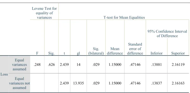
§ 2mm:
Figure 3 - Box-plot graph representing the volumetric loss on control group (natural socket healing) and experimental group (grafted socket healing). There is a greater loss on the control group.
Group EXPERIMENTAL
CONTROL
LO
S
Table 2 – Group statistics for tissue loss at 2 mm.
Group Statistics
Group N Mean Standard Deviation Mean Standard Error
Loss
Control 8 3.5125 .97459 .34457
Experimental 8 2.3625 .91016 .32179
Table 3 – Testing of independent samples for tissue loss at 2 mm.
p-value = 0.029 <0.05. For 2 mm, on average, the control group had a significantly higher loss than the experimental group.
Testing of independent samples Levene Test for
equality of
variances T-test for Mean Equalities
F Sig. t gl (bilateral) Sig. difference Mean
Standard error of difference 95% Confidence Interval of Difference Inferior Superior Loss Equal variances assumed .248 .626 2.439 14 .029 1.15000 .47146 .13881 2.16119 Equal variances not assumed 2.439 13.935 .029 1.15000 .47146 .13837 2.16163
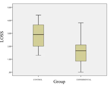
§ 3mm:
Figure 4 - Box-plot graph representing the volumetric loss on control group (natural socket healing) and experimental group (grafted socket healing). There is a greater loss on the control group.
Table 4 – Group statistics for tissue loss at 3 mm.
Group Statistics
Group N Mean Standard Deviation Mean Standard Error
Loss Control 8 2.8500 1.07968 .38173
Experimental 8 1.6250 1.14984 .40653
Table 5 – Testing of independent samples for tissue loss at 3 mm.
p-value = 0.045 <0.05. For 3 mm, on average, the control group had a significantly higher loss than the experimental group.
Testing of independent samples Levene Test for
equality of
variances T-test for Mean Equalities
F Sig. t gl (bilateral) Sig. difference Mean
Standard error of difference 95% Confidence Interval of Difference Inferior Superior Loss Equal variances assumed .006 .941 2.197 14 .045 1.22500 .55766 .02894 2.42106 Equal variances not assumed 2.197 13.945 .045 1.22500 .55766 .02850 2.42150 LO S S Group EXPERIMENTAL CONTROL
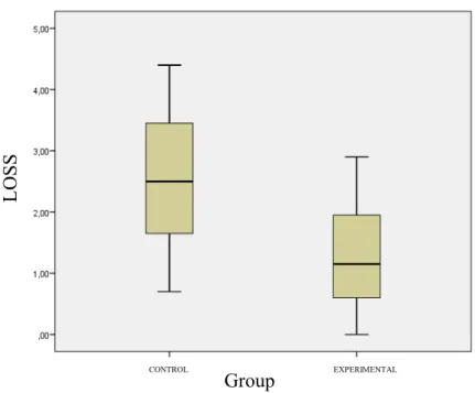
§ 4mm:
Figure 5 - Box-plot graph representing the volumetric loss on control group (natural socket healing) and experimental group (grafted socket healing). There is a greater loss on the control group.
Table 6 – Group statistics for tissue loss at 4 mm.
Group Statistics
Group N Mean Standard Deviation Mean Standard Error
Loss
Control 8 2.5375 1.24778 .44116
Experimental 8 1.2875 .95833 .33882
Table 7 – Testing of independent samples for tissue loss at 4 mm.
p-value = 0.041 <0.05. For 4 mm, on average, the control group had a significantly higher loss than the experimental group.
Testing of independent samples Levene Test for
equality of
variances T-test for Mean Equalities
F Sig. t gl (bilateral) Sig. difference Mean
Standard error of difference 95% Confidence Interval of Difference Inferior Superior Loss Equal variances assumed .681 .423 2.247 14 .041 1.25000 .55626 .05695 2.44305 Equal variances not assumed 2.247 13.126 .042 1.25000 .55626 .04946 2.45054 LO S S Group EXPERIMENTAL CONTROL
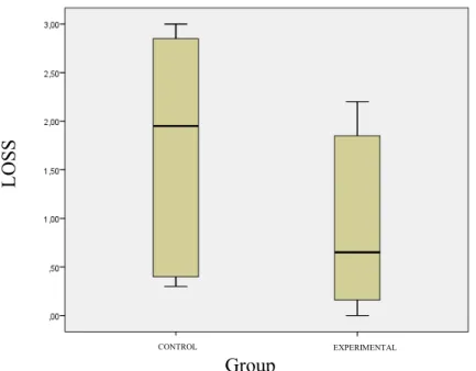
§ 5mm:
Figure 6 - Box-plot graph representing the volumetric loss on control group (natural socket healing) and
experimental group (grafted socket healing). There is a greater loss on the control group but with no statistical differences.
Table 8 – Group statistics for tissue loss at 5 mm.
Group Statistics
Group N Mean Standard Deviation Mean Standard Error
Loss
Control 8 2.2750 1.23491 .43661
Experimental 8 1.1375 .95310 .33697
Table 9 – Testing of independent samples for tissue loss at 5 mm.
p-value = 0.058 > 0.05. For 5 mm, there are no significant differences between the loss of the control group and the loss of the experimental group.
Testing of independent samples Levene Test for
equality of
variances T-test for Mean Equalities
F Sig. t gl Sig. (bilateral) Mean difference Standard error of difference 95% Confidence Interval of Difference Inferior Superior Loss Equal variances assumed .716 .412 2.062 14 .058 1.13750 .55152 -.04539 2.32039 Equal variances not assumed 2.062 13.155 .059 1.13750 .55152 -.05256 2.32756 LO S S Group EXPERIMENTAL CONTROL
LO S S Group EXPERIMENTAL CONTROL § 6mm:
Figure 7 - Box-plot graph representing the volumetric loss on control group (natural socket healing) and experimental
group (grafted socket healing). There is a greater loss on the control group but with no statistical differences.
Table 10 – Group statistics for tissue loss at 6 mm.
Group Statistics
Group N Mean Standard Deviation Mean Standard Error
Loss
Control 8 1.7125 1.19216 .42149
Experimental 8 .9400 .90092 .31852
Table 11 – Testing of independent samples for tissue loss at 6 mm.
p-value = 0.166> 0.05. For 6 mm, there are no significant differences between the loss of the control group and the loss of the experimental group.
Testing of independent samples Levene Test for
equality of
variances T-test for Mean Equalities
F Sig. t gl (bilateral) Sig. difference Mean
Standard error of difference 95% Confidence Interval of Difference Inferior Superior Loss Equal variances assumed 1.226 .287 1.462 14 .166 .77250 .52831 -.36062 1.90562 Equal variances not assumed 1.462 13.029 .167 .77250 .52831 -.36859 1.91359
6. Discussion
The findings in the present study suggest that there is a statistically significant difference between test and control groups regarding bone resorption, soft tissue healing and collapse on the alveolar socket after bone regeneration compared to non-regenerated alveolar sites. Thus, the null hypothesis (H0) of the present study is discarded.
In alloplastic grafts, HA resembles the inorganic component of human bone and is mostly used due to its osteoconductive properties, hardness and acceptability by bone. (19) TCP is gradually replaced by new bone due to its bioresorbability. BCP have a longer degradation rate than that of TCP and are stable enough to provide bone formation. The higher porosity and more suitable pore size an interconnected pore structure has, the more bone cells transforms into new bone tissue. In addition, a steady and gradual dissolution of BCP can help create a rich environment in calcium and phosphorus for osteogenic precursor cell adhesion, differentiation, production of bone matrix, and finally, ossification. (38) Therefore, the use of Adbone®BCP is advantageous whenever there is indication to graft an alveolar socket, preserving the dimension of the alveolar ridge.
The present study showed that the placement of an alloplastic graft in fresh extraction sockets may prevent ridge reduction in extraction sites. This finding is supported by previous studies that showed that bone grafting in combination with collagen membrane placement significantly limited the resorption of hard tissue after tooth extraction. (38-39) Araújo et al found that, during healing of a xenograft, the particles of the graft material became integrated with the bone crest and further enhanced its dimensions. This is in agreement with findings presented by Nevins et al, who reported that sockets treated with bovine bone mineral showed a reduction of less than 20% of the buccal plate, whereas control sites, i.e. not grafted, showed a reduction of more than 20%. (41)
The results from measurements at 2 mm, 3 mm and 4 mm shown a significantly higher loss on the control group, having p-values of 0.029, 0.045 and 0.041 respectively, in comparison to those on the experimental group, thus having a statistically significant difference (p<0.05). On the other hand, measurements at 5 mm and 6 mm, being the two most apical measurements of the extraction socket, show no significant differences between tissue loss of the control group and that of the experimental group. This suggests that all the alveolar ridge alterations following a tooth extraction might not have happened yet due to the short follow-up on this investigation. Follow-up appointments were made at days 7, 14 and after 3 months of the tooth extraction.
In a later and long-term study, Trombelli et al. (2008) examined socket healing in biopsies sampled during a 6‐month period from human volunteers. In later phases of healing, it was observed that the process by which woven bone was replaced by lamellar bone and marrow, was slow and exhibited great individual variation. At 6 months of healing, only a limited number of specimens had woven bone been replaced with bone marrow and trabeculae of lamellar bone.
In a 12-month prospective study, Schropp analysed 46 premolar and molar extraction sockets from 46 patients and found a 50% loss in ridge with an average 6.1 mm of horizontal loss. However, two thirds of this loss of bone volume occurred within the first 3 months. (15) Therefore, it can be assumed that tissue modelling following tooth extraction in humans is a rather rapid process, while the subsequent woven bone being remodelled into lamellar bone and marrow, is slow and may take years to be completed. However, it must be taken into account that this methodology was done by raising flaps which can alter the final results.
On the test group, the buccal plate resorbed until reaching the graft wall whereas the control group would have a greater resorption, given the fact that it had no stimuli. Thus, we speculate that a longer-follow up might have statistically significant differences between test and control groups in the apical portion of the socket site.
Other studies have suggested that the amount of ridge contraction varies within the socket itself. Araújo and Lindhe stated that in the apical and middle portions of the socket site, minor dimensional alterations occurred, while in the coronal portion of the ridge the reduction of the hard tissue volume was much more significant. (9,15) Hence, these finding are in accordance with those on the present study given that the more apical measurements have less statistically significant differences compared to those on the control group.
Comparing differences between jaws, mandible resorbs more than maxilla. (4) However, the direction of resorption is opposite in the maxilla, as the buccal wall of the alveolar socket tends to resorb more rapidly after dental extraction and the ridge gradually becomes represented by the previous palatal wall (centripetal resorption). (15) The amount of tissue loss that occurs in these processes varies considerably from subject to subject and from site to site (12) in the same individual.
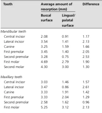
On the following table (table 12), it is noticeable the existing differences among the amount of resorption between different sites on the maxilla and mandible.
Table 12 – Average amount of resorption of tooth extraction in different tooth areas (Source: Lindhe(12))
In the study of Pietrokovski & Massler, in 1967, the amount of resorption was greater along the buccal surface than along the lingual or palatal surface in every specimen examined, although the absolute amounts and differences varied very widely. This caused a shift in the centre of the edentulous ridge towards the lingual or palatal side of the ridge with a concomitant decrease in arch length in the mandible as well as the maxillae. In another study, the molar teeth site had a greater value of reabsorption, but it was more critical in the anterior region due to aesthetics demands. (9,16)
In this context, it is important to acknowledge that the buccal bone plate in the frontal tooth region in humans is frequently (>80% of sites) <1 mm wide. (12) Hence, it can be anticipated that tooth loss in this part of the dentition may result in noticeable dimension alterations (horizontal as well as vertical) of the ridge and that this in turn may cause aesthetic concerns. (12)
On the present study, we included teeth on the anterior maxilla, from last premolar to the contralateral last premolar (5 to 5). On the control group, two central incisors were included, as well as 1 lateral incisor, 1 canine and four pre-molars. On the test group, we included two lateral incisors, one canine and five pre-molars. Knowing that different types of teeth may have different resorption rates, the comparison between test and control groups may have that bias since we had different clinical situations.
The extraction of single as well as multiple teeth induces a series of adaptive changes in the soft and hard tissues that result in an overall regression of the edentulous site(s). (12) The repair process results in reduced height and width of the residual ridge. The reduction in the alveolar bone ridge is much greater when multiple adjacent teeth are extracted, rather than a single tooth. (41)
As the buccal bone has been suggested to be completely composed of bundle bone, a significantly higher osteoclastic activity may occur on this surface of the extraction socket as compared with the lingual or palatal surface, given the osteoclastic activity occurring 16 weeks post-extraction. (11)
Misch et col and Araújo et col speculated that constriction of the blood clot within the alveolus may significantly contribute in bone remodelling process following tooth extraction. This comes in accordance with Al-Hezaimi et al who attributed the difference in bone loss to a reduction in the blood supply during healing of multiple adjacent extraction sites. (11) It is also hypothesized that extraction of contiguous teeth is associated with more severe alveolar bone resorption as compared with when a single tooth is extracted.
In 2012, Mansour Al-Askar showed that on a single-tooth extraction, a slightly higher remodelling on the buccal side compared with the palatal/lingual side occurred whereas on the extraction of two adjacent teeth showed an equivocal bone remodelling on buccal and lingual/palatal side. When extracting three adjacent teeth, the Micro-CT analysis demonstrated a more significantly pronounced remodelling (p < .001) on the lingual/palatal side compared with the buccal side having more bone resorption than the other two groups. (11) The author concluded that extraction of contiguous teeth causes a more extensive bone remodelling compared with extraction of a single tooth.
In another study, ridge preservation surgery results in a similar pattern of bone remodelling in the horizontal and vertical dimensions of the edentulous ridge after single and multiple adjacent teeth extractions (41), thereby suggesting that regardless the number of teeth extracted, the socket healing with grafting regenerative procedure reduced bone resorption. However, it only had 4-months follow-up.
Our sample had both single and contiguous extractions, regardless the groups, which might have compromised the results given that we were not able to have the same amount of both types of extractions on control and test groups. However, our results show that when grafting the socket, whether it had adjacent teeth or not, there is a lower resorption of the ridge.