license), applicable to the online version of the article only. Distribution permitted for non-commercial purposes only.
Amyloid Spells and High Blood
Pressure: Imminent Danger?
Andre Caetanoa Miguel Pintoa Sofia Caladoa, b Miguel Viana-Baptistaa, b
aDepartment of Neurology, Hospital Egas Moniz, Centro Hospitalar de Lisboa Ocidental,
and bCEDOC – Faculdade de Ciências Médicas, Universidade Nova de Lisboa,
Lisbon, Portugal
Key Words
Cerebral amyloid angiopathy · Elderly patients · Cortical and leptomeningeal vessels
Abstract
We present the case of a 71-year-old male, admitted after a generalized tonic-clonic seizure, with a history of recurrent left arm and face paresthesias, associated with sulcal cortical sub-arachnoid hemorrhages. During the next 48 h, he remained agitated with a high blood pres-sure profile; he also suffered a cardiac arrest in relation to a severe left fronto-parietal and a smaller right parietal parenchymal hemorrhage that developed over the subarachnoid hem-orrhage locations. There were no intracranial vascular abnormalities. Three months later, an MRI revealed disseminated superficial siderosis. He was discharged with a modified Rankin scale of 4. He died 1 month later of unknown cause. A diagnosis of probable cerebral amy-loid angiopathy was assumed. Patients with pathologically proven cerebral amyamy-loid angiopa-thy that present with transient focal neurological symptoms in relation to cortical bleeds, the so-called ‘myloid spells’ seem to be at an increased risk of future parenchymal hemorrhages. Avoiding antiplatelet agents in these cases has been proposed. Our case suggests that these patients should be monitored closely in the hyperacute phase , and tight blood pressure control should be considered as the immediate risk of bleeding may be high, even without a
definitive diagnosis of cerebral amyloid angiopathy. © 2015 S. Karger AG, Basel
Introduction
Cerebral amyloid angiopathy (CAA) is characterized by the deposition of beta amyloid proteins within the cortical and leptomeningeal vessels. It is a common pathological finding
in elderly patients, regardless of concomitant cognitive impairment, and 10–40% of elderly brains have pathological evidence of amyloid deposition [1]. The most frequent presentation of CAA in older patients is intracranial hemorrhage, usually recurrent, sparing regions af-fected by hypertensive hemorrhage and mainly affecting the cerebral or cerebellar cortex and the cortico-subcortical regions (reflecting locations of amyloid deposition) [2, 3]. One of the landmarks in CAA history was the development of the Boston criteria by Greenberg and collaborators in 1996. These criteria were created to improve the diagnostic accuracy of CAA, to provide an easier method for clinical decision-making, and to facilitate a diagnosis during lifetime (without the need for postmortem pathological evaluation) by allowing for probable and possible diagnosis [2]. These criteria reflect the main defining characteristics of CAA (age >55 years, presentation with multiple parenchymal hematomas and typical pathological findings), but they also accommodate the classical imaging findings of cerebral chronic microbleeds, which represent asymptomatic, small, spontaneous bleedings caused by CAA [2].
Recently, superficial siderosis and cortical subarachnoid hemorrhage (cSAH) in patients with transient focal neurological symptoms (TFNS), the so-called amyloid spells have been related to the phenotype of CAA, and several studies suggest that these findings might pre-dict intracerebral hemorrhages caused by CAA in the long run [4–6]. We report a patient who suffered a massive intracerebral hemorrhage 48 h after presenting to the emergency department with TFNS and cSAH.
Case Report
A 71-year-old male with a recent history of recurrent episodes of left arm and face par-esthesias was admitted to our department after a first generalized convulsive seizure. His past medical history disclosed hypertension, hypercholesterolemia and peripheral arterial disease, but no known epilepsy, cerebrovascular events or cognitive impairment. His usual medication included bisoprolol, sinvastatin and clopidogrel. On admission, his blood pres-sure was 180/100 mm Hg. A general examination was unremarkable, and a neurological examination revealed mild left facial palsy and left visual and tactile extinction. A CT of his brain showed right parietal and left frontal sulcal cortical subarachnoid hemorrhages (fig. 1a, b). An electroencephalogram showed no evidence of paroxysmal activity. His antithrom-botic medication was stopped, and the patient was started on phenytoin (100 mg t.d.s.) and perindopril (5 mg o.d.). Despite his treatment, the patient remained confused and agitated and his blood pressure values ranged from 160–200 (systolic) and 70–97 mm Hg (diastolic). On day 2 of his hospitalization, the patient suffered a cardiac arrest and was resuscitated. Afterwards, global aphasia and right hemiplegia were noted. A brain CT revealed a large left frontoparietal and a circumscribed right parietal parenchymal hemorrhage (PH) (fig. 1c, d). His cervical and intracranial CT angiograms were normal. The patient’s blood pressure was controlled with a low dose of bisoprolol and furosemide, and then, there were no further events. A brain MRI at 3 months revealed disseminated superficial siderosis without any microbleeds on T2* (fig. 1e, f). The patient was discharged at 3 months with a modified Ran-kin Scale score of 4 but died 1 month later of unknown causes and without any autopsy be-ing performed on him. CAA was the assumed cause of death.
Discussion
Despite usually presenting as a PH, CAA is also a recognized and important cause for spontaneous cSAH [7–9]. On the other hand, there have also been several reports of patients without the classical CAA findings where blood susceptibility imaging revealed only a super-ficial siderosis, presumed to be the result of recurrent asymptomatic cortical bleeds [7, 8, 10, 11], suggesting that cSAH can occur not only as a secondary phenomenon in the context of a deep cerebral hemorrhage but also as a primary event [11, 12].
Some interest has been found in patients with atypical TFNS who have positive or nega-tive transient symptoms (hemianesthesia or hemiparesis), the so-called 'amyloid spells' [4,
10, 13, 14]. In these patients, blood susceptibility imaging revealed the presence of
superfi-cial siderosis, a finding which has been strongly associated with the CAA pathology [11] and which presumably reflects previous spontaneous cSAH. There is no clear explanation for these TFNS, but it is believed that the presence of superficial siderosis could be the catalyst for an epileptic manifestation or even a migraine aura-like cortical spreading depression (as proposed by some authors) [13]. The finding of superficial siderosis in CAA-proven patients, associated with presentations as TFNS, supports the claim that some patients with CAA do not have the classical clinical picture of PH and cerebral microbleeds on MRI, but they can present with TFNS and have isolated superficial siderosis as an imaging marker [13, 14], thus reinforcing the need for a revision of the Boston criteria [10]. However, when patients present with TFNS, CAA is usually disregarded as a possible cause, because most of these patients do not have a cognitive decline, and most of them have other cardiovascular risk factors for transient neurological episodes (ischemic events being the main suspect) [13]. Additionally, most of these patients do not undergo susceptibility-weighted MRI. Although the natural history of these patients is not clearly established, several studies suggest that they are at an increased risk of intracerebral bleeding within the next days/months [5, 6, 14, 15]. Curiously, when these PHs occur, they tend to occur in the same location as a pre-existing superficial siderosis (as it has occurred in our patient) [5, 12]. It has been suggested that in these patients, the first evidence of bleeding may occur as a cSAH, which could be seen as a warning sign for future PHs [5, 12]. The case reports describing these events show a variable latency period between cSAH and PH (range 7–32 days) [5, 12], but clearly, and this case seems to have the shortest latency period, the risk can be present since an initial presentation of cSAH. Avoiding antithrombotics has been emphasized before [4, 14], but vigilance and strict blood pressure control should also be considered due to the risk of de-veloping severe PHs. Further studies are needed to understand the natural history of pa-tients with amyloid spells and cSAH, but lowering the risk of future bleeds seems to be of utmost importance.
In summary, CAA patients are at a higher risk of PHs and cSAH. Some of these patients can present with transient focal neurological symptoms and have evidence of superficial siderosis on susceptibility-weighted MRI, which would probably reflect previous spontane-ous symptomatic or even asymptomatic cSAH. These patients should be tightly monitored, especially regarding their blood pressure profile and because they are at an increased risk of PHs, which can occur in the hyper-acute phase.
References
1 Jellinger KA: Alzheimer disease and cerebrovascular pathology: an update. J Neural Transm 2002;109:813– 836.
2 Knudsen KA, Rosand J, Karluk D, Greenberg SM: Clinical diagnosis of cerebral amyloid angiopathy: validation of the Boston criteria. Neurology 2001;56:537–539.
3 Yamada M: Cerebral amyloid angiopathy: an overview. Neuropathology 2000;20:8–22. 4 Charidimou A, Law R, Werring DJ: Amyloid 'spells' trouble. Lancet 2012;380:1620.
5 Katoh M, Yoshino M, Asaoka K, Asaoka K, Aoki T, Imamura H, Kashiwazaki D, Takano K, Aida T: A restricted subarachnoid hemorrhage in the cortical sulcus in cerebral amyloid angiopathy: could it be a warning sign? Surg Neurol 2007;68:457–460.
6 Linn J, Wollenweber F, Lummel N, Bochmann K, Pfefferkorn T, Gschwendtner A, Bruckmann H, Dichgans M, Opherk C: Superficial siderosis is a warning sign for future intracranial hemorrhage. J Neurol 2013;260:176-181.
7 Raposo N, Viguier A, Cuvinciuc V, Calviere L, Cognard C, Bonneville F, Larrue V: Cortical subarachnoid haemorrhage in the elderly: a recurrent event probably related to cerebral amyloid angiopathy. Eur J Neurol 2011;18:597–603.
8 Khurram A, Kleinig T, Leyden J: Clinical associations and causes of convexity subarachnoid hemorrhage. Stroke 2014;45:1151–1153.
9 Renou P, Tourdias T, Fleury O, Debruxelles S, Rouanet F, Sibon I: Atraumatic nonaneurysmal subarachnoid hemorrhages: a diagnostic workup based on a case series. Cerebrovasc Dis 2012;34:147–152.
10 Linn J, Halpin A, Demaerel P, Ruhland J, Giese AD, Dichgans M, van Buchem MA, Bruckmann H, Greenberg SM: Prevalence of superficial siderosis in patients with cerebral amyloid angiopathy. Neurology
2010;74:1346–1350.
11 Linn J, Herms J, Dichgans M, Brückmann H, Fasi G, Freilinger T, Wiesmann M: Subarachnoid hemosiderosis and superficial cortical hemosiderosis in cerebral amyloid angiopathy. AJNR Am J Neuroradiol
2008;29:184–186.
12 Takeda S, Hinokuma K, Yamazaki K, Onda K, Miyakawa T, Ikuta F, Arai H: The hemorrhage caused by sporadic-type cerebral amyloid angiopathy occurs primarily in the cerebral sulci. Neuropathology 2012;32:38–43.
13 Charidimou A, Baron J-C, Werring DJ: Cerebral amyloid angiopathy and transient focal neurological episodes. Cerebrovasc Dis 2013;36:245–246.
14 Charidimou A, Peeters A, Fox Z, Gregoire SM, Vandermeeren Y, Laloux P, Werring DJ: Spectrum of transient focal neurological episodes in cerebral amyloid angiopathy: multicentre magnetic resonance imaging cohort study and meta-analysis. Stroke 2012;43:2324–2330.
15 Charidimou A, Peeters AP, Jäger R, Fox Z, Vandermeeren Y, Laloux P, Baron JC, Werring DJ: Cortical superficial siderosis and intracerebral hemorrhage risk in cerebral amyloid angiopathy. Neurology 2013;81:1666–1673.
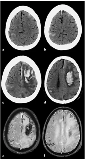
Fig. 1. a, b A CT scan performed on admission with 2 circumscribed cortical subarachnoid hemorrhages, a frontal left and a right parietal one. c, d A CT scan 48 h after admission and after cardiac resuscitation with evidence of PHs, a large left frontoparietal and a circumscribed right parietal one, over the original cSAH location. e, f A brain MRI performed 3 months after admission, revealing disseminated supratentorial superficial siderosis on the T2 gradient echo sequence.