BJRS
RADIATION SCIENCES
08-01A (2020) 01-13ISSN: 2319-0612 Accepted: 2020-04-09
Comparative evaluation of gamma cameras
performance for internal monitoring of workers
exposed to
131I in nuclear medicine services
Oliveira S. M., Dantas A. L. A., Dantas B. M
Instituto de Radioproteção e Dosimetria - IRD, CEP - 22783-127, Rio de Janeiro - RJ, Brazil [email protected]
ABSTRACT
Among the radionuclides handled in Nuclear Medicine Services (NMS), 131I is one of the most used for both
diagnostic and therapy proposes. This practice represents a risk of incorporation these radionuclides by workers. The International Atomic Energy Agency (IAEA) recommends the implementation of an internal monitoring program on workers potentially exposed to annual effective doses higher than 1 mSv, as for example, those who handle 131I for therapy. In Brazil there are approximately 90 NMS authorized to handle 131I for
therapy and there are available only five laboratories qualified to provide internal monitoring services. This scenario turns impossible to attend all possible demand for internal monitoring if such regulation is required by the Brazilian Nuclear Energy Commission (CNEN). This work presents a simple and inexpensive method for in
vivo routine thyroid monitoring of 131I using gamma cameras available in the clinics itself and compares their
performance for the proposed method. It has been verified that all equipment evaluated in this work present enough sensitivity for such application.
1. INTRODUCTION
In the nuclear medicine practice, a variety of radionuclides in the form of unsealed sources are routinely handled for diagnostic and therapy purposes. This practice represents a significant risk of external and internal exposure for workers who handle these radionuclides.
The external exposure is predominant in SMN, however, depending on the scenario, internal exposure may be also significant as a consequence of intake of radionuclides via inhalation and ingestion. Based on international criteria of evaluation, the permanent risk of intakes of radionuclides requires the implementation of a routine monitoring plan, aiming to control and limit internal doses [1].
National and international regulations appoint the Radiation Safety Officer (RSO) of the facility as responsible for managing the Radiation Protection Programme (RPP). The RSO shall implement, based on monitoring results, the necessary measures to keep exposure levels as low as possible [2]. The RPP should consider the risks of external and internal occupational exposures and includes, when necessary, a routine monitoring plan with specific methodologies that allows identification and quantification of intakes as well as the estimation of the committed effective doses of the workers [3].
Currently, in Brazil, approximately 90 Nuclear Medicine Services (NMS) are authorized by the CNEN to handle 131I for therapy purposes [4], resulting in a significant number of workers routinely exposed to internal exposures.
Iodine is a volatile element, and its isotope 131I is still one of the most frequently used radionuclides in NMS. Because of its physical, chemical and radioactive properties, 131I presents a high risk of intake and, consequently, internal doses. It should be also considered in this scenario the high activities routinely handled for therapy purpose.
Although the IAEA recommends implementing an internal monitoring program of such group of workers, in Brazil there are not enough qualified laboratories available to offer internal dosimetry services and attend the possible demand of internal monitoring in this field. Consequently, it would represent an impeditive high cost to the medical institutions if the Brazilian Nuclear Regulatory Board would apply the requirements of internal monitoring [5].
In order to overcome this situation, the Institute for Radiation Protection and Dosimetry (IRD), a branch of the CNEN, has developed several studies over the last 15 years proposing the use of devices available in the NMS itself for use in 131I occupational monitoring [6]. Among such
equipment, the gamma camera stands out to present the highest sensitivities for the proposed application. This work presents a performance comparison of gamma cameras available in NMS in Brazil for 131I in vivo monitoring purpose.
2. MATERIALS AND METHODS
2.1. Materials
2.2.1 Gamma Cameras
Eight models of gamma camera available in seven different facilities were evaluated in this work. Table 1 presents the models intended to be applied in internal routine monitoring.
Table 1: Gamma cameras evaluated in seven Nuclear Medicine Services. Manufacturer Model Facility (NMS)
Phillips BrightView XCT A GE Millennium MG B GE Discovery MN/CT 670 C Siemens e.cam 180 D Elscint Apex SPX-6 E Siemens Symbia E GE Millennium MG F Siemens e.cam 180 G 2.2.2 Neck-Thyroid Phantom
The neck-thyroid phantom (Figure 1) used for the calibration of the detectors was developed at the In Vivo Monitoring Laboratory of the IRD. The neck-thyroid phantom is made of
polyurethane-base tissue equivalent material. A filter paper simulating a human thyroid is spiked with a known amount of a 133Ba liquid standard source [5].
It is a conventional practice to use a 133Ba standard source as a surrogate for measuring 131I
since the energy and yield of photons emitted by 133Ba are very similar to the photon emissions from 131I. Furthermore, the half-life of 133Ba is long (10.5 years) compared with 131I (8 days), so that
a certified calibration standard containing 133Ba can be used for many years.
After being sealed with a plastic film, the filter paper is fixed in the proper position with an acrylic part and inserted in the neck phantom. The phantom used in this work contained originally 29771 ± 214 Bq of 133Ba in 28/April/2004, certified by the National Laboratory of Metrology of Ionizing Radiations (LNMRI) of IRD.
Figure 1: Neck-thyroid phantom produced in the In Vivo Monitoring Laboratory of IRD [5].
2.2. Methods
The methodology consisted basically in determining the calibration factors for measurement of
131I in the thyroid, calculation of the minimum detectable activities and the corresponding minimum
2.2.1 Calibration of the Detection System
Step 1 – Calculation of 131I Equivalent Activity: The 133Ba activity content in the phantom
was corrected between fabrication and calibration dates. The equivalent 131I activity is calculated as shown in Eq. (1) by multiplying the activity of 133Ba added to the phantom by the ratio between the
sum of 133Ba and 131I photon yields in the region of interest for the measurement, using the
parameters provided in Table 2:
(1)
Where:
131I Eq Ac = is the equivalent activity of 131I, in Bq;
A (133Ba) = is the activity of 133Ba present in the phantom, in Bq;
Σ (133Ba) = is the sum of emission intensities of 133Ba; and
Σ (131I) = is the sum of emission intensities of 131I.
Table 2: Energy photons and emission intensity for 133Ba and 131I [7]. Energies 133Ba (keV) Emission Intensity
276.39 0.071
302.85 0.183
356.01 0.620
383.85 0.089
Energies 131I (keV) Emission Intensity
284.30 0.061
364.48 0.817
The 131I equivalent activity of the phantom used in this work is in the order of 14000 Bq. This
value must be corrected according to the date of calibration of the equipment.
Step 2 – Determination of Measurement Setup (Figure 2): The standard geometry
The count time was determined according to each detector sensitivity for a time after intake of 1 and 7 days, considering a weekly generic monitoring frequency, resulting in 48 monitoring periods per year.
Step 3 – Calculation of Calibration Factor (CF): The calibration factor is given by:
CFcpm/Bq = (NC/T) / 131I Eq Ac (2)
Where:
NC is the Net counts;
T is the Count Time (min); and
131I Eq Ac = Equivalent activity of 131I, corrected for the date of the calibration in the facility.
Figure 2: Geometries used in this work.
a) Measurement geometry for gamma-camera calibration. b) Measurement geometry for worker monitoring.
2.2.2 Evaluation of Sensitivity
The evaluation of the sensitivity of the method for its application in routine internal monitoring is based on the calculation of the Minimum Detectable Activity (MDA), in Bq, Minimum Detectable Intake (MDI), in Bq and Minimum Detectable Effective Dose (MDED), in mSv.
The MDA of the method is given by equation 3 [8]:
/ CF . T (3)
Where:
N is the total counts of the background of a non-exposed subject, when the calibration is made with
only one measurement; and
CF is the calibration factor, and T is the count time (min).
The Minimum Detectable Intake (MDI), a function of the MDA and the exposure scenario, is given by:
(4)
Where:
MDA is the minimum detectable activity (Bq); and
m(t) is the retention fraction in the compartment of interest for inhalation or ingestion (Bq/Bq).
The last parameter to be calculated is the Minimum Detectable Effective Dose, which is based on the MDI, and is given by:
Where:
MDI is the minimum detectable intake (Bq); and e(g)ina/ ing is the dose coefficient (mSv/Bq).
In order to be considered useful for internal dosimetry purposes, the technique should, at least, be able to detect an activity that would result in an annual effective dose below 1 mSv for the most likely internal exposure scenario [9]. The values of “m(t)” and “e(g)” are available in the publication 78 of the ICRP [10] and can also be generated for specific exposure scenarios and times through the software Activity Internal Dose Estimate (AIDE) [11].
The number of monitoring periods is determinant to evaluate the sensitivity of the proposed method to be applied on a routine internal monitoring in a specific NMS. This evaluation can be made by the following equation:
(6)
Where:
MDED is the minimum detectable effective dose (mSv); and
n is the number of annual monitoring periods (e.g., 48 when workers are monitored once a week).
3. RESULTS AND DISCUSSION
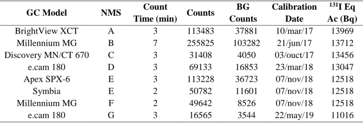
Table 3 shows the total counts and background count results, count times, calibration dates and its respective 131I equivalent activities present in the neck-thyroid phantom in the time of calibration of gamma cameras evaluated.
Table 3: Calibration parameters and counts results of the detectors analyzed. GC Model NMS Count
Time (min) Counts
BG Counts Calibration Date 131I Eq Ac (Bq) BrightView XCT A 3 113483 37881 10/mar/17 13969 Millennium MG B 7 255825 103282 21/jun/17 13712 Discovery MN/CT 670 C 3 31408 4050 03/ouct/17 13456 e.cam 180 D 3 69133 16853 23/mar/18 13047 Apex SPX-6 E 3 113228 36723 07/nov/18 12518 Symbia E 2 50782 11601 07/nov/18 12518 Millennium MG F 2 49642 8526 07/nov/18 12518 e.cam 180 G 3 16565 3544 22/may/19 11016
Table 4 shows the results for CF and MDA for each gamma camera evaluated at the different facilities using the neck-thyroid phantom. These calibrations were made in a single measurement of the neck-thyroid phantom and the background, using equation 3 to calculate the MDA.
Table 4: CF and MDA of the gamma cameras analyzed.
GC Model NMS CF (Cpm/Bq) MDA (Bq) BrightView XCT A 1.804 167 Millennium MG B 1.589 134 Discovery MN/CT 670 C 0.678 146 e.cam 180 D 1.336 151 Apex SPX-6 E 2.037 146 Symbia E 1.603 149 Millennium MG F 1.642 131 e.cam 180 G 0.359 257
The parameter MDI depends on the exposure scenario and the time elapsed between intake and in vivo measurement. In this work it was used the retention fraction values for 1 and 7 days between
131I intake by inhalation and in vivo measurement (Table 5). Such values were obtained through the
software AIDE. The MDED was calculated based on the MDI, considering the dose coefficient for inhalation associated to the corresponding intake scenario adopted in this study, as shown in
Table 5. The inhalation incorporation scenario was chosen because it is the most likely when handling iodine in liquid form, which is widely used in nuclear medicine therapies.
Table 5: Retention fractions and dose coefficient for inhalation obtained with the software AIDE. m(t) (Bq/Bq) e(g) (mSv/Bq)
1 day 7 days
1.98 x 10⁻⁵
0.229 0.139
Table 6 shows the results for MDI and MDED of the GCs evaluated, considering 1 and 7 days after intake by inhalation.
Table 6: MDI and MDED of the detectors evaluated for 1 and 7 days after intake by inhalation. GC Model NMS MDI (Bq) MDED (mSv)
1 day 7 days 1 day 7 days
BrightView XCT A 730 1203 0.014 0.024 Millennium MG B 587 966 0.012 0.019 Discovery MN/CT 670 C 636 1047 0.013 0.021 e.cam 180 D 658 1084 0.013 0.020 Apex SPX-6 E 637 1049 0.013 0.021 Symbia E 652 1075 0.013 0.021 Millennium MG F 517 940 0.011 0.019 e.cam 180 G 1122 1848 0.022 0.037
In order to evaluate the sensitivity of the detectors, it was simulated a weekly generic monitoring period, totalizing 48 annual monitoring periods. The choice of monitoring frequency depends on the routine of each NMS, it can be altered by the number of patients treated, the amount of 131I activities handled and the 131I physical form, for example. It was observed that the frequency required for NMS participants is lower than the generic frequency used in this study. Table 7 shows the annual sensitivities obtained for a weekly monitoring period.
Table 7: Annual sensitivities (mSv) for 48 monitoring periods per year. GC Model NMS Count time
(min) Sensibility (mSv) 1 day 7 days BrightView XCT A 3 0.69 1.14 Millennium MG B 7 0.56 0.92 Discovery MN/CT 670 C 3 0.60 1.00 e.cam 180 D 3 0.57 0.95 Apex SPX-6 E 3 0.61 1.00 Symbia E 2 0.62 1.02 Millennium MG F 2 0.54 0.89 e.cam 180 G 3 1.07 1.76
The gamma cameras evaluated have significant differences in manufacturing characteristics and time of operation. Some of them are in operation for about 20 years, such as the Elscint Apex SPX-6 (A) and the GE Millennium MG (B/F), while others, such as the GE Discovery MN/CT 670 are much newer. However, despite of the time of use, all gamma cameras presented enough sensitivity for occupational monitoring in a reasonable short count time. The exception is the Siemens e.cam 180 of NMS “G”, which does not present enough sensitivity for the time and generic monitoring frequency simulated in this work. However, this fact does not prevent the use of this equipment for the proposed application. The sensitivity values obtained may be increased simply by optimizing two basic measurement parameters: (i) reducing distance between detector and neck-thyroid phantom and (ii) increasing measurement count time, as done with the model GE Millennium MG of NMS “B”, which was 7 minutes ok. In order to avoid disturbing the routine of the NMS, it is proposed to use the minimum count time, but keeping the necessary sensitivity of the method for its application in occupational monitoring.
The counting distance (12 cm) was optimized to be the minimum distance that provides a reasonable comfort to the subject during measurement. This distance may be altered, but decreasing it may cause difficulties during the measurement procedure. Furthermore, the monitoring plan would be more effective if the measurements are schedule according to the radionuclide handling of the facility, when it is more likely to detect significant intakes by the workers.
Significant differences in sensitivity were observed between equipment of the same model of different NMS. The GE Millenium MG of NMS “B”, for example, presents less sensitivity than the same model of NMS “F”, which reached the minimum sensitivity required in a shorter counting time. This can be explained by the equipment maintenance conditions: the equipment of NMS “F” presents better general conditions and integrity of the crystals of its detectors. These conditions were observed through visual evaluation and the results of equipment quality control tests. The same was observed between the model Siemens e.cam 180 of NMS “D” and “G”, which can also be explained by their different maintenance conditions.
It is important to emphasize that the results presented in this work are valid for a generic weekly monitoring frequency resulting in 48 monitoring periods per year. Thus, it should be kept in mind that the monitoring plan to be effectively implemented on each NMS would depend on the service routine, especially the radionuclide handling schedule.
4. CONCLUSIONS
The gamma camera performance can be evaluated by analyzing the MDA of the detection system as well as the count time required for the system to reach the minimum sensitivity to be considered useful for internal monitoring purpose. This study evaluated modern and old gamma camera models, and despite of the differences on time of manufacture between the models, all gamma cameras can be used to implement the proposed methodology. In most cases, the appropriate sensitivity was achieved setting the count time to 3 minutes, which may be reduced or increased according to the NMS routine in which the methodology is implemented. This can be considered a feasible and short count time.
It should also be highlighted that the proposed methodology is easy, simple and inexpensive, since the measurements are performed by the staff of the clinic and there is no need to purchase new equipment for the implementation of the monitoring plan.
Finaly, this study provides a methodology to be implemented in routine in order to comply with requirements established by the National Regulatoy Board for individual monitoring of workers who handle 131I in Nuclear Medicine Services.
REFERENCES
[1] IAEA - International Atomic Energy Agency. Assessment of Occupational Exposure due to
Intakes of Radionuclides. Safety Guide No. RS-G-1.2, 1999.
[2] CNEN - Comissão Nacional de Energia Nuclear. Diretrizes Básicas de Radioproteção.
Norma CNEN-NE-3.01. Rio de Janeiro: RJ, 2011.
[3] IAEA - International Atomic Energy Agency. Direct methods for measuring radionuclides in
the human body. Safety Series n. 115, 1996.
[4] CNEN - Comissão Nacional de Energia Nuclear. Instalações Autorizadas. Available at: <http://www.cnen.gov.br/instalacoes-autorizadas>. Last accessed: 01 Sep. 2019.
[5] DANTAS, B. M.; CARDOSO, J. S.; DANTAS, A. L. A.; LUCENA, E. A.; RAMOS, M. A. P., SÁ, M. S., ALONSO, T. C.; SILVA, T. V.; OLIVEIRA, C. M.; LIMA, F. F.; OLIVEIRA, M. L.; LACERDA, I. V. B.; FAJGEL, A. Intercomparação Nacional de Medição In Vivo de
Iodo-131 na Tireoide, Projeto TC IAEA BRA 9055. Scientia Plena, 9(8), 2013.
[6] Dantas, B.M., Lucena, E.A., Dantas, A.L.A., Oliveira, S.M. Avaliação das exposições ocupacionais internas em medicina nuclear: dificuldades e alternativas. Revista Brasileira de Física Médica. 2019;13(1):122-127.
[7] SAR, 2020, Sistema de Avaliação Radiológica. Available at: <http://www.ird.gov.br/ear>. Last accessed: 02 Apr. 2020.
[8] HPS - Health Physics Society. Performance Criteria for Radiobioassay, N13.30, 1996.
[9] IAEA - International Atomic Energy Agency. Methods for Assessing Occupational Radiation
Doses due to Intakes of Radionuclides. Safety Report Series No. 37, Vienna, 2004.
[10] ICRP - International Commission on Radiological Protection. Individual Monitoring for
Internal Exposure of Workers. ICRP Publication 78, Vol. 27/3-4, 1998.
[11] BERTELLI, L.; MELO, D. R.; LIPSZTEIN, J.; CRUZ-SUAREZ, R. AIDE. Internal

![Figure 1: Neck-thyroid phantom produced in the In Vivo Monitoring Laboratory of IRD [5]](https://thumb-eu.123doks.com/thumbv2/123dok_br/18275817.881039/4.892.309.582.530.853/figure-neck-thyroid-phantom-produced-vivo-monitoring-laboratory.webp)
![Table 2: Energy photons and emission intensity for 133 Ba and 131 I [7] . Energies 133 Ba (keV) Emission Intensity ](https://thumb-eu.123doks.com/thumbv2/123dok_br/18275817.881039/5.892.263.632.713.899/table-energy-photons-emission-intensity-energies-emission-intensity.webp)