Faculdade de Farmácia
BIMODAL PROBES FOR IMAGING OF PROSTATE CANCER
Sofia Margarida Alves Batanete
Dissertation supervised by Professor Doutor António Rocha Paulo and
co-supervised by Professor Doutor António J. I. Alfaia
MSc Course in Pharmaceutical and Medicinal Chemistry
Faculdade de Farmácia
BIMODAL PROBES FOR IMAGING OF PROSTATE CANCER
Sofia Margarida Alves Batanete
Dissertation supervised by Professor Doutor António Rocha Paulo and
co-supervised by Professor Doutor António J. I. Alfaia
MSc Course in Pharmaceutical and Medicinal Chemistry
i
Acknowledgements
First of all, I thank Dr. António Paulo for accepting my participation in this project of the Radiopharmaceutical Sciences Group (RSG) of the Center for Nuclear Sciences and Technologies (C2TN) and for his enormous scientific knowledge, his demand and the guidance
of the work that led to the conclusion of this thesis. I also thank Francisco Silva for his availability for the introduction and integration in laboratory work, the co-supervision of the project and the knowledge wisely transmitted. I thank my co-adviser, Professor António Alfaia, for the encouragement given to this work.
To all the professionals in this group, I thank you, above all, for the hospitality and kindness with which you received me and my colleagues from the Chemical Synthesis laboratories, thank you for your warm inclusion, support and good working environment. Thank you all for your willingness to perform and assist in the interpretation of the ESI-MS spectras, as well as the NMR spectras and the tips provided for their interpretation. I also thank Elisa Palma and Polatom investigator Arek, for sharing their experience for the synthesis of pyrazolyl - diamine derivatives and the PSMA inhibitor, respectively.
A special thanks goes to Dr. Paula Campello for having received me in her laboratory and for all the knowledge transmitted, as well as for the encouragement, availability and for all the practical suggestions and advices.
Last but not least, I thank my family (especially!) and my friends for the unconditional support given to me along this course. Thank you so much for your encouragement and your ability to give me the perseverance and patience needed to continue this work, even when frustration set in and giving up seemed like the easy way out.
ii
Abstract
Prostate cancer (PCa) is the most common malignancy found in men affecting one in six and is the second leading cause of cancer-related death in men. Recently, new radioactive probes targeted at the prostate-specific membrane antigen (PSMA) have been introduced, which have revealed a high potential for PCa detection and staging based on nuclear imaging techniques, like Single-Photon Emission Computed Tomography (SPECT) or Position Emission Tomography (PET). In particular, very promising results were obtained for complexes prepared with the 99mTc and 68Ga radiometals and functionalized with a PSMA inhibitor, which includes
glutamic acid (Glu) and lysine (Lys) as amino acids linked by an urea bond.
The PET or SPECT probes allow the detection of PCa and its metastases through the acquisition of whole body images. However, the use of similar fluorescent probes is more favorable in intraoperative procedures for a more selective excision of tumor tissue. To this end, encouraging results have been described for PSMA inhibitors functionalized with indocyanine green (ICG) dyes. The design of PSMA-specific dual probes containing either a PET or SPECT radionuclide or a fluorophore for optical imaging may simultaneously profit from the advantages of nuclear and optical medical imaging modalities.
The main goal of this thesis was the design, synthesis and biological evaluation of bimodal probes, with PET / SPECT and Near‑infrared fluorescence (NIRF) valences for PCa detection, starting from the base structure of 99mTc and 68Ga complexes functionalized with PSMA
inhibitors that have already shown clinical relevance and that should be functionalized with fluorophores of the ICG type.
In this work it was devised the synthesis of bimodal probes for PCa imaging, wherein the general structure included a PSMA inhibitor (PSMA11), a chelating agent, like (N,N′-Bis[2-hydroxy-5-(carboxyethyl)-benzyl]ethylenediamine-N,N- diacetic acid) (HBED-CC) or pyrazolyl diamine, and a derivative of the fluorophore IR820 (aromatic and aliphatic). Concerning the required organic precursors, a solid phase synthesis strategy allowed the synthesis of the Glu-urea-Lys scaffold needed for PSMA inhibition, while two synthetic approaches were tried out to obtain an aliphatic and an aromatic derivative of IR820. The synthesis of two ligands, suitable for further coupling to the fluorophore and PSMA inhibitor, was successfully achieved in this work: HBED-CC, a two-fold carboxylic acid substituted HBED, was synthesized as the chelating agent for 68Ga, while for labeling with 99mTc and for SPECT imaging a
iii
Keywords:
Prostate Cancer PSMA Radiopharmaceuticals Fluorescent Probes Bimodal Probesiv
Resumo
O cancro da próstata (PCa) é a neoplasia maligna mais comum em homens, afetando um em cada seis, sendo a segunda principal causa de morte por cancro em homens. Recentemente, foram introduzidas novas sondas radioactivas direcionadas ao antigénio de membrana específico da próstata (PSMA), que revelaram um elevado potencial para detecção e estadiamento do PCa com base em técnicas de imagiologia nuclear, como a Tomografia por Emissão de Fotão Único (SPECT) ou a Tomografia por Emissão de Positrões (PET). Em particular, foram obtidos resultados muito promissores para complexos preparados com os radioisótopos 99mTc e 68Ga e funcionalizados com um inibidor de PSMA, cuja estrutura incluí
ácido glutâmico (Glu) e lisina (Lys) como aminoácidos ligados por uma ligação de ureia. As sondas radioactivas para PET ou SPECT permitem a detecção do cancro da próstata e das suas metástases, mediante aquisição de imagens de corpo inteiro. No entanto, o uso de sondas congéneres fluorescentes, de preferência na zona do infravermelho próximo (NIR), é mais favorável em procedimentos intraoperativos que visem a excisão, o mais selectiva possível, do tecido tumoral. Para este efeito, foram descritos resultados encorajadores para inibidores do PSMA funcionalizados com fluoróforos do tipo “indocyanine green” (ICG). O desenho de sondas duais específicas para o PSMA, contendo um radionuclídeo para imagiologia PET ou SPECT ou um fluoróforo para imagiologia óptica, poderá permitir aproveitar simultaneamente as vantagens inerentes às modalidades nucleares e ópticas de imagem médica.
O principal objetivo desta tese foi a concepção, síntese e avaliação biológica de sondas bimodais (PET/SPECT e NIRF) para detecção do cancro da próstata, partindo da estrutura base de complexos de 99mTc e 68Ga funcionalizados com inibidores do PSMA com relevância
clínica e que deveriam ser funcionalizados com fluoróforos do tipo ICG.
Relativamente à seleção do radionuclídeo para a síntese das sondas duais, várias possibilidades estão disponíveis para diagnóstico, terapia ou teranóstica. O 99mTc é o
radioisótopo mais utilizado em SPECT, enquanto o 68Ga é um radioisótopo com crescente
importância para PET, o que justifica seu o interesse na oncologia nuclear, principalmente na detecção do PCa. Vale a pena ressaltar que um dos complexos de 68Ga direcionados ao
PSMA já foi clinicamente testado em vários centros europeus de Medicina Nuclear e conduziu a resultados promissores na detecção de PCa e das suas metástases quando comparado a outras sondas PET ou outras técnicas de imagem. Relativamente à valência de imagem óptica das sondas bimodais, a mesma seria conseguida através da conjugação a um fluoróforo
v
NIRF, a heptametina IR820, cuja estrutura apresenta semelhanças com a ICG (único fluoróforo NIRF aprovado pela FDA para uso clínico). O IR820 é estruturalmente semelhante à ICG, mas apresenta maior estabilidade em solução devido à presença de grupos de ácido sulfónico nas duas cadeias laterais que permitem ainda uma melhor solubilidade em água e menor toxicidade através da rápida depuração in vivo. O aumento da estabilidade in vitro do IR820 traduz-se igualmente num aumento da estabilidade in vivo, permitindo obter imagens ao longo de um maior período de tempo. Por estes motivos, o IR820 foi selecionado como o cromóforo NIRF a ser avaliado neste trabalho na concepção das sondas bimodais.
Neste trabalho, foi estudada a síntese de sondas bimodais para imagiologia do PCa, em que a estrutura geral visava a inclusão de um inibidor do PSMA (PSMA11), um agente quelante bifuncional (HBED-CC ou pirazolilo - diamina) e um derivado do fluoróforo IR820 (aromático e alifático). Em relação aos precursores orgânicos que foram objecto de estudo, a síntese em fase sólida permitiu obter o scaffold Glu-ureia-Lys necessário para a inibição do PSMA, apesar de ter sido também testada, previamente, a síntese em fase líquida para obtenção do mesmo
scaffold, sendo que a mesma não conduziu a resultados promissores uma vez que não foi
possível sintetizar o análogo da lisina desejado. Duas abordagens sintéticas foram testadas para obter um derivado alifático e aromático do IR820. A funcionalização do fluoróforo - IR820 - para posterior acoplamento aos ligandos bifuncionais foi testada com recurso a dois linkers contendo uma função amina: 4-aminotiofenol e hexametildiamina. A síntese dos derivados do IR820 envolveu a modificação do ciclohexenil central com um cloro reactivo na posição meso para obtenção dos derivados do IR820. Foi efectuada com sucesso a síntese de dois ligandos bifuncionais. Sintetizou-se o (N,N′-Bis[2-hidroxi-5-(carboxietil)-benzil]etilenodiamina-N,N- ácido diacético) (HBED-CC), um derivado do HBED substituído por dois ácidos carboxílicos que actua como um ligando bifuncional eficiente para o radionuclídeo 68Ga. Um complexo de
Fe(III) do ligando HBED-CC foi também sintetizado neste trabalho, o qual pode ser usado diretamente para acoplamento do inibidor do PSMA ou da sonda fluorescente, sem necessidade de inclusão de grupos protectores adicionais. Espera-se que a posterior descomplexação do ferro conduza à obtenção dos produtos desejados. Tendo em vista a marcação com 99mTc e para estudos SPECT foi sintetizado um agente quelante do tipo
pirazolilo-diamina. Inicialmente, foi testada a formação de um éster ativado na posição 4- do anel pirazolilo, para a síntese de um composto que ao possuir uma função éster ativado deverá reagir com os grupos amina dos fluoróforos estudados neste trabalho, sendo esperada a formação de uma ligação amida estável.
Em relação ao acoplamento entre o ligando pirazolilo-diamina e o derivado aromático do fluoróforo IR820, a síntese proposta não se mostrou eficaz. Esta falha poderá ser explicada pela menor reatividade do derivado aromático do IR820, comparativamente ao derivado
vi
alifático. Também é importante considerar que o éster ativado testado poderá hidrolisar na presença de água residual e mesmo durante o armazenamento, de acordo com a experiência dos investigadores do Grupo de Ciências Radiofarmacêuticas (RSG) do C2TN/IST que
desenvolveram inicialmente o ligando do tipo pirazolilo-diamina.
Durante o desenvolvimento deste trabalho, foram testadas várias técnicas para caracterização e identificação estrutural dos compostos estudados, nomeadamente TLC, ESI-MS, HPLC e RMN de 1H e 13C. Como métodos de purificação recorreu-se a técnicas cromatográficas em
coluna (SiO2 ou Al2O3) e também à técnica de HPLC. Deve ter-se em consideração que foram
encontradas várias dificuldades na obtenção dos produtos no seu estado puro, apesar de terem sido estudados diferentes métodos de purificação.
A necessidade de otimização das sínteses propostas, para maximização dos rendimentos e selecção dos métodos de purificação mais adequados, levou ao atraso na obtenção dos compostos previstos neste trabalho. Por estes motivos, não foi possível obter os compostos finais propostos. No entanto, foi possível obter vários dos precursores necessários para a síntese das sondas bimodais pretendidas. No futuro, e aproveitando a estratégia de síntese desenvolvida para o derivado alifático do IR820, deve-se considerar a otimização dos métodos de síntese e purificação propostos para efectivamente realizar com sucesso a síntese das sondas bimodais e avançar para a sua avaliação biológica, nomeadamente a avaliação dos complexos duais de 99mTc e 68Ga em células de PCa e em modelos animais com tumor
induzido. No futuro, assim que os ligandos finais e os complexos radiomarcados estejam disponíveis, será crucial realizar essa avaliação biológica para verificar qual o potencial dos complexos previstos como sondas bimodais para a imagiologia e detecção de PCa.
Palavras chave:
Cancro da próstata PSMA Radiofármacos Sondas Fluorescentes Sondas bimodaisvii
Table of contents
Acknowledgements i
Abstract and keywords ii
Resumo e palavras-chave iv
Abbreviations x
List of Tables xiii
List of Figures xiii
List of Schemes xv
1. Introduction 1
1.1. PCa incidence, diagnosis and therapy 2
1.2. PSMA and its targeting 6
1.3. Radioligands for PSMA targeting 9
1.4. NIRF Probes for PSMA – targeting 15
1.5. NIRF and Nuclear imaging dual probes for PSMA targeting 20
1.6. Aim 22
2. Synthesis and Characterization of Organic precursors: PSMA inhibitors and fluorescence
dyes 25
2.1. Urea based PSMA Inhibitor 27
2.1.1. Liquid-phase synthesis 28
2.1.2. Solid-phase synthesis 35
2.2. Synthesis and characterization of IR820 derivatives 38
2.2.1. Aromatic derivative of IR820 38
viii
3. Synthesis and characterization of bifunctional chelators and its functionalization 46
3.1. HBED-CC derivatives 47
3.1.1. [Fe(HBED-CC)]- 49
3.1.2. (HBED-CC)TFP2 50
3.2. Pyrazolyl - diamine derivatives 53
3.2.1. IR-820-S-Ph-Nh-Pz 56
4. Final considerations and future perspectives 59
5. Experimental 62
5.1. Solvents, reagents and purification and characterization techniques 63
5.1.1. Solvents and Reagents 63
5.1.2. Thin Layer chromatography (TLC) 63
5.1.3. Flash chromatography 63
5.1.4. High Pressure Liquid Chromatography (HPLC) 64
5.2. Chemical Synthesis 66
5.2.1. Synthesis of 4-methoxybenzyl
N2-(((9H-fluoren-9-yl)methoxy)carbonyl)-N6-(tert-butoxycarbonyl)lysinate (9) 66
5.2.2. Attempted synthesis of 2-Amino-6-tert-butoxycarbonylamino-hexanoic
acid 4-methoxybenzyl ester (1) 67
5.2.3. Synthesis of bis-4-metoxybenzyl-L-glutamate.HCL (2) 68 5.2.4. Solid-phase synthesis of the Glu-urea-Lys scaffold 69
5.2.5. Synthesis of IR820-S-Ph-NH2 (20) 71
5.2.6. Synthesis of HXMDA-BOC (22) 72
5.2.7. Synthesis of IR820-Hxmda-BOC (23) 72
5.2.8. Attempted synthesis of IR820-Hxmda (24) 73
ix
5.2.10. Synthesis of [Fe(HBED-CC)]- (28) 74
5.2.11. Synthesis of [Fe(HBED-CC)]TFP2 (29) 75
5.2.12. Attempted synthesis of (HBED-CC)]TFP2 (30) 76
5.2.13. Synthesis of ethyl-4 - ((2- (tert-butoxycarbonylamino) ethyl) (2- (4- (2- (2,5-dioxopyrrolidinyloxy) 2-oxoethyl) -3,5-dimethylpyrazole) ethyl) amino)
butanoate (37) 76
5.2.14. Attempted synthesis of IR-820-S-Ph-NH-Pz ligand (38) 77
x
Abbreviations
A
ACN: Acetonitrile
Ahx: 6-aminohexanoic acid Alloc: allyloxycarbonyl B BOC: tert-butyloxycarbonyl BFC: Bifunctional chelator BFLs: Bifunctional ligands BCR: Biochemical recurrence BODIPY: Borondipyrromethane C
C2TN: Center for Nuclear Sciences and
Technologies CT: Computed tomography D d: Days DCC: Dicyclohexylcarbodiimide DCM: Dichloromethane DIPEA: N,N-Diisopropylethylamine DMF: Dimethylformamide DOTA: (1,4,7,10-tetraazacyclododecane-1,4,7,10-tetraacetic acid) E
EC: Electronic Capture
EDC: Ethyl(dimethylaminopropyl)
carbodiimide
EDDA: N,N' ethylenediamine diacetic acid ESI-MS: Electrospray ionization - Mass
Spectrometry
EtOAc: Ethyl acetate
Et3N: Triethylamine
EtOH: Ethanol
F
Fmoc: Fluorenylmethyloxycarbonyl FOLH1: Folate hydrolase
FDA: Foods and Drugs Administration
G
Glu: Glutamic acid or glutamate GCPII: Glutamate carboxypeptidase II
H
xi
HBED-CC:
(N,N′-Bis[2-hydroxy-5- (carboxyethyl)-benzyl]ethylenediamine-N,N- diacetic acid)
HCl: hydrogen chloride or hydrochloride
HBTU: Hexafluorophosphate
benzotriazole tetramethyl uronium
HPLC: High performance liquid
chromatography
I
ICG: Indocyanine green IT: Isomeric transition
L
Lys: Lysine
LET: Linear energy transfer
M
mCRPC: Metastatic castration-resistant
prostate cancer
MeOH: Methanol
MRI: Magnetic resonance imaging Min: Minutes
N
NAAG: N-acetyl aspartyl glutamate
NIRF: Near‑infrared
NIRF: Near‑infrared fluorescence
NHS: N-Hydroxysuccinimide NM: Nuclear medicine
NMR: Nuclear Magnetic Resonance
P
PCa: Prostate cancer
PET: Position Emission Tomography PMB: p-methoxybenzyl
PSA: Prostate-specific antigen
PSMA: Prostate-specific membrane
antigen R RN: Radionuclide rt: Retention time RT: Room temperature RSG: Radiopharmaceutical Sciences Group S
SAAC: Single-amino-acid chelator
SPECT: Single-Photon Emission
xii T
TEA: Triethylamine TFA: Trifluoroacetic acid TFP: Tetrafluorophenol THF: Tetrahydrofuran
TLC: Thin-layer chromatography TMS: Trimethylsilane
TsOH: p-Toluenesulfonic acid
U
xiii
List of Tables
Table 1.1. Radiopharmaceuticals in use or tested for diagnosis and therapy of PCa 3 Table 1.2. Physical characteristics of some medical RNs for imaging and therapy 10 Table 1.3. Characteristics of 68Ge/68Ga and 99Mo/99mTc generators 12
Table 1.4. Glu-ureido-based PSMA radioligands of clinical relevance 13 Table 1.5. Some NIR fluorophore-labeled PSMA targeting probes 19 Table 1.6. PSMA dual probes with both nuclear and optical imaging valences 21 Table 2.1. Triphosgene’s reaction conditions for a urea bond formation 27
List of Figures
Figure 1.1. (A) 68Ga-PSMA-11 PET/CT scans of a PCa patient. (B) 68Ga-PSMA-11 PET/CT
scans of another PCa patient 4
Figure 1.2. Enzymatic actions of PSMA 6
Figure 1.3. Representation of a cross-section of PSMA 8
Figure 1.4. Some examples of small molecules for PSMA targeting 9 Figure 1.5. General structure of a Urea-based PSMA radioligand 9 Figure 1.6. (A) Examples of commercially available 99Mo/99mTc and 68Ge/68Ga generators,
respectively. (B) Schematic presentation of the cross section of both column-based generators 12 Figure 1.7. Some examples of newly developed NIR fluorescent dyes 16
Figure 1.8. Chemical structure of some cyanine dyes 17
Figure 1.9. Proof-of-concept fluorescence-guided surgery studies in tumor-bearing mice and
healthy pigs 21
Figure 1.10. (A) Generic chemical structure of the dual probes. (B) BFCs for 68Ga complexation
xiv
Figure 2.1. Mechanism for the formation of the isocyanate intermediate using triphosgene (A) and mechanism for the urea bond formation between the corresponding isocyanate and an
amine (B) 28
Figure 2.2. 1H NMR spectrum of compound 9 in CDCl
3 30
Figure 2.3. ESI-MS spectrum (positive mode) of compound 9 32 Figure 2.4. 1H NMR spectrum of the collected fractions from the purification devised to obtain
compound 1 in CDCl3(*) 32
Figure 2.5. 1H NMR spectrum of compound 2 in CDCl
3 33
Figure 2.6. 13C NMR spectrum of compound 2 in CDCl
3 34
Figure 2.7. ESI-MS spectrum (positive mode) of compound 2 35 Figure 2.8. (A) ESI-MS spectrum (positive mode) of compound 18. (B) Isotopic abundances
simulation for compound 18 37
Figure 2.9. ESI-MS spectrum (negative mode) of compound 20 39 Figure 2.10. HPLC chromatogram of IR820-S-PH-NH2 (20) 39
Figure 2.11. 1H NMR spectrum in MeOH of compound 20 40
Figure 2.12. 1H NMR spectrum of compound 22 in CDCl
3 42
Figure 2.13. ESI-MS spectrum (positive mode) of compound 22 43 Figure 2.14. ESI-MS spectrum (negative mode) of compound 23 44 Figure 2.15. 1H NMR spectrum in MeOH of compound 23 44
Figure 3.1. Mechanism of the electrophilic addition between an imine and a phenolic
compound 47
Figure 3.2. ESI-MS Spectrum (positive mode) of compound 27 48 Figure 3.3. ESI-MS spectrum (negative mode) of compound 28 50 Figure 3.4. ESI-MS spectrum (negative mode) of compound 29 51 Figure 3.5. Analytical HPLC chromatogram of the collected fraction in the reaction attempted
xv
Figure 3.6. ESI-MS spectrum (positive mode) of the collected fraction in the reaction attempted
to obtain compound 30 52
Figure 3.7. 1H NMR spectrum of the collected fraction in the reaction attempted to obtain
compound 30 53
Figure 3.8. Mechanism of formation of NHS-activated esters in the presence of carbodiimides 54 Figure 3.9. ESI-MS Spectrum (positive mode) of compound 37 56 Figure 3.10. Mechanism of formation of an amide group with release of NHS 57 Figure 3.11. ESI-MS spectrum (positive mode) of the crude from the reaction studied to obtain
compound 38 58
Figure 3.12. Preparative HPLC chromatogram of the crude from the reaction studied to obtain
compound 38 58
List of Schemes
Scheme 2.1. Generic strategies used for the synthesis of the organic precursors 26 Scheme 2.2. Reactions devised for the synthesis of the Glu-urea-Lys scaffold starting from the
protected amino acids 1 and 2 29
Scheme 2.3. Synthesis of protected Lys 30
Scheme 2.4. Synthesis of protected Glu 33
Scheme 2.5. Solid-phase synthesis of the Glu-urea-Lys scaffold 36 Scheme 2.6. Synthesis of the aromatic derivative of IR820 (20) 38 Scheme 2.7. Synthesis the aliphatic derivative of IR820 (24) 42
Scheme 3.1. Synthesis of HBED-CC (27) 48
Scheme 3.2. Synthesis of [Fe(HBED-CC)]- (28) 49
Scheme 3.3. Synthesis of [Fe(HBED-CC)TFP2] (29) and (HBED-CC)TFP2 (30) 50
xvi
1
2
1. Introduction
1.1. PCa incidence, diagnosis and therapy
PCa is the most common malignancy found in men (1-2), affecting one in six (3) and is the second
leading cause of cancer-related death in men(1),(4). According to a study published in 2018
about the overall cancer incidence and mortality patterns in Europe, PCa was the fourth most common cancer (450,000 cases), being only surpassed by female breast (523,000 cases), colorectal (500,000 cases) and lung (470,000 cases) cancers(5).The occurrence of metastases
is one of the major causes of morbidity and mortality in PCa patients(6) The early detection of
these metastatic or recurrent lesions is of high clinical relevance for staging, prognosis, and therapy management(6-7).
Due to the high incidence of PCa and the resistance to androgen therapy and chemotherapy of the advanced disease, extensive research is focused on the development of many types of technologies both for the high resolution visualization/detection of PCa lesions and for cost effective and selective therapies(8).
PCa is a complex and biologically heterogeneous disease and cannot be fully assessed with conventional imaging alone(1). In the radiology arena, a variety of imaging technologies,
including ultrasound (US), computed tomography (CT), and magnetic resonance imaging (MRI) are widely used, where imaging obtained by primary external energy sources is utilized for detection and evaluation of structural/anatomical abnormalities(8). However, these
techniques also show the limitations of current cancer imaging and cannot reliably delineate the occurrence, the location, and the biochemical status of PCa cancer and its metastases(9).
Several nuclear medicine (NM) procedures have also been developed in the past for imaging PCa. The advantage of the NM procedures is that these methods assess, noninvasively, the functional and not just anatomical abnormalities as is evaluated in conventional imaging methods, allowing the monitorization of the metabolic and molecular characteristics of cancer cells(8). The NM imaging approaches utilize short-lived nuclear tracers and acquire signals
emanating from the body after administration of imaging agents that target cancer-specific alterations, including glucose, amino acid and fatty acid metabolism, receptor status, cellular proliferation, tumor hypoxia and blood flow(9). So far, several radiopharmaceuticals have been
3
Tracer Principle Application
18F-FDG Glucose metabolism Diagnosis, PET
11C-acetate Fatty acid de novo synthesis Diagnosis, PET 11C-choline
18F-choline 18F-methylcholine
18F-ethylcholine
Phospholipid biosynthesis Diagnosis, PET
18F-FACBC Amino acid transport; synthetic
leucine analogue
Diagnosis, PET
111In-J591 PSMA ligand; antibody Diagnosis, SPECT
64Cu-J591 PSMA ligand; antibody Diagnosis, PET
89Zr-J591 PSMA ligand; antibody Diagnosis, PET
99mTc-MIP-1404 PSMA ligand; small-molecule
inhibitor
Diagnosis, SPECT
123I-MIP-1072 123I-MIP-1095
PSMA ligand; small-molecule inhibitor
Diagnosis, SPECT
18F-DCFBC PSMA ligand; small-molecule
inhibitor
Diagnosis, PET
68Ga-PSMA-HBED-CC
(PSMA-11)
PSMA ligand; small-molecule inhibitor
Diagnosis, PET
90Y-J591 PSMA ligand; antibody Therapy
177Lu-J591 PSMA ligand; antibody Therapy
131I-MIP-1466 PSMA ligand; small-molecule
inhibitor
Therapy
177Lu-PSMA-617 PSMA ligand; small-molecule
inhibitor
Therapy Table 1.1. Radiopharmaceuticals in use or tested for diagnosis and therapy of PCa.
4
Nuclear molecular imaging with SPECT and PET is poised to fill the need of the noninvasive detection of the multiple molecular and cellular processes that are active in PCa patients(1).
Furthermore, the hybrid imaging techniques, such as SPECT/CT and PET/CT, combine functional and morphological information leading to high diagnostic accuracy(1), as they
combine the innate advantages of the fused imaging technologies in a synergetic way as the CT modality provide anatomical details and also attenuation correction to the functional data from nuclear molecular imaging modalities(11-12).Nowadays, both hybrid imaging techniques
are used in the clinical routine, not only as the primary staging tool in PCa cancer, but also in patients with suspected disease recurrence(1). Also, these imaging techniques are useful for
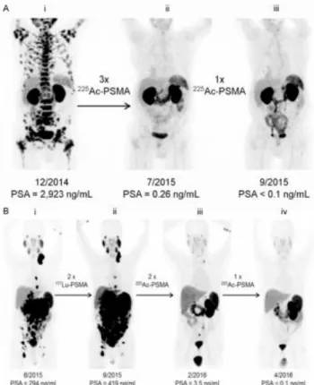
restaging patients with PCa before and after RN therapy, as shown in figure 1.1. [adapted from
(13-14)].
Figure 1.1. (A) 68Ga-PSMA-11 PET/CT scans of a PCa patient. Pretherapeutic tumor spread (i),
restaging 2 months after third cycle of 225Ac-PSMA-617 (ii), and restaging 2 months after one
additional consolidation therapy (iii). (B) 68Ga-PSMA-11 PET/CT scans of another PCa patient. In
comparison to initial tumor spread (i), restaging after 2 cycles of β-emitting 177Lu-PSMA-617
presented progression (ii). In contrast, restaging after second (iii) and third (iv) cycles of 225
5
Over the past decade, optical imaging has also emerged as a real-time, sensitive, high resolution and noninvasive modality for visualization, localization, and measurement of bioactive molecules in vivo(15). The optical signals can provide molecular information of
biological tissues and are related to the tumor anatomical structure as well as the tumor metabolism and biochemistry(16). NIRF imaging is an attractive novel modality for cancer
detection and acquisition of real-time pathophysiological information(17). It was demonstrated
to be a feasible and practicable method for PCa detection in addition to the predominant modalities for clinical detection and diagnosis of this pathology(17).
There are several therapeutic options for PCa which include surgery, chemotherapy, cryotherapy, brachytherapy with radioactive seeds, and the use of external radiation which is now being highly promoted with the use of proton therapy machines using conformal targeting technologies(8).
It is described that approximately 30%–40% of patients will fail primary treatment, and a rising prostate-specific antigen (PSA) level will herald the onset of biochemical recurrence (BCR) (2).
After potential salvage procedures (radiation and surgery), patients usually undergo androgen deprivation therapy(2). This treatment is typically followed by an increase in the
prostate-specific antigen (PSA) level after 2–8 y; this increase indicates the onset of metastatic castration-resistant PC (mCRPC)—the lethal form of the disease(2). Reactivation of androgen
receptor signaling occurs in early mCRPC; therefore, second-line agents targeting the androgen receptor signaling axis have extended survival in clinical trials(2). Treatment with
other systemic agents, including taxane-based chemotherapy (e.g., docetaxel), the immunotherapeutic agent sipuleucel-T and the bone-seeking -emitter 223RaCl
2, has been
shown to improve overall survival and quality of life(2). Nevertheless, patients with mCRPC
have a poor prognosis and a median survival of 19 months(2).
However, there is lack of effective imaging methods for early diagnosis, localization and identification of metastases, choice of postoperative treatment and prevention of postoperative recurrence(18). Accurate staging in primary PCa and early assessment of BCR for tailoring initial
and subsequent treatment strategies are unmet clinical needs(2). Once mCRPC is diagnosed,
effective therapy for improving overall survival and quality of life is desperately needed(2).
PSMA ligands are currently being investigated intensively, as they hold promise for extending the frontier in PCa imaging and RN therapy(2). Therefore, thanks to certain recent
developments in radiopharmaceutical chemistry, the role of NM for the diagnosis as well as therapy of PCa is expected to significantly grow in the future(8).
6
1.2. PSMA and its targeting
Among the markers of PCa, PSMA is the most well-established(15),(19). PSMA corresponds to a
highly specific prostate epithelia cell membrane antigen(20) and, recently, has emerged as one
of the most extensively investigated and exploited targets for molecular imaging and RN therapy of PCa(21). PSMA represents a highly valuable molecular marker in PCa, for both
diagnostic and therapeutic application, due to the following reasons: i) strong upregulation in poorly differentiated, metastatic, and hormone-refractory PCa; ii) low basal expression in nonprostatic tissues; iii) direct correlation between PSMA expression levels and androgen independence, metastasis, and PCa progression(21).
PSMA is a type II transmembrane glycoprotein that contains 750 amino acids and has a molecular weight of 100 kDa(1-3),(10),(18),(22). It has a unique structure containing three distinct
parts; a 707-amino acid extracellular region, a cell membrane part of 24 amino acids and a cytoplasmic tail which contains 19 amino acids(1),(8).PSMA is a key player in prostate
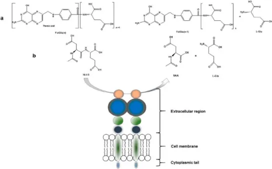
carcinogenesis and disease progression, glutamatergic neurotransmission and folate absorption(1) (see figure 1.2.). These various functions and the tissue distribution of the protein
result in different designations(1). The name which is also frequently used for this enzyme is
glutamate carboxypeptidase II, or GCPII(1),(3),(8),(14),(22-23). Furthermore, in central nervous
system, it metabolizes the brain neurotransmitter N-acetyl aspartyl glutamate (NAAG), and is named NAALADase(1),(8),(14). In the proximal small intestine, it removes γ-linked glutamates
from poly-g-glutamated folate, which is reflected in its name, folate hydrolase FOLH1(1),(18).
Figure 1.2. Enzymatic actions of PSMA. (a) Glu is released from folate polyglutamate resulting in the release of folic acid; (B) NAAG is hydrolyzed to aspartate (NAA) and L-Glu. Adapted from(1).
7
PSMA expression and localization in the normal human prostate are associated with the cytoplasm and apical side of the epithelium surrounding the prostatic ducts(2). PSMA is
enzymatically active only in its dimeric form(2). Its function for prostate cells is still unknown(2)
but it has been described that PSMA undergoes constitutive receptor-mediated endocytosis via clathrin-coated pits(1),(3),(6),(14). Dysplastic and neoplastic transformation of prostate tissue
results in the transfer of PSMA from the apical membrane to the luminal surface of the ducts(2).
PSMA possesses the criteria of a promising target for PCa, i.e., abundant and restricted (to prostate) expression at all stages of the disease, presentation at the cell surface but not shed into the circulation as it is not secreted and is membrane bound, and association with enzymatic or signaling activity(23).
It is described that PSMA is significantly over expressed (100–1000 fold) on nearly all PCa cells(2), compared to the physiologic levels found in other tissues such as , small intestine, or
brain(24) and its expression is further increased in advanced stages and in mCRPC(2). Also,
after binding to the active center of the extracellular domain, PSMA ligands are internalized and subsequent endosomal recycling leads to enhanced tumor uptake, retention, and subsequent high image quality for diagnostic procedures and a high local dose for therapeutic applications(2).
Available tools for PSMA targeting include monoclonal antibodies and a variety of small molecule or peptide ligands, each of which has advantages and limitations associated with its use(3).
Antibody-based probes, like the FDA-approved imaging agent for targeting PSMA in PCa – ProstaScint - are attractive because of the high level of specificity for the target and the picomolar range affinities that can often be achieved(3). The major disadvantages of antibodies
are the slow target binding and background clearance in an appropriate time frame for diagnostic imaging, reduced utility for image-guided surgical approaches and their potentially immunogenic behavior (20). In contrast, small molecule probes can often be generated with
nanomolar affinity ranges and rapid clearance rates that not only facilitate tissue penetration but also minimize potential toxicity resulting from exposure times(3).
In the search for PSMA targeting agents with more favorable characteristics compared to antibody molecules(25), the development of highly PSMA-specific small peptides referred to as
PSMA ligands or PSMA inhibitors was pursued. PSMA inhibitors bind to the active center located in the extracellular domain and are subsequently internalized(2). The recognition of the
8
development of these small molecules(2), which is based on modifications of NAALADase
inhibitors and on the known crystal structure of PSMA, as discussed below.
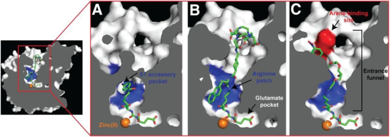
The PSMA internal inhibitor–binding cavity can be roughly divided into 3 continuous parts: the S19 Glu recognition pocket (S1’), the dinuclear zinc(II) active site, and an irregularly shaped entrance funnel connecting the active site to the external surface of PSMA (see figure 1.3.)(26).
Crystal structure investigations also indicated that, besides the electrostatic interactions of urea and carboxylic groups at the active Zn(II)-containing center of PSMA, there are lipophilic interactions resulting from a hydrophobic pocket (S1) located next to the active site(24).
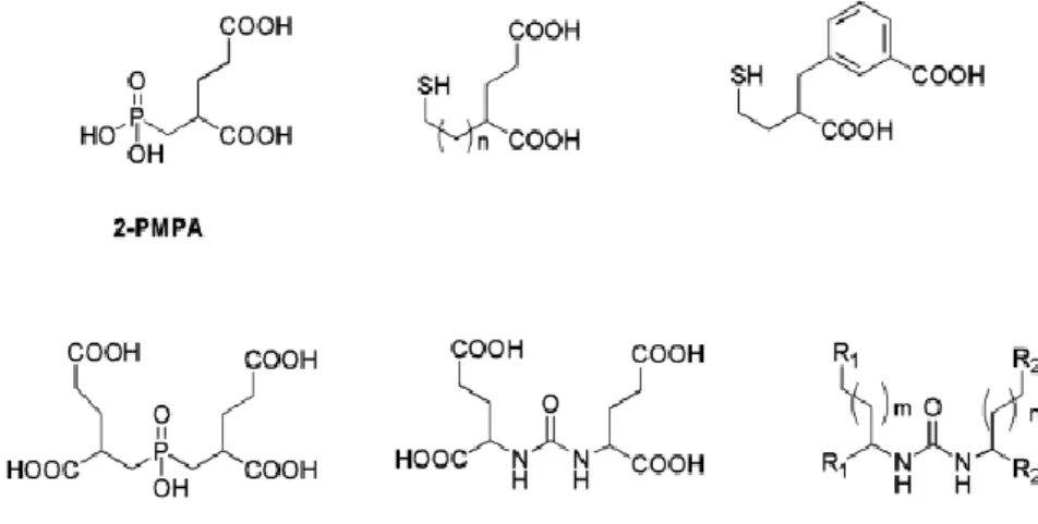
Taking into consideration the PSMA structure, the design of small-molecule PSMA ligands includes, as common features, the presence of pentanedioic acid groups as glutamate mimics and a zinc-binding group to interact with the catalytic zinc atom at the PSMA active site(27).
Since 2-PMPA (see figure 1.4.) was firstly reported as a potent PSMA inhibitor in 1996, many efforts have been devoted to generate other molecules with inhibitory action towards PSMA(1).
The main strategy for the discovery of those inhibitors is to find zinc-binding groups to be linked to a glutamate moiety (or its mimics)(1). Different functionalities with affinity for zinc, including
phosphonates, phosphates, phosphoramidates, thiols and ureas, were identified and evaluated(1), as exemplified in figure 1.4. In the field of radiopharmaceutical development, a
great number of urea-based PSMA inhibitors have been synthesized and modified accordingly in order to be labeled with a variety of RNs(1), as discussed in the next section.
Figure 1.3. Representation of a cross-section of PSMA. The internal inhibitor-binding cavity presents a S1’ Glu recognition pocket, dinuclear zinc(II) active site and irregularly shaped entrance funnel, where an arginine patch, a S1 accessory hydrophobic pocket and an arene-biding site are significant structural features for inhibitor design. Zinc ions are shown as orange spheres and some PSMA ligands are shown as stick representations: DCIBzL (A) (PDB code 3D7H), PSMA-617 (B) (unpublished data) and ARM-P4 (C) (PDB code 2XEG). Adapted from(26).
9
1.3. Radioligands for PSMA targeting
Low molecular-weight peptidomimetic Glu-ureido–based PSMA inhibitors are the most studied class of targeting molecules in the design of radioligands suitable for PCa imaging by nuclear techniques or for RN therapy(1),(26). The designed and evaluated radioligands usually consist of
3 components: i) the binding motif (e.g. Glu-urea-Lys, the most widely used scaffold); ii) a linker; iii) a radiolabel-bearing moiety, comprising a chelator molecule for labeling with radiometals or a prosthetic group for labeling with radiohalogens(2) (see figure 1.5).
Figure 1.5. General structure of a Urea-based PSMA radioligand. Adapted from(2).
10
Regarding the selection of the RN, several possibilities are available either for diagnosis, therapy or theranostic. Theranostic is a new and emerging approach that provides a transition from conventional medicine to personalized and precision medicine, as it combines diagnostic and therapeutic applications in a single agent (or related ones), allowing targeted diagnosis, drug delivery and treatment response monitoring(28). For imaging purposes, besides a relatively
physical short half-life, the isotope must be a pure gamma emitter or positron emitter, i.e. have no particle emission such as alpha and beta particles (these particles increase the radiation dose to the patient while not providing any diagnostic information)(29). However, for therapy
purposes a suitable range of the physical half-life for therapeutic RNs is between 6 h and 7 days(30), because a very short physical half-life limits the delivery flexibility and is very
impractical, while a long half-life will retain the radiation dose in the patients and expose surrounding tissues(30). α- and β- emitters are preferable for therapy purposes, as α and β
particles present high-linear energy transfer (LET) that leads to its fully deposition within a small range of tissue (usually in mm) not allowing the radiation to penetrate through the patient’s body(30). However, some β-emitting RNs also decay with γ-radiation, which can be
advantageous if the energy and intensity are within the diagnostic range, as it also allows the visualization of the radiopharmaceutical distribution within the patient’s body using gamma scintigraphy methods(30). The physical characteristics of some relevant medical RNs(30-37) are
shown in table 1.2.
RN Use Half-life Main types of
decay Emax (MeV)
99mTc SPECT imaging 6 h IT 0.141
123I SPECT imaging 13.2 h EC 0.16
111In SPECT imaging 67.9 h EC 0.17/0.25
201Tl SPECT imaging 73.1 h EC 0.17
67Ga SPECT imaging 78.3 h EC 0.09/0.19/0.30
11C PET imaging 20.3 min + 0.97
13N PET imaging 9.97 min + 1.20
11
15O PET imaging 2.03 min + 1.74
18F PET imaging 110 min + 0.64
64Cu PET imaging and
therapy 12.7 h + (17.5%) EC (43.5%) - (38.5%) 0.653 1.675 0.579
68Ga PET imaging 68.1 min
+ (87.7%) EC (8.9%) (3.2%) 1.899 2.921 1.077 124I PET imaging 4.18 d + (25.6%) EC (74.4%) 1.532/2.135 0.603/1.691 131I Therapy 8 d - (89.9%) (7.27%) 0.606 0.364 177Lu Therapy 6.7 d - (79.3%) (11.6%) 0.498 0.177 225Ac Therapy 10 d α 5.7
Some medical RNs are easily obtained in situ through elution of a RN generator, being readily available for the preparation of radiopharmaceuticals. For example, 99mTc is obtained daily in
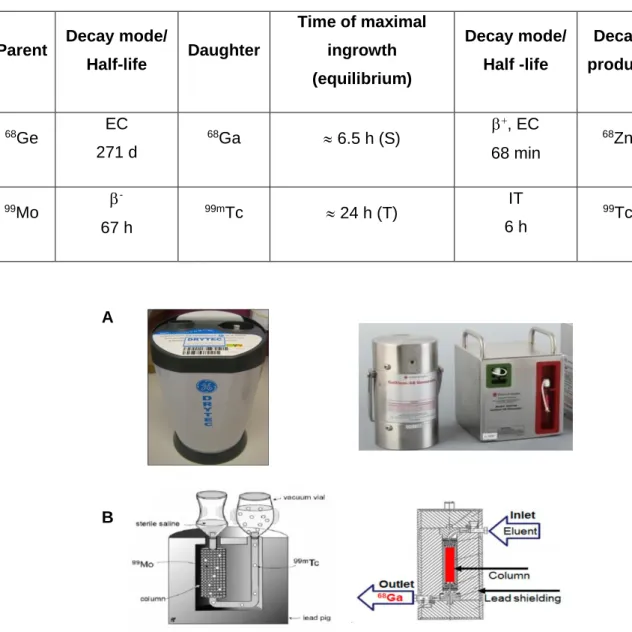
NM centers from commercially available 99Mo/99mTc generators (see table 1.3. and figure
1.6.)(29),(31),(38). 99mTc still is the most important RN used in conventional NM. The parent 99Mo,
which has a longer half-life (67 hours), continually produces 99mTc that presents a short
half-life (6 hours) suitable for SPECT imaging(29),(31),(37-38).
68Ga is a positron emitter that can be also obtained, in situ, by elution of a 68Ge/68Ga generator
(see table 1.3. and figure 1.6.)(29),(31),(39),(40-41). 68Ga presents a physical half-life of 68 min (29),(40-41), suitable for many PET exams and, for this reason, is assuming a growing importance in
diagnostic imaging studies and in theranostic applications when combined with a therapeutic radiometal (e.g. 177Lu)(1),(8),(41).
12
A variety of urea-based PSMA inhibitors have been labeled with different medical RNs, bearing in most cases the Glu-urea-Lys as scaffold(8),(10),(26),(42). The resulting radioligands can be
synthesized in a simple and standardized fashion and, in some cases, have already entered clinical settings and are being used either for SPECT or PET imaging and/or therapy purposes(26). Some examples of clinically relevant urea-based PSMA radioligands are listed in
table 1.4.
Parent Decay mode/
Half-life Daughter Time of maximal ingrowth (equilibrium) Decay mode/ Half -life Decay product 68Ge EC 271 d 68Ga 6.5 h (S) +, EC 68 min 68Zn 99Mo -67 h 99mTc 24 h (T) IT 6 h 99Tc
Table 1.3. Characteristics of 68Ge/68Ga and 99Mo/99mTc generators.
Figure 1.6. (A) Examples of commercially available 99Mo/99mTc and 68Ge/68Ga generators, respectively.
(B) Schematic presentation of the cross section of both column-based generators. Adapted from (39), (41).
A
13
Cpd. Chemical Structure Class RN Ref.
MIP-1095 Theranostic 123I 124I 131I (25- 26),(43-44) DCFBC Diagnostic PET 18F (25-26),(45) DCFPyL Diagnostic PET 18F (25- 26),(46-47) MIP-1404 Trofolastat Diagnostic SPECT 99mTc (22),(25-26)
PSMA I&T Theranostic
68Ga; 177Lu; 111In
(26),(48-49)
14 PSMA-617 Theranostic 68Ga; 177Lu; 64Cu 225Ac (13),(26), (50-51) PSMA-11 Diagnostic PET 68Ga (24-26) PSMA-1007 Diagnostic PET 18F (26)(52)
PSMA I&S Diagnostic
SPECT
99mTc (21)(26)
Cpd. = compound; Ref. = reference
Concerning the chelator choice and modifications, Eder et al. compared the pharmacokinetics and targeting characteristics of the same Glu-urea-Lys binding motif coupled to either the acyclic HBED-CC chelator or the macrocyclic DOTA (1,4,7,10-tetraazacyclododecane-1,4,7,10-tetraacetic acid) chelator(24). HBED-CC shows fast 68Ga-complexation kinetics and
affords complexes with high in vitro and in vivo stability(7),(24),(53-55). DOTA is a chelator that is
suitable for the labeling of biomolecules with a variety of trivalent radiometals, which includes
68Ga and its therapeutic counterpart 177Lu(55). This study showed that the 68Ga-radioconjugate
obtained using HBED-CC as the bifunctional chelator (BFC) exhibited reduced non-specific accumulation and higher specific internalization in PSMA-expressing LNCaP cells, when compared with the DOTA congener(24). These results might be due to the lipophilicity of the
15
with urea-based ligands and one representing a lipophilic pocket(7),(24). This idea is supported
by the suggestion that the binding site has a pocket that interacts with hydrophobic aromatic groups(7),(24).
Worldwide, 68Ga-PSMA-11 ([68Ga]Glu-urea-Lys(Ahx)-HBED-CC) (see table 1.4.) is currently
the most prominent radioligand for the PET imaging of PSMA-positive PCa. 68Ga-PSMA-11
was clinically introduced several years ago and is commonly used for diagnostic imaging of PCa in the clinics, especially in Europe(26). This PSMA inhibitor exhibits the Glu-
NH-CO-NH-Lys pharmacophore and HBED-CC as an efficient 68Ga chelator. The success of 68
Ga-PSMA-11 reflects the favorable physical properties of 68Ga, as mentioned above, which allow
high-resolution PET images with the option of accurate quantification.
Because 99mTc is the preferred RN for developing SPECT radiopharmaceuticals, efforts have
been made to obtain 99mTc-labelled PSMA inhibitors suitable for PCa imaging(25). The more
encouraging results were obtained for a Glu-urea-Glu pharmacophore, MIP-1404 (see table 1.4.), functionalized with a single-amino-acid chelator (SAAC) containing imidazolyl rings as coordinating units(56). The resulting conjugate is labeled with 99mTc based on the so-called
tricarbonyl chemistry(22),(25). The SAACs allows facile labeling with 99mTc, forms a robust 99mTc(I)
tricarbonyl complex, and was designed to minimize hepatobiliary uptake(25),(56). Preclinical
studies showed that 99mTc-MIP-1404 has high tumor uptake and high tumor-to-blood ratios in
PCa-tumor bearing mice, early after injection of 99mTc-MIP-1404, leading to SPECT images
with excellent tumor-to-background contrast(25). These favorable features revealed that this
PSMA ligand is feasible for 99mTc-SPECT/CT detection of PCa lesions(25). It is expected that 99mTc-MIP-1404 (Trofolastat) will be launched on the market as the first low-molecular-weight
SPECT PSMA radioligand, as a phase 3 clinical trial with this tracer is already under way(44),(57).
Besides this promising SPECT PSMA radioligands, also an 99mTc urea-based PSMA
radioligand for imaging and surgery - PSMA I&S (see table 1.4.) - is also in clinical development(21).
1.4. NIRF Probes for PSMA-targeting
Cancer near infrared (NIR) molecular imaging relies greatly on the development of stable, highly specific and sensitive fluorescent probes(16). In particular, it has been dedicated
immense attention to NIRF imaging with probes emitting within the wavelength range of 700-1000 nm, owing to its low absorption and autofluorescence from organisms and tissues in this NIR spectral range(16). This can minimize background interference, improve tissue depth
16
function in the NIR spectral range, with large Stokes shift for minimum interference between absorption and emission spectra(16). Moreover, it must display high molar absorption coefficient
and quantum yield for intense fluorescence, sufficient chemical and photostability in solvents, buffers or biological conditions for imaging or detection, and good water solubility to avoid dye aggregation in aqueous environment(16). Additionally, a promising NIR probe should have
suitable chemical functionalities for bioconjugation with specific ligands for targeting purpose(16), to obtain minimal background and enhanced sensitivity(58).
Current NIR probes generally comprise two categories: inorganic and organic molecules. The organic probes present more favorable features than the inorganic ones, which show some disadvantages, such as potential cytotoxicity of their heavy metals, small-scale and expensive preparation(16). So, NIR organic dyes have attracted increasing attention in biomedical
applications as they show improved photophysical properties and easy availability by large-scale chemical synthesis, leading to promising clinical implications for optical imaging(16). In
the past few years, several classes of NIR organic dyes were developed and evaluated: cyanine dyes, squaraines, phthalocyanines, porphyrin derivatives and BODIPY (borondipyrromethene) analogues (see figure 1.7.).
Figure 1.7. Some examples of newly developed NIR fluorescent dyes. (A) Cyanine dyes: small organic molecules with two aromatic nitrogen-containing heterocycles with a delocalized charge linked by a polymethine bridge. (B) Squaraines derivatives: consist of an oxocyclobutenolate core with aromatic or heterocyclic components at both ends of the molecules. (C) Phthalocyanines: two-dimensional 18π-electron aromatic porphyrin derivatives, consisting of four bridged pyrrole subunits linked together through nitrogen atoms. (D) BODIPY dyes: have a general structure of 4,40-difluoro- 4-bora-3a, 4a-diaza-s-indacene. Adapted from(16).
A B
17
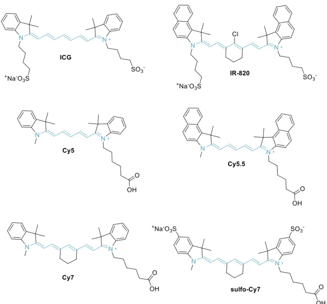
One of the most prominent classes of NIR probes corresponds to cyanine dyes (also called as polymethine cyanine dyes) (see figures 1.7. and 1.8.). The monomethine and trimethine cyanines (Cy3) generally show absorption in the visible region, but the extension of the chromophore by one vinylene moiety causes a bathochromic shift of about 100 nm(16). Thus,
pentamethine (Cy5) derivatives can reach a near-infrared region (>700 nm), and heptamethine cyanines (Cy7) may show absorption beyond 1000 nm(16). Some cyanine dyes (such as Cy5,
Cy5.5, Cy7 and their derivatives) are among the most common NIR fluorescent dyes with high molar absorption coefficient and fluorescence quantum yield(16).
However, many cyanine dyes suffer from poor photostability, low quantum yield, high plasma protein binding rate, undesired aggregation and mild fluorescence in aqueous solution(16). To
overcome these limitations several modifications have been studied, such as:
Figure 1.8. Chemical structure of some cyanine dyes: first digit identifies the number of carbon atoms between the indolenine groups and the suffix .5 is added for benzo-fused cyanines.
18
a) introduction of a rigid cyclohexenyl substitution in the middle of polymethine linker to increase the photostability and fluorescence quantum yield(16);
b) introduction of electron-donating groups on N-position of 3H-indolenine and substitution of the central chlorine atom at the cyclohexene ring by electron-donor groups to improve the photochemical stability(16),(59);
c) introduction of carboxylic or sulfonic acid groups to increase Stokes shift, fluorescence quantum yield and water solubility(16),(60).
Cyanine dyes have gained much attention in biomedical applications because of their optimal spectral, chemical, and biological properties(61). Moreover, it is worthwhile to mention the
excellent safety profile of ICG dye, which has been approved by the FDA almost for over 50 years for ophthalmic angiography and to determine cardiac output and liver blood flow and function(9).
Besides ICG, other heptamethine indocyanine dyes have emerged as potential tools for in vivo tumor diagnostic and therapeutic applications(16), due to their excellent photophysical
properties and ease of synthetic modifications(61). ICG can accumulate in normal tissues and
emit fluorescence signals with high intensity during some surgical procedures, largely compromising the potential use of ICG for tumor visualization as it lacks tumor-targeting capability, similarly to other conventional NIRF polymethine cyanine organic dyes(60).
As clinically approved NIR dyes (ICG and methylene blue) are contrast agents with no tumor specificity, efforts have been made to develop agents that are designed to recognize biochemical features of the tumor cells in a specific manner, leading to effective targeting of tumor cells(3). Particularly promising results have been obtained in the targeting of PSMA,
which is overexpressed in primary and metastatic PCa as mentioned above(3). This comprised
NIRF probes carrying a variety of cancer specific ligands (such as humanized anti-PSMA antibodies, peptide ligands or small-molecule PSMA inhibitors)(3),(16),(62). These target specific
NIRF probes (see table 1.5.) are expected to increase the signal-to-background ratios, presenting clinical utility for image-guided surgery and tumor resections(3),(16), as they allow a
precise discrimination between tumor and healthy tissue leading to a better outcome of PCa patients.
19
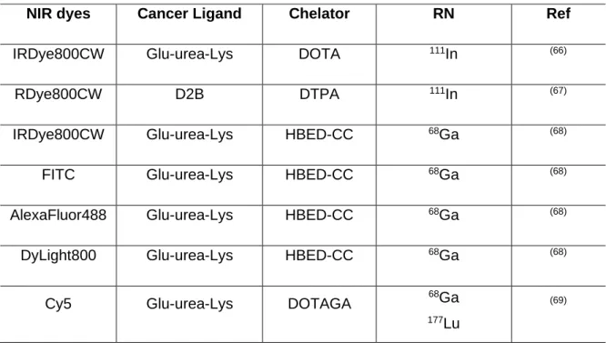
Table 1.5. Some NIR fluorophore-labeled PSMA targeting probes.
NIR dyes PSMA
ligands Chemical Structure Ref.
Cy5.5 CTT-54.2 (63) IRDye800CW YC-27 (3),(64) IRDye800CW PSMA-1 ´ (15) Cy5.5 PSMA-1 (15) ICG PSMA-MB (65)
20
1.5. NIRF and Nuclear imaging dual probes for PSMA targeting
Intense efforts have been directed toward the development of dual PSMA-targeted probes for a variety of clinical applications ranging from diagnostic imaging using SPECT, PET, MRI, or optical methods toward innovative theranostic and therapeutic concepts(21).
Dual modality agents are expected to fulfill the unmet needs for specific imaging of PCa, which to some extent is inadequately assessed by conventional methods(9). Several dual modality
imaging agents are being developed to take advantage of the sensitivity of nuclear imaging, such as SPECT or PET, and the high sensitivity and resolution of NIRF for possible use in pre- and intraoperative applications, respectively(3),(66).
Nuclear imaging is an attractive modality for the detection and characterization of disease because it is non-invasive, quantitative, provides dynamic real-time data, and allows the diagnosis and follow-up of patients undergoing therapy(9). In fact, for staging and preoperative
assessment of tumor burden the RN component could initially be used to map disseminated lesions or lesions within the prostate at sub-centimeter resolution(66).
Optical imaging modalities could aid in image-guided surgery to more accurate real-time delineation of tumor margins precisely during and following resection, using a minimally invasive laparoscopic instrument equipped with a NIRF detection system (see figure 1.9.)(66),(67).
The NIRF approach shares common features with nuclear imaging techniques, such as the potential use of tracer administration of the probe, which enables the development of combinational use of both techniques for cancer imaging(9). NIRF alone has inherent
shortcomings such as its sensitivity and resolution being severely influenced by position and depth of the imaging probes in the body(9). The positron- or gamma-emitting properties of the
dual probe can overcome the shortcomings of NIRF and provide high sensitivity and deep-tissue spatial resolution for initial detection of primary tumors and their metastatic lesions, allowing also a sensitive intra-operative localization of tumor lesions using a gamma probe(9),(67). Consequently, both nuclear and NIRF imaging might complement each other,
merging the strength of both techniques, and dual-modality image-guided surgery may overcome limitations of the currently used single-modality imaging techniques(67),(68). Some
21
NIR dyes Cancer Ligand Chelator RN Ref
IRDye800CW Glu-urea-Lys DOTA 111In (66)
RDye800CW D2B DTPA 111In (67)
IRDye800CW Glu-urea-Lys HBED-CC 68Ga (68)
FITC Glu-urea-Lys HBED-CC 68Ga (68)
AlexaFluor488 Glu-urea-Lys HBED-CC 68Ga (68)
DyLight800 Glu-urea-Lys HBED-CC 68Ga (68)
Cy5 Glu-urea-Lys DOTAGA 68Ga
177Lu
(69)
Table 1.6. PSMA dual probes with both nuclear and optical imaging valences.
Figure 1.9. Proof-of-concept fluorescence-guided surgery studies in tumor-bearing mice and healthy pigs. (A) 68Ga-Glu-urea-Lys-HBEDCC-IRDye800CW (0.5 nmol) was injected in mice (LNCaP tumor
xenograft) for small-animal PET imaging, followed by ex vivo fluorescence detection after 120 min (IMAGE1 S system). (B) In healthy pigs, after pre-imaging acquisition of background fluorescence with da Vinci FireFly system, Glu-urea-Lys-HBED-CC-IRDye800CW (30 μg/kg) was intravenously injected. Fluorescence-guided prostatectomy accompanied by in vivo and ex vivo fluorescence detection was performed 1 h after injection. Adapted from(68).
22
1.6. Aim
As mentioned above, new PSMA radioactive probes have been developed recently and revealed high potential for detection and staging of PCa based on nuclear imaging techniques (SPECT or PET)(1-2),(25-26). In particular, very promising results were obtained for complexes
prepared with the 99mTc and 68Ga radiometals and functionalized with a PSMA inhibitor
containing the Glu and Lys amino acids(1-2),(10),(25-26). 99mTc is the most widely used radioisotope
in SPECT while 68Ga is a radioisotope with increasing importance in PET, which justifies its
interest in nuclear oncology, namely in the detection of PCa. It is noteworthy that one of the
68Ga complexes directed to PSMA has already been clinically tested in numerous European
NM Centers and has led to better results in the detection of PCa and its metastases when compared to other PET tracers or other imaging techniques(25),(42),(70-71).
Radioactive probes for PET or SPECT allow the detection of PCa and its metastases by acquisition of whole body images. However, the use of fluorescent congeners is more favorable in intraoperative procedures aiming at the most selective excision of tumor tissue. For this purpose, encouraging results have been described for PSMA inhibitors functionalized with ICG fluorophores(19),(65). The design of specific dual probes for PSMA, containing a RN for
PET or SPECT imaging or a fluorophore for optical imaging, profits from the inherent advantages of both nuclear and optical medical imaging modalities(9),(67).
Thus, the aim of the work was the design, synthesis and biological evaluation of bimodal probes (PET / SPECT and NIR) for the detection of PCa, starting from the base structure of complexes of 99mTc and 68Ga functionalized with PSMA inhibitors that already showed clinical
relevance, followed by further functionalization with indocyanine green (ICG) fluorophores. To tackle this goal, the following steps were thought:
1) Functionalization of bifunctional ligands (BFLs); with PSMA inhibitors and ICG derivatives;
2) Synthesis and characterization of 99mTc and 68Ga complexes with the BFLs;
3) Biological evaluation of the 99mTc and 68Ga complexes in cells of prostate carcinoma
and in mice with induced tumor.
As one of the objectives of the work was to develop two bimodal probes for PSMA with SPECT and PET image capabilities, it was intended to synthesize a BFC for radiolabeling with 99mTc
and another one for radiolabeling of 68Ga, respectively. As referred above, in the design of the
PSMA probe, the bifunctional ligands must present three components: the binding motif, a chelating unit and a linker spacer that connects the binding motif to the chelator. In this work,
23
the same binding motif (Glu-urea-Lys) and linker was used for the development of both dual probes, while two different chelators were tested. HBED-CC was the chelator chosen for labeling with 68Ga, as it was already described in literature as an efficient chelator of 68Ga(24),(40),(55). Also, a pyrazolyl - diamine BFC was selected for radiolabeling with 99mTc, one
of the most preferred RNs for SPECT imaging purposes. The pyrazolyl-diamine chelators have been introduced several years ago by the team of the Radiopharmaceutical Sciences group from C2TN/IST and, since then, were successfully applied for the 99mTc-labeling of a variety of
targeting biomolecules, spanning from bioactive peptides to small organic molecules and bis(phosphonates)(72-74). For both dual probes, the optical image’s capability would be added
through the conjugation to a NIRF heptamethine dye. As known, ICG is the only FDA-approved NIRF cyanine agent for clinical use(9),(60),(75). However, ICG can make normal tissues emit
fluorescence signals with high intensity during some surgical procedures and, consequently, largely compromises its potential use for tumor visualization(60). IR820 is structurally similar to
ICG but the presence of sulfonic acid groups in both side chains allows better water solubility and lower toxicity through a quick clearance in vivo(60). IR820 is also a better alternative than
ICG when stability of the dye solution over time is a concern or when a consistent peak emission wavelength is desired(76). The increased in vitro stability of IR820 translates into an
increased stability in vivo as well, thus allowing for longer image collection times(76). For these
reasons, IR820 was selected as the NIRF chromophore to be evaluated in this work in the design of the dual probes.
Figure 1.10. represents the generic chemical structure of the proposed PSMA - bimodal probes studied in this work for both nuclear and optical imaging, as well as the BFCs selected for the labeling with two radioisotopes with clinical relevance for PET (68Ga) and SPECT (99mTc).
24
Figure 1.10. (A) Generic chemical structure of the dual probes. (B) BFCs for 68Ga complexation
(HBED-CC) and 99mTc coordination (pyrazolyl - diamine).
Figure 10. (A) Generic chemical structure of the dual probes. (B) Bifunctional chelators for
25
2. Synthesis and Characterization of Organic
precursors: PSMA inhibitors and fluorescence dyes
26
2. Synthesis and Characterization of Organic precursors:
PSMA inhibitors and fluorescence dyes
In this chapter the synthesis of the organic precursors (urea-based inhibitor and NIRF derivatives) for conjugation with the BFLs will be described, either for labeling with 99mTc or 68Ga. In Scheme 2.1., the different steps needed for the synthesis of the proposed compounds
are presented.
The synthesis of urea-based inhibitor involved the formation of an urea bond between two amino acids, Glu and Lys (see scheme 2.1.A). It was necessary to block or protect some functional groups of the reacting amino acids to prevent possible undesired side reactions during the urea bond formation, as detailed below. The desired urea-based inhibitor contains a flexible aminohexanoyl moiety as a spacer / linker that is accommodated at the spacious entrance funnel in the active site of PSMA, which is important for the binding affinity.
As for the synthesis of fluorophore derivatives, the functionalization of the NIRF dye – IR820- was attempted using two amino-containing linkers (see scheme 2.1.B), suitable for further coupling to the BFCs. This comprised the conjugation of the fluorophore with 4-aminothiophenol or with an hexamethylenediamine moiety. In both cases it was intended to obtain precursors with a terminal amine for conjugation with a carboxylic acid of the BFLs, through the formation of an amide bond.
Scheme 2.1. Different steps needed for the synthesis of the organic precursors: urea-based inhibitor (A) and NIRF derivatives (B).