Corresponding author: Drª Laura Francisca Campo Polanco e-mail: [email protected]
Received 17 April 2018 Accepted 21 June 2018
Major Article
Strongyloidiasis in humans: diagnostic efficacy of four
conventional methods and real-time
polymerase chain reaction
Laura Francisca Campo-Polanco
[1], José Mauricio Hernández Sarmiento
[2],
Miguel Antonio Mesa
[3], Carlos Jaime Velásquez Franco
[3], Lucelly López
[2],
Luz Elena Botero
[1]and Lina Andrea Gutiérrez Builes
[1][1]. Grupo Biología de Sistemas, Escuela de Ciencias de la Salud, Facultad de Medicina, Universidad Pontificia Bolivariana, Medellín, Colombia. [2]. Grupo Salud Publica, Escuela de Ciencias de la salud, Facultad de Medicina, Universidad Pontificia Bolivariana, Medellín, Colombia.
[3]. Sección Reumatología, Clínica Universitaria Bolivariana, Universidad Pontificia Bolivariana, Medellín, Colombia.
Abstract
Introduction: Strongyloides stercoralis is an intestinal parasitic nematode that causes hyperinfection and/or a dissemination
syndrome in hosts, which is often difficult to diagnose. This study aims to compare the diagnostic efficacy of four conventional
methods used to diagnose strongyloidiasis with real-time polymerase chain reaction (qPCR) to detect S. stercoralis in fecal samples. Methods: We analyzed 143 fecal samples collected from Colombian regions with varying degrees of risk for intestinal
infections caused by S. stercoralis to assess the validity, performance, overall efficiency, and concordance of the qPCR using a direct stool test, modified Ritchie concentration technique, agar plate culture, and Harada-Mori technique as reference tests.
Results: While four fecal samples were positive for S. stercoralis using conventional methods, 32 were positive via qPCR.
The diagnostic sensitivity of the qPCR was 75% [95% confidence interval (CI): 20.07-100%], whereas its specificity, negative predictive value, negative likelihood ratio, and Youden’s J index were 78.42% (95% CI: 71.22-85.62%), 99.09% (95% CI: 96.86-100%), 0.32 (95% CI: 0.06-1.74), and 0.53, respectively. In addition, the estimated kappa index between the qPCR and the conventional methods was 0.12 (95% CI: -0.020-0.26). Conclusions: The diagnostic sensitivity of qPCR to detect
strongyloidiasis is analogous to that of conventional parasitology methods, with an additional advantage of being capable of
identifying the parasite DNA at low sample concentrations.
Keywords: Strongyloides stercoralis. Intestinal diseases. Helminthology. Molecular diagnostic techniques.
INTRODUCTION
Strongyloides stercoralis is an intestinal nematode that is
commonly detected in the tropical and subtropical parts of the world, with a prevalence of 10-40% in tropical countries1,2.
Alternating stages of parasitic life, autoinfection cycles, and free
life account for the high complexity of the biological cycle of this
nematode3,4, which is further exacerbated by high humidity and
temperatures of 20-37°C required for its proliferation5,6. In some cases, socioeconomic and environmental factors often converge
with high-risk factors, such as extreme poverty, inadequate ecological sanitation, poor excreta disposal, and soil organic debris5,7, which are all associated with a higher prevalence of
intestinal infections caused by S. stercoralis. In addition, in people
with an impaired cellular immune response, the occurrence of autoinfection cycles of strongyloidiasis might cause hyperinfection and/or the dissemination of larval stages toward other organs,
increasing the risk of potentially fatal complications6,8,9.
Typically, strongyloidiasis is diagnosed by identifying the
larval stages using methods such as the direct stool test, which is
the routinely performed test in clinical laboratories because of its simplicity and fast sample-processing rate. However, the direct stool test exhibits low performance, efficiency, and diagnostic
certainty for detecting S. stercoralis10, frequently leading
to false-negatives11. Other available methods for detecting
S. stercoralis are the modified Ritchie concentration technique, isolation or culture on an agar plate, and the Harada–Mori
technique and Baermann methods for larval separation5,12,13. Although the diagnostic sensitivities of these methods are
higher, these procedures are laborious and time-consuming7,14.
Lately, the polymerase chain reaction (PCR) has been proposed as a valid, reliable, and rapid alternative for the
advantages, including the ability to precisely detect the genetic
material of S. stercoralis, even if its deoxyribonucleic acid (DNA) is a free molecule in the analyzed sample, and that its
identification does not depend on the viability of the parasite22.
This study aims to compare the diagnostic efficacy of four
conventional methods used to diagnose strongyloidiasis with real-time PCR (qPCR) to detect S. stercoralis in fecal samples
collected from Colombian regions with varying degrees of risk for intestinal infections caused by S. stercoralis.
METHODS
Study population
In this study, we collected fecal samples from 143 men and women, using non-probabilistic or convenience sampling, in groups where the Universidad Pontificia Bolivariana School of Health Sciences performed community work. Adult habitants of the rural areas of Chocó and the peri-urban areas of the Chocó
and Antioquia departments for >18 year and who consented
to participate in the study were included. They were divided into three groups based on geographical zones. The first group comprised of indigenous people from Emberá Dobida and Emberá
Chamí ethnic communities living in rural areas of the Department
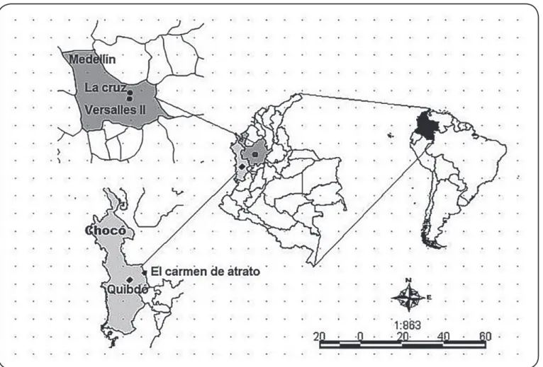
FIGURE 1: Amap of the sampled areas in Antioquia and Chocó, Colombia (represented using the DIVA-GIS© software, http://www.diva-gis.org/).
DIVA-GIS: Graphic Information System.
of Chocó as well as inhabitants of two villages in the municipality of Carmen de Atrato (Chocó) where a health services network was not available (Figure 1). The second group comprised of residents
of outlying neighborhoods in Quibdó and Medellín (Colombia),
where some essential sanitation services, such as pipe water and
excreta disposal, were available (Figure 1). Finally, the third group comprised of patients with rheumatic diseases who were undergoing immunosuppressive treatment at Clínica Bolivariana
(Medellín, Colombia). Individuals who received antiparasitic treatment 3 months before the study period were excluded.
Ethical considerations
All procedures in this diagnostic test evaluation study and data collection methods were in accordance with the fundamental ethical principles regulating the ethical conduct
and governed by the Nuremberg Code (1947), the Helsinki Declaration (enacted in 1964, amended in Korea in 2008 and at the 64th ICH’s General Assembly held in Fortaleza, Brazil, in
2013), and the national standard 008430 for health research. In addition, the study protocol was approved by the Universidad Pontificia Bolivariana’s Health Research Committee (October 21, 2013). We obtained written informed consent from all
Sample collection and processing using conventional parasitology methods
We collected spontaneous emission fecal samples and divided those into three aliquots as follows: a) first aliquot with
no added preservatives for setting up tests requiring viability to generate a positive result (e.g., agar plate culture and
Harada-Mori technique); b) second aliquot with 10% formalin added
for preservation, for tests requiring no viability (e.g., direct stool test and modified Ritchie concentration technique); and c) third aliquot with storage at -20°C and use in the qPCR. All stool samples were assessed using four conventional parasitology
methods – direct stool test, modified Ritchie concentration technique, agar plate culture, and Harada-Mori technique – recommended by the World Health Organization because a unique diagnostic method that could be used as a reference
test for the detection of S. stercoralis was unavailable23. Before
carrying out the study, the magnitude of the interobserver variability was estimated through a concordance study that facilitated the evaluation of the correlation power between the observers involved in the test readout. This translated into reliability and validity of the results obtained from the readouts
of different parasitology methods and into the minimization of
measurement errors originating in observer variability. In the end, kappa index values higher than 0.70 were obtained for most of the identified intestinal parasites, which suggest a good
correlation among the analysts24-26.
Molecular analysis and qPCR based on 18S rRNA gene sequences
A formerly standardized qPCR technique was used for the molecular analysis of the fecal samples tested in this study.
Primers reported by Verweij JJ et al.17, and a TaqMan probe
(Custom TaqMan®, MGB (minor groove binder) Probes,
Applied Biosystems®, Thermo Fisher Scientific; Massachusetts, United States), marked with FAMTM fluorochrome, as previously
described16,were employed in the amplification of the 18S
ribosomal ribonucleic acid (rRNA) gene of S. stercoralis. In all tests, an exogenous DNA or IPC Internal Positive control (TaqMan® Exogenous Internal Positive control, Applied
Biosystems®, Thermo Fisher Scientific; Massachusetts, United
States) was introduced as an internal positive control of the
reaction to evaluate the presence of inhibitors in the fecal samples, and ultrapure water was used as a negative control. The qPCR conditions were established by evaluating the sensitivity
and analytical specificity of serial dilutions of samples containing S. stercoralis larvae as well as the occurrence of
cross-reactions with other parasites and amplification inhibitors. The threshold cycle (Ct) value obtained with the standardized qPCR was less than or equal to 29.99 in the positive samples. A Ct value between 30.00 and 34.99 was considered
indeterminate, assuming that only samples with a very low parasitic load would show those values27.
We randomly selected a PCR product from one of the positive samples and one from the positive controls to validate the S. stercoralis rRNA 18S gene-specific sequence19. Consequently, we randomly selected seven positive samples
(Ct of 23-29.99), three with Ct values between 30-34.99, and three positive controls. Subsequently, bidirectional sequencing
of the PCR product was performed in Macrogen (Maryland,
USA). We edited and aligned the obtained sequences using the
Geneious software ver 7.1.728. Further, we verified the identity
using the Basic Local Alignment Search Tool (BLASTn) for the analysis of the consensus sequence in GenBank (blast.ncbi. nlm.nih.gov) and conducted the maximum likelihood molecular
phylogeny analysis using the MEGA (Molecular Evolutionary
Genetics Analysis) software ver. 6. In addition, we used the
Bayesian information criterion29 and Akaike information
criterion to select a molecular evolution model. The dendrogram estimation was performed by a heuristic search and a bootstrap
resampling with 1,000 pseudo values. Furthermore, we used the same molecular evolution model previously selected for partial
sequences of the ribosomal RNA 18S region with the MrBayes software ver. 3.2.2, with the plug-in available in Geneious
ver. 7.1.728.
Statistical analysis
We conducted a survey to identify epidemiological variables
and hygiene-sanitary conditions, and further analyzed the
obtained data with Statistical Package for the Social Sciences
(SPSS) ver. 2430, both to elucidate the epidemiology and
parasitology data by a descriptive analysis of quantitative variables and frequency analysis. Epidemiological data were presented as absolute numbers and percentages, and parasitological data were presented as absolute numbers. In addition, we calculated the sensitivity, specificity, positive and negative predictive values, likelihood ratio, and the kappa index, while comparing the results obtained with the conventional parasitological methods to those with the qPCR. A kappa index closer to 1, calculated using the Epidat 4.1 software31,
indicated an almost perfect match between the analyzed tests24,
and these results were presented in percentages. Furthermore,
we performed an adjustment to estimate these same parameters
considering the prevalence of this parasitic infection, as reported
by Colombia’s Ministry of Health (1.5%, according to the
national survey of intestinal parasitism)32, and using the Bayes
theorem in the Bayesian analysis module of Epidat 4.1.
RESULTS
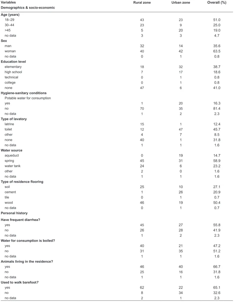
Characterization of the study population
Among the 143 fecal samples, we collected 72 from the rural
zone of Chocó, 57 from the outlying areas of Quibdó and Medellín
Cities, and 14 from individuals receiving immunosuppressive treatment. Table 1 summarizes the hygiene and sanitary, or
housing conditions of individuals in rural and urban zones. All participants undergoing immunosuppressive therapy had drinking water, sewerage, pets, and did not walk barefoot.
Conventional parasitology methods
The modified Ritchie concentration technique detected 4/4
positive samples for S. stercoralis (three were from the rural
TABLE 1: Epidemiological characteristics of the study population.
Variables Rural zone Urban zone Overall (%)
Demographics & socio-economic
Age (years)
18–29 43 23 51.0
30–44 23 9 25.0
>45 5 20 19.0
no data 3 3 4.7
Sex
man 32 14 35.6
woman 40 42 63.5
no data 0 1 0.8
Education level
elementary 18 32 38.7
high school 7 17 18.6
technical 0 1 0.8
college 0 1 0.8
none 47 6 41.0
Hygiene-sanitary conditions
Potable water for consumption
yes 1 20 16.3
no 70 35 81.4
no data 1 2 2.3
Type of lavatory
latrine 15 1 12.4
toilet 12 47 45.7
other 4 7 8.5
none 40 1 31.8
no data 1 1 1.6
Water source
aqueduct 0 19 14.7
spring 45 31 58.9
water tank 24 6 23.2
other 2 0 1.6
no data 1 1 1.6
Type of residence flooring
soil 25 10 27.1
cement 1 26 20.9
tile 0 1 0.7
wood 46 19 50.4
no data 0 1 0.7
Personal history
Have frequent diarrhea?
yes 45 27 55.8
no 26 28 41.9
no data 1 2 2.3
Water for consumption is boiled?
yes 40 21 47.2
no 31 35 51.2
no data 1 1 1.6
Animals living in the residence?
yes 46 40 66.7
no 25 16 31.8
no data 1 1 1.6
Used to walk barefoot?
yes 62 22 65.1
no 8 34 32.6
diagnosis of strongyloidiasis were negative for all samples,
except for the isolation on agar plates, in which one of the four positive samples by the modified Ritchie concentration method could be detected with a sensitivity and specificity of 25% (95% CI: 4.6-69.9%) and 100% (95 CI: 97%-100%), respectively.
Table 2 summarizes the parasitological results based on groups and detection methods.
Molecular analysis and qPCR based on 18S rRNA gene sequences
Among the 143 samples, 32 were positive (Ct value <
29.99) according to the qPCR. Of these 32 positive samples, three corroborated the parasitology diagnosis by conventional
methods (one sample was from the outlying area of Medellín and the other two were from the rural zone of Chocó). Among
the remaining 29 positive samples, two were from the rural zone of Chocó, 24 from the La Cruz neighborhood in Medellín, and
three from patients undergoing immunosuppressive treatment.
In addition, we classified 16 samples from Chocó, 6 from
TABLE 2: The number of positive samples for each parasite, stratified according to the group of subjects analyzed and the diagnostic method used.
Diagnostic method
Parasite 1 2 3 4 5 1 2 3 4 5 1 2 3 4 5
rural zone outlying zone Immunosuppressive treatment
Nematodes
Strongyloides stercoralis 0 3 0 0 6 0 1 1 0 23 0 0 0 0 3
1: direct stool test; 2: modified Ritchie concentration technique; 3: agar plate culture; 4: Harada-Mori technique; 5: qPCR. qPCR: real-time polymerase chain reaction.
TABLE 3: Estimates for the qPCR operative characteristics.
Parameter
Values obtained from test comparison
(%)
95% CI
Values adjusted using Bayes's
Theorem
Sensitivity 75.0 20.07–100.0 75.0
Specificity 79.1 72.02–86.25 78.4
Positive predictive value 9.4 0.0–21.04 5.2
Negative predictive value 99.1 96.89–100.0 99.5
Percentage of false positives 20.6 14.5–28.5 –
Percentage of false negatives 25.0 4.6–69.9 –
Accuracy 79.2 71.5–85,3 –
Diagnostic odds ratio 11.5 1.15–115.55 –
Youden’s index 0.5 – –
LR(+) 3.6 1.87–6.90 –
LR(-) 0.3 0.06–1.73 –
Pre-test likelihood (prevalence) 2.8 – –
Validity index 79.0 72.0–86.04 –
qPCR: real-time polymerase chain reaction; 95% CI: 95% confidence interval; LR+: positive likelihood ratio; LR-: negative likelihood ratio; LR: likelihood ratio.
the outlying area of Medellín, and 8 from patients receiving immunosuppressive therapy as undetermined (Ct value = 30-34), and 81 samples displayed negative results (Table 2).
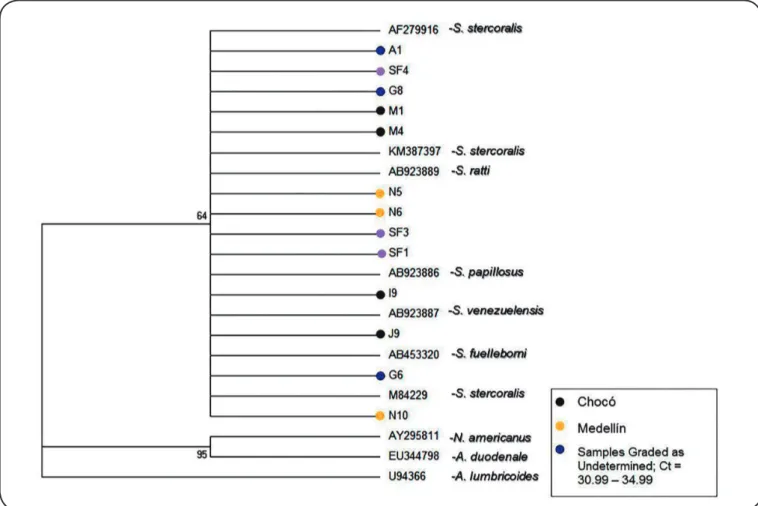
The analysis of the sequences obtained for the rRNA 18S rRNA 101-bp fragment found in sample I9 and the SF3-positive control amplified by the qPCR revealed an identity percentage of 95% and 96%, respectively, compared with the partial sequence of the same marker reported for S. stercoralis in GenBank (access code: M89229; analyzed size: 50bp). Meanwhile, the BLASTn analysis of the sequences obtained for the 18S rRNA 244-bp fragment revealed a percentage of identity with the
FIGURE 2: Dendrogram obtained by consensus partial sequences of the ribosomal RNA 18S gene from this study reported at the National Center for
Biotechnology Information using the maximum likelihood analysis based on the Jukes Cantor model. The value on the branches indicates the bootstrap percentage (1,000 iterations). Ct: Cycle threshold; RNA: ribonucleic acid.
Diagnostic evaluation of the qPCR
In this study, the qPCR standardized and tested for the
diagnosis of S. stercoralis exhibited 75% sensitivity
(20.07-100%), 78.42% specificity (71.22-85.62%), 9.09% positive predictive value (0.0-20.41%), and 99.09% negative predictive value (99.86-100%). Regarding the matches between the results obtained with conventional parasitological methods,
such as direct stool test, modified Ritchie concentration
technique, agar plate culture, and Harada-Mori technique,
with the qPCR, there was low agreement, since the estimated
kappa index was 0.12 (95% CI: -0.020-0.26). No statistically significant differences were observed in the results obtained in the diagnostic assessment by means of contingency tables and
Bayesian analysis (Table 3).
DISCUSSION
In this study, the sensitivity and specificity values obtained by the qPCR were lower than those reported previously, ranging 88.9-100% and 94.8-100%, respectively18,19,21,33. However,
the fact that studies reporting 100% sensitivity for the PCR
have tested this technique only in samples from people with
gastrointestinal symptoms and high parasitic burdens both
for S. stercoralis and other pathogens18,19,33 is noteworthy; this
suggests that results obtained in such studies could neither be extrapolated to the general population nor comparable with data
collected in this study.
Regarding other operative features tested compared to
qPCR, the diagnostic certainty expressed in the obtained
predictive values suggests that the performance of qPCR was higher for identifying people who did not have strongyloidiasis.
However, the test presents problems for the detection of true
positives. Some studies have reported a good correlation
between molecular methods, such as PCR, and conventional
parasitology methods used for the detection of S. stercoralis, primarily the agar plate culture17,33.
The consensus observed between results from the qPCR
and the conventional methods tested in this study was low, coinciding only in the diagnosis of three out of four positive
results identified with traditional parasitology techniques (kappa index = 0.12). This may be explained by the fact that, in this study, the qPCR tests detected a larger number of positive
samples for S. stercoralis than conventional parasitological
diagnostic methods. These findings corroborate the results by
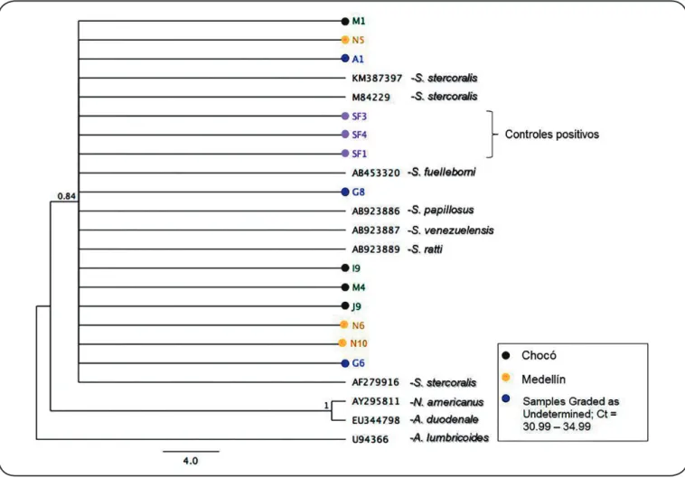
FIGURE 3: Dendrogram obtained by consensus partial sequences of the ribosomal RNA 18S gene from this study reported at the National Center for
Biotechnology Information using the Bayesian Inference based on the JC model. The value on the branches indicates the probability a posteriori.RNA: ribonucleic acid.
that were not previously identified by the reference methods
(agar plate culture, direct stool test, and Ritchie concentration technique)18, suggesting higher efficacy of PCR than that of
conventional diagnostic methods for strongyloidiasis, in people
with low parasitic burden. The higher number of positive samples by the qPCR can be attributed to the fact that viability and low parasitic burden were not indispensable for the specific
detection of this nematode, as opposed to reference diagnostic
tests for this parasitic infection, where the larval viability, amount of sample analyzed, and parasitic burden play an
essential role in classifying a patient as positive or negative12.
However, in this particular study, one of the samples that tested positive by conventional methods was not detected by the qPCR, which also influenced the low concordance observed between the different techniques. This finding was similar to that reported by Schar et al.21, who reported that the number of
samples detected by PCR (38/218) was lower than the amount classified as positive by jointly using the Baermann’s method
and agar plate culture (41/218)21. Likewise, Sharifdini et al.34
reported that their tested PCR failed to detect 13 positive cases
by microscopy, suggesting a potential presence of inhibitors in
fecal samples.
Furthermore, several factors can explain the occurrence of
false-negative results from the qPCR, including preservation,
storage, and the presence of inhibitors in fecal samples35.
Nsubuga et al.36 analyzed ape fecal samples and reported that
the collection and storage temperature of fecal samples played an essential role in DNA recovery36, with lower amounts of genomic DNA isolated from samples collected and stored during warm periods of the year37,38.
Notably, fecal matter is a complex biological sample where the presence of amplification inhibitors might be associated even with an individual’s diet, as demonstrated by Monteiro et al.39.
They characterized the presence of endogenous inhibitors of
PCR and reported that multiple polysaccharides derived from
the diet were potent inhibitors to the amplification39. In studies
that assessed several DNA isolation protocols in fecal samples
depending on the performance and efficiency of the PCR40-42,
treatment of samples using physical methods, such as sonication
or incubation, at temperatures of 50-100°C using enzymes, such as proteinase K, enhanced the efficiency and performance of
DNA isolation from fecal samples are required to improve the
performance and efficiency of the tested qPCR.
In this study, primers and probes used for the amplification
of the S. stercoralis ribosomal RNA 18S gene in the qPCR
revealed high species-specificity, evidenced by the analysis of DNA sequences obtained from positive control amplicons and
sequenced samples. Although the phylogenetic analyses did
not establish a correlation between the analyzed Strongyloides specimens and those obtained in this study, the topologies of the phylogenetic trees suggest a close relationship between parasites from Chocó and Medellín and those reported in GenBank. These results support the high specificity of the qPCR method, even highlighting a correlation between samples classified as indeterminate (Ct value = 30.99-34.99) and Strongyloides identification, when the parasitic burden was low. Hence, we
recommend standardization of a protocol for concentrating the
parasitic forms before the isolation of genomic DNA to enhance the concentration of the isolated DNA and the efficiency and
linearity of the qPCR.
Concurrently with the results obtained in this study, Schar
et al.21 reported that the concentration of parasite DNA in
the sample plays an essential role in precisely classifying an individual as negative or positive for S. stercoralis infection21.
In addition, the infection stage of a person affects the precise classification, which perhaps directly affects the performance of the qPCR by increasing the Ct value if low concentrations of DNA are isolated from the sample. Hence, it is possible
that samples positive for S. stercoralis (detected by the qPCR only) were from patients with a chronic parasitic infection
characterized by an intermittent excretion of larvae, fluctuations in the parasitic burden, and requiring a multi-sample analysis to detect the parasite using traditional tests, thereby demonstrating
the advantage of the qPCR for the detection of low parasitic
burdens. Of note, this is suggested because these results were elicited from people who have lived for over 5 years in the outlying neighborhoods of Medellín (La Cruz and Versalles II), but were native to the Chocó and Urabá zones (Department
of Antioquia).
In such scenarios, some researchers have implemented
Bayesian analyses to estimate the prevalence of S. stercoralis
and assess the operative characteristics of methods for the
detection of this nematode in the absence of a gold standard, suggesting that parameters such as the sensitivity, specificity, and
predictive values of the tests evaluated remain unaffected43,44.
In this study, the Bayesian analysis of these parameters yielded values analogous to those obtained in the contingency table or 2 × 2 table, assuring with this statistical approach that the tested qPCR method can detect 99% of uninfected people.
Finally, based on the analysis of the sequences obtained for the RNA18S ribosomal gene, Pakdee et al.45 reported
that S. stercoralis populations from different geographical
areas (Thailand and Japan) presented genetic divergence45.
In Colombia, genetic differentiation among S. stercoralis populations has not been evaluated at the national level; this is one of the main limitations for evaluating the results obtained
in this study, as the genetic diversity of the parasite and how
this could affect the diagnostic sensitivity of the method were
not considered. Therefore, other studies that can elucidate
the genetic structure of the circulating populations of this parasite are warranted, since they could affect the operational characteristics of the qPCR tested in this study.
In conclusion, this study deduces that although the sensitivity
of the tested qPCR was similar to that of the conventional diagnostic methods for strongyloidiasis, it offered the advantage
of specifically detecting low concentrations of S. stercoralis
DNA in fecal samples, especially in cases of chronic phases of
the infection. These enhanced features favor the establishment of
effective antiparasitic therapies primarily for patients with failed
cellular immune responses, whose risk for the development of fatal
complications, such as hyperinfection syndrome and dissemination of the parasite, is higher. Of note, the linearity of the method is an
essential parameter that should be determined to assess the ability of the test to be used as a quantitative method, as well as to evaluate
whether performing a method for the concentration of parasitic
forms before genomic DNA isolation increases the likelihood of
detecting the parasite in cases of chronic infections owing to low parasitic loads46. Furthermore, this aims to evaluate the behavior
of the test in different epidemiological and clinical contexts that might provide a better insight into the efficacy of this test.
Acknowledgments
We thank the PIRAGUAS Group of the Universidad Pontificia Bolivariana for their support during the field trips to the Department of Chocó and collection of fecal samples, and the Madre Teresa de Calcuta Foundation, led by Father Miguel Perez, and the people of the La Cruz neighborhood for their valuable collaboration at sample collection.
Conflict of interest
The authors declare that there is no conflict of interest.
Financial support
This work was funded by the Centro de Investigación para el Desarrollo y la Innovación (CIDI) at the Universidad Pontificia Bolivariana through the projects with filing numbers: 175-11/11 935A and 252B-08/14-44.
REFERENCES
1. Schar F, Trostdorf U, Giardina F, Khieu V, Muth S, Marti H, et al.
Strongyloides stercoralis: global distribution and risk factors. PLoS
Negl Trop Dis. 2013;7(7):e2288.
2. Buonfrate D, Mena MA, Angheben A, Requena-Mendez A,
Munoz J, Gobbi F, et al. Prevalence of strongyloidiasis in Latin America: a systematic review of the literature. Epidemiol Infect. 2015;143(3):452-60.
3. Botero D, Restrepo M. Human parasitosis. 5th edition. Medellín:
Colombia; 2012. 735p.
4. Becerril MA. Medical parasitology. 3rd edition. Mexico: 2011. 401p.
5. Kozubsky L, Archelli S. Considerations on the biology and
the diagnosis of Strongyloides stercoralis. Acta Bioquim Clin Latinoam. 2004;38(3):333-8.
6. Montes M, Sawhney C, Barros N. Strongyloides stercoralis: there
7. Beknazarova M, Whiley H, Ross K. Strongyloidiasis: a disease
of socioeconomic disadvantage. Int J Environ Res Public Health. 2016;13(5). pii: E-517.
8. McDonald HH, Moore M. Strongyloides stercoralis hyperinfection.
N Engl J Med. 2017;376(24):2376.
9. Mobley CM, Dhala A, Ghobrial RM. Strongyloides stercoralis in solid organ transplantation: early diagnosis gets the worm. Curr
Opin Organ Transplant. 2017;22(4):336-44.
10. Campo PL, Gutiérrez LA, Cardona AJ. Infection by Strongyloides stercoralis: meta-analysis on evaluation of conventional diagnostic
methods (1980-2013). Rev Esp Public Health. 2014;581-600.
11. Mendes T, Minori K, Ueta M, Miguel DC, Allegretti SM. Strongyloidiasis current status with emphasis in diagnosis and drug
research. J Parasitol Res. 2017;2017:ID: 5056314.
12. Siddiqui AA, Berk SL. Diagnosis of Strongyloides stercoralis
infection. Clin Infect Dis. 2001;33(7):1040-7.
13. Agrawal V, Agarwal T, Ghoshal UC. Intestinal strongyloidiasis: a
diagnosis frequently missed in the tropics. Trans R Soc Trop Med Hyg. 2009;103(3):242-6.
14. Ketzis JK. Limitations to the adoption of a standardized
Strongyloides stercoralis diagnostic method: Case study in the
Caribbean. Acta Trop. 2017;170:178-83.
15. Sitta RB, Malta FM, Pinho JR, Chieffi PP, Gryschek RC, Paula
FM. Conventional PCR for molecular diagnosis of human
strongyloidiasis. Parasitology. 2014;141(5):716-21.
16. Basuni M, Muhi J, Othman N, Verweij JJ, Ahmad M, Miswan N, et al. A pentaplex real-time polymerase chain reaction assay for detection of four species of soil-transmitted helminths. Am J Trop Med Hyg. 2011;84(2):338-43.
17. Verweij JJ, Canales M, Polman K, Ziem J, Brienen EA, Polderman AM, et al. Molecular diagnosis of Strongyloides stercoralis in
faecal samples using real-time PCR. Trans R Soc Trop Med Hyg. 2009;103(4):342-6.
18. Moghaddassani H, Mirhendi H, Hosseini M, Rokni M, Mowlavi G,
Kia E. Molecular diagnosis of Strongyloides stercoralis infection
by PCR detection of specific DNA in human stool samples. Iran J Parasitol. 2011;6(2):23-30.
19. Janwan P, Intapan PM, Thanchomnang T, Lulitanond V, Anamnart
W, Maleewong W. Rapid detection of Opisthorchis viverrini
and Strongyloides stercoralis in human fecal samples using a
duplex real-time PCR and melting curve analysis. Parasitol Res. 2011;109(6):1593-601.
20. Sultana Y, Jeoffreys N, Watts MR, Gilbert GL, Lee R. Real-time polymerase chain reaction for detection of Strongyloides stercoralis
in stool. Am J Trop Med Hyg. 2013;88(6):1048-51.
21. Schar F, Odermatt P, Khieu V, Panning M, Duong S, Muth S, et al. Evaluation of real-time PCR for Strongyloides stercoralis and
hookworm as diagnostic tool in asymptomatic schoolchildren in Cambodia. Acta Trop. 2013;126(2):89-92.
22. Wong SS, Fung KS, Chau S, Poon RW, Wong SC, Yuen KY.
Molecular diagnosis in clinical parasitology: When and why? Exp Biol Med (Maywood). 2014;239(11):1443-60.
23. World Health Organization (WHO). Basic laboratory methods in
medical parasitology.Geneva: WHO; 1991. 114p.
24. Cerda L J, Villarroel del P L. Evaluación de la concordancia
inter-observador en investigación pediátrica: Coeficiente de Kappa. Rev Chil Pedriatr. 2008;79(1):54-8.
25. Cortés-Reyes E, Rubio-Romero JA, Gaitán-Duarte H. Métodos estadísticos de evaluación de la concordancia y la reproducibilidad de
pruebas diagnósticas. Rev Colomb Obstet Ginecol. 2010;61(3):247-55.
26. Campo-Polanco LF, Botero LE, Gutiérrez LA, Cardona Arias JA.
Reproducibilidad del examen directo de heces y de la concentración formoléter y validez del examen directo de heces para el diagnóstico de parásitos intestinales. Archivos de Medicina. 2015;11(4):1-9.
27. Campo-Polanco LF, Hernandez-Sarmiento JM, Botero-Palacio LE, Gutiérrez-Builes LA. Estandarización de una reacción en cadena de la polimerasa en tiempo real (qPCR) para la detección de Strongyloides stercoralis en muestras de materia fecal. Med Laboratry. 2016;22.
Available from: http://www.edimeco.com/medicina-laboratorio/2016/ otros-articulos/item/413-volumen-22-07-07-2016.
28. Kearse M, Moir R, Wilson A, Stones-Havas S, Cheung M, Sturrock
S, et al. Geneious Basic: an integrated and extendable desktop
software platform for the organization and analysis of sequence
data. Bioinformatics. 2012;28(12):1647-9.
29. Tamura K, Stecher G, Peterson D, Filipski A, Kumar S. MEGA 6: Molecular Evolutionary Genetics Analysis version 6.0. Mol Biol Evol. 2013;30(12):2725-9.
30. International Business Machines Corporation (IBM Corp). Released
2016. IBM SPSS Statistics for Windows, Version 24.0. Armonk, NY: IBM Corp; 2016.
31. World Health Organization-Pan American Health Organization
(WHO-PAHO). Epidata: epidemiological data analysis software
http://dxsp.sergas.es; 2014.
32. Ministerio de Salud y Protección Social. MinSalud. Encuesta
nacional de parasitismo en población escolar. Fase II. Colombia 2012-2014;p.35.
33. Rayan HZ, Solimán R, Metwally NG. Detection of Strongyloides
stercoralis in fecal samples using conventional parasitological
techniques and real-time PCR: a comparative study. Parasitol
United J. 2012;5:27-34.
34. Sharifdini M, Mirhendi H, Ashrafi K, Hosseini M, Mohebali M,
Khodadadi H, et al. Comparison of nested polymerase chain reaction
and real-time polymerase chain reaction with parasitological methods for detection of Strongyloides stercoralis in human fecal
samples. Am J Trop Med Hyg. 2015;93(6):1285-91.
35. Ramos F, Zurabian R, Moran P, Ramiro M, Gomez A, Clark CG, et al. The effect of formalin fixation on the polymerase chain
reaction characterization of Entamoeba histolytica. Trans R Soc
Trop Med Hyg. 1999;93(3):335-6.
36. Nsubuga AM, Robbins MM, Roeder AD, Morin PA, Boesch C, Vigilant L. Factors affecting the amount of genomic DNA extracted from ape faeces and the identification of an improved sample storage method. Mol Ecol. 2004;13(7):2089-94.
37. Wilke H, Robertson LJ. Preservation of Giardia cysts in stool
samples for subsequent PCR analysis. J Microbiol Methods. 2009;78(3):292-6.
38. Cardona S, Eck A, Cassellas M, Gallart M, Alastrue C, Dore J, et al.
Storage conditions of intestinal microbiota matter in metagenomic analysis. BMC Microbiol. 2012;12:158.
39. Monteiro L, Bonnemaison D, Vekris A, Petry KG, Bonnet J, Vidal R, et al. Complex polysaccharides as PCR inhibitors in feces:
Helicobacter pylori model. J Clin Microbiol. 1997;35(4):995-8.
40. Demeler J, Ramunke S, Wolken S, Ianiello D, Rinaldi L, Gahutu JB, et al. Discrimination of gastrointestinal nematode eggs from crude
fecal egg preparations by inhibitor-resistant conventional and real-time PCR. PLoS One. 2013;8(4):e61285.
41. Adamska M, Leonska-Duniec A, Maciejewska A, Sawczuk M,
methods from cysts of Giardia intestinalis measured by PCR and
TaqMan real time PCR. Parasite. 2010;17(4):299-305.
42. Nunes CM, Lima LG, Manoel CS, Pereira RN, Nakano MM, Garcia JF. Fecal specimens preparation methods for PCR diagnosis of
human taeniosis. Rev Inst Med Trop Sao Paulo. 2006;48(1):45-7.
43. Joseph L, Gyorkos TW, Coupal L. Bayesian estimation of disease
prevalence and the parameters of diagnostic tests in the absence of a gold standard. Am J Epidemiol. 1995;141(3):263-72.
44. Dendukuri N, Joseph L. Bayesian approaches to modeling the
conditional dependence between multiple diagnostic tests.
Biometrics. 2001;57(1):158-67.
45. Pakdee W, Thaenkham U, Dekumyoy P, Sa-Nguankiat S, Maipanich W, Pubampen S. Genetic differentiation of Strongyloides stercoralis
from two different climate zones revealed by 18S ribosomal DNA sequence comparison. Southeast Asian J Trop Med Public Health. 2012;43(6):1333-8.
46. Asher AJ, Waldron LS, Power ML. Evaluation of a PCR protocol for sensitive detection of Giardia intestinalis in human faeces. Parasitol