DM
June | 2017
Nádia Sofia Henriques Nunes
MASTER IN APPLIED BIOCHEMISTRYNitrile based PAMAM dendrimers
functionalized with [RuCp(PPh
3)
2]
+moiety for anticancer applications
MASTER DISSERTATIONNádia Sofia Henriques Nunes
MASTER IN APPLIED BIOCHEMISTRYNitrile based PAMAM dendrimers
functionalized with [RuCp(PPh
3)
2]
+moiety for anticancer applications
MASTER DISSERTATIONSUPERVISOR João Manuel Cunha Rodrigues
Nitrile based PAMAM dendrimers functionalized with [RuCp(PPh3)2]+ moiety for anticancer applications
ACKNOWLEDGMENTS
I would like to express my sincere gratitude to my supervisor, Professor João Rodrigues for all the support, patience, understanding, supervision, willingness, and share of knowledge and the review of this master thesis. I would like to extend my thanks to my co-supervisor Professor Helena Tomás for the encouragement and kindness. The given encouragement, ideas, useful critiques, and constructive suggestions helped me to keep improving my work. In addition, I would like to acknowledge for the opportunity to work in cancer research and nanotechnology, a dream that I had for a while, namely through my valued internship, poster and oral communications.
I am particularly grateful for all the precious assistance, share of knowledge, motivation and friendship of my lab colleagues of Molecular Materials Research Group (MMRG), Dr.ª Cláudia Camacho, Dr.ª Dina Maciel, Dr.ª Nilsa Oliveira, Dr.ª Carla Alves, Dr.ª Mara Gonçalves and Dr.ª Rita Castro.
To my lab colleague and friend Dr.ª Ana Olival for all the appreciated support and share of ideas in the NMR characterization which helped me to improve the quality of my work.
To the laboratory technicians from Chemistry Department, Paula Andrade and Paula Vieira for all the kindness and readiness in providing some lab materials and reagents that I needed for this project.
I would also like to acknowledge the Madeira Chemistry Research Centre (CQM), Chemistry Department and University of Madeira (UMa) for disclosing the facilities to carry out this work.
I would like to extend my thanks to the Fundação para a Ciência e a Tecnologia (FCT) because this work would not have been possible without the partial support of this agency through the CQM Strategic Project PEst-OE/QUI/UI0674/2015-2016, and the NMR and MS Portuguese Networks (PTNMR-2015-2016, RNEM-2015-2016). The partial support of ARDITI – Agência Regional para o Desenvolvimento da Investigação Tecnologia e Inovação – through the project M1420-01-0145-FEDER-000005 – Centro de Química da Madeira – CQM+ (Madeira 14-20), was highly appreciated.
To my dearest friends, Ana Morna, Sofia Ferreira, Carolina Cardoso, André Delgado, Liliana Rodrigues, Jackie Ferreira, Daniel Huang, João Leça and Jorge Gonçalves for all the relentless and caring support, companionship, understanding, enthusiasm and cherished
Nitrile based PAMAM dendrimers functionalized with [RuCp(PPh3)2]+ moiety for anticancer applications
moments that helped me to overcome all the encountered obstacles and to celebrate the achievements in the progress of this work and life.
I am everlastingly thankful for all the love, care and believe that my grandparents, Maria Isabel Henriques and Martinho Henriques, and my aunt, Fátima Célia Henriques, and cousin, Cassandra Gomes, deposited on me through all my life and work.
Finally, but not less important at all, I would like to express my sincere gratitude to my mum, Zita Henriques, for all the endless love, comprehension, inspiration, care and for always believing in my potential, not only professionally but also emotionally, which helped me to always move forward. I find it hard to express by words all my sincere appreciation and love. Thank you!!
Nitrile based PAMAM dendrimers functionalized with [RuCp(PPh3)2]+ moiety for anticancer applications
L
IST OF PUBLICATIONS
Part of the results and findings of this work were presented in the following:
Oral communications:
Maciel D., Camacho C., Nunes N., Muñoz-Fernández Mª.A., Tomás H., Rodrigues J.; Metallodendrimers as anticancer and antiviral drug candidates; The 10th International Dendrimer Symposium (IDS10), August 5-9, Weihai, China, 2017 (invited talk).
Nunes N., Rodrigues J., Tomás H.; Generation 0 and 1 of new PAMAM dendrimers with different terminal groups. Synthesis and characterization; 3rd CQM Annual Meeting, April 01-02, Funchal, Madeira Island-Portugal, 2016.
Nunes N., Camacho C., Rodrigues J.; Synthesis and functionalization of PAMAM dendrimers with different surface groups; 2nd CQM Annual Meeting, January 30-31,
Funchal, Madeira Island-Portugal, 2015.
Poster communications:
Nunes N., Rodrigues J., Tomás H.; PAMAM dendrimers as a platform for the preparation of low-generation of ruthenium metallodendrimers; MAD-Nano16: Madeira International Conference on Emerging Trends and Future of Nanomaterials for Human Health, November 17-20, Funchal, Madeira Island-Portugal, 2016.
Olival A., Nunes N., Rodrigues J.; Low generation dendrimers with different surface groups. Characterization by nuclear magnetic resonance; 1st Joint French-Portuguese NMR congress, April 19-23, Lisbon, 2016.
Nunes N., Camacho C., Rodrigues J.; Synthesis and functionalization of PAMAM dendrimers with different surface groups; INCB: Indo-Portuguese Workshop on Emerging Trends of Nanotechnology in Chemistry and Biology, February 12-13, New Delhi, India, 2016.
Nitrile based PAMAM dendrimers functionalized with [RuCp(PPh3)2]+ moiety for anticancer applications
RESUMO
Os dendrímeros são moléculas hiperramificadas que se apresentam com diferentes grupos terminais na sua estrutura e que, por exemplo, podem ser conjugados com complexos de metais de transição. Esta estratégia conduz à preparação de metalodendrímeros com propriedades de interesse e diversas aplicações, incluindo as terapêuticas. Os complexos de Ruténio (II) são potenciais fármacos anticancerígenos que poderão vir a substituir os fármacos derivados da
cisplatina por serem dotados de uma variedade de propriedades mais vantajosas, como por
exemplo: uma cinética de permuta dos ligandos biocompatível, múltiplos estados de oxidação acessíveis, a capacidade de interagir com o DNA e/ou com proteínas e baixa toxicidade.
O objetivo principal desta dissertação de mestrado consistiu na preparação e caracterização de novos tipos de metalodendrímeros tendo como base os dendrímeros PAMAM: G0/G1-CN, G0/G1-(CNRu(η5-C5H5)(PPh3)2)𝑥(CF3SO3)𝑥, G0/G1-CO2tBu e G0/G1-OH. Os polinitrilos –
G0-(CN)4 e G1-(CN)8 – sintetizados a partir de G0/G1-PAMAM foram utilizados na preparação
de nitrilo-metalodendrímeros de ruténio G0-(CNRu(η5-C5H5)(PPh3)2)4(CF3SO3)4 e
G1-(CNRu(η5-C5H5)(PPh3)2)8(CF3SO3)8, respetivamente. A estratégia de síntese aplicada foi
adaptada a partir da metodologia previamente desenvolvida pelo Grupo de Materiais Moleculares do CQM e as técnicas de caracterização utilizadas para cada um dos dendrímeros e metalodendrímeros foram as técnicas de RMN (1H, 13C, 31P, HSQC) e de IV (com transformada de Fourier). G0/G1-PAMAM foram caracterizados adicionalmente por espetroscopia de massa. Todos os compostos foram sintetizados com sucesso, com bons rendimentos (77% – 94%), e os resultados obtidos confirmaram a sua estrutura. Os hidroxilo-dendrímeros – G0-(OH)8 and G1-(OH)16 – foram sintetizados a partir dos ésteres G0-(CO2tBu)8
e G1-(CO2tBu)16, respetivamente, segundo a adaptação da metodologia de N. Jayaraman et al.
(1, 2). A sua caracterização foi realizada por espetroscopia de RMN (1H, 13C, COSY e HSQC)
e de IV (com transformada de Fourier). Apenas os dendrímeros G0-(OH)8 e G0-(CO2tBu)8 foram
caracterizados por espetroscopia de massa. Os resultados adquiridos validaram a estrutura de todos os dendrímeros sintetizados que foram obtidos com rendimentos elevados (83% – 93%). Uma das principais linhas de trabalho futuro será a análise da atividade anticancerígena destes novos nitrilo-metalodendrímeros e a comparação destes resultados com a cisplatina.
Nitrile based PAMAM dendrimers functionalized with [RuCp(PPh3)2]+ moiety for anticancer applications
ABSTRACT
Dendrimers are hyperbranched molecules having different terminal groups that can be conjugated with, for example, transition-metal complexes leading to new and interesting compounds with a variety of interesting therapeutic applications. In the last decade, Ruthenium (II) complexes revealed to be potential alternatives to the clinically used antitumor Platinum-based drugs due to several characteristics, e.g. biocompatible ligand exchange rates, redox-accessible oxidation states, covalent binding with DNA and/or proteins and low toxicity.
The main goal of this master thesis was to prepare and characterize new PAMAM dendrimers based: G0/G1-CN, G0/G1-(CNRu(η5-C5H5)(PPh3)2)𝑥(CF3SO3)𝑥, G0/G1-CO2tBu
and G0/G1-OH. The polynitrile dendrimers – G0-(CN)4 and G1-(CN)8 – synthesized from the
G0/G1-PAMAM, were used in the preparation of the nitrile ruthenium-based metallodendrimers G0-(CNRu(η5-C
5H5)(PPh3)2)4(CF3SO3)4 and G1-(CNRu(η5-C5H5)(PPh3)2)8(CF3SO3)8,
respectively. The applied synthetic strategy was adapted from the reported methodology previously developed by the Molecular Materials Research Group of CQM (Madeira Chemistry Research Centre) and the structural characterization techniques used for each dendrimer/metallodendrimer were the NMR (1H, 13C, 31P, HSQC) and FTIR. G0/G1-PAMAM were also characterized by MS. All the compounds were successfully synthesized, with good yields (77% – 94%), and the characterization data have confirmed their adequate structure.
The hydroxyl moieties – G0-(OH)8 and G1-(OH)16 – were synthesized from the ester
compounds – G0-(CO2tBu)8 and G1-(CO2tBu)16 – respectively, through the adaptation of the
reported methodology of N. Jayaraman et al (1, 2).These compounds were characterized by NMR (1H, 13C, COSY and HSQC) and FTIR spectroscopy and only the G0-(OH)
8 and
G0-(CO2tBu)8 were characterized by MS. The obtained results have showed that all the dendrimers
were properly synthesized with very good yields (83% – 93%). The main future goal is to analyse the anticancer activity of these new metallodendrimers G0/G1-(CNRu(η5
-C5H5)(PPh3)2)𝑥(CF3SO3)𝑥 and to compare it with cisplatin.
Nitrile based PAMAM dendrimers functionalized with [RuCp(PPh3)2]+ moiety for anticancer applications
Table of Contents
PART I – INTRODUTION……...……….………...1
1. METAL-BASED ANTICANCER DRUGS ... 3
1.1. RUTHENIUM COMPLEXES ... 7
1.1.1. CYC LOPENTADIENYL RU (II) C OMPLEXES ... 10
2. METALLODENDRIMERS AS ANTICANCER DRUGS ... 12
2.1. DENDRIMERS IN DRUG DE LIVERY APPLICATIONS ... 12
2.1.1. PAMAM DENDRIM ER S ... 15
2.2. METALLODENDRIMERS ... 16
2.2.1. POLY NITRILES C OMPOUN DS AS CORE LIGANDS FOR THE PR EPARATION O F M ETALLODENDR IM ERS ... 17
2.2.2. RUTHENIUM METALLODEND R IMER S ... 18
3. SCOPE AND OBJECTIVES ... 20
PART II – DEVELOPED METHODOLOGY…….……….………...23
4. ORGANOMETALIC COMPOUNDS ... 25
4.1. MATERIALS AND METHODS ... 25
4.1.1. REAGENTS AND SOLVENTS ... 25
4.1.2. SAM PLES PR EPAR ATION ... 26
4.1.3. SYNTHESIS OF POLY-NITRILE DENDR IM ER S G0/G1-CN ... 26
4.1.4. SYNTHESIS OF STAR TING ORGANOMETALLIC COM POUND [RUCP(PPH3)2CL] ... 27
4.1.5. SYNTHESIS OF METALLODENDR IMERS G0/G1-(CNRUCP(PPH3)2)X(CF3SO3)X ... 28
4.1.6. CHAR AC TER IZATION ... 29
4.2. RESULTS AND DISCUSSION ... 30
5. ORGANIC COMPOUNDS ... 71
5.1. MATERIALS AND METHODS ... 71
5.1.1. REAGENTS AND SOLVENTS ... 71
5.1.2. SAM PLES PR EPAR ATION ... 71
5.1.3. SYNTHESIS OF POLY-ESTER DENDRIM ER S G0/G1-CO2TBU ... 71
5.1.4. SYNTHESIS OF POLY-HYDROXYL DENDR IM ERS G0/G1-OH ... 72
5.1.5. CHAR AC TER IZATION ... 73
5.2. RESULTS AND DISCUSSION ... 74
PART III – CONCLUSIONS AND PERSPECTIVES…….……….…………...91
6. CONCLUSIONS ... 93
Nitrile based PAMAM dendrimers functionalized with [RuCp(PPh3)2]+ moiety for anticancer applications
8. REFERENCES ... 96
9. ATTACHMENT ... 118
9.1. CHARACTERIZATION SPEC TRA ... 118
9.1.1. G0/G1-PAMAM ... 118 9.1.2. G0/G1-CN... 119 9.1.3. G0/G1--(CNRUCP(PPH3)2)X(CF3SO3)X... 121 9.1.4. G0/G1-CO2TBU ... 121 9.1.5. G0/G1-OH ... 125
List of Tables
Table 1 - 1H and 13C-NMR data, of the corresponding spectra represen ted in fig. 18a) and b) for G0-PAMAM.CDCl3 was the NMR solvent. The 1H chemical shifts values are averages. 33 Table 2 - Main characteristic bands (cm- 1), obtained by FTIR, for each functional group of
G0/G1-PAMAM. The type of vibration is also showed. 35 Table 3 - 1H and 13C-NMR data, of the corresponding spectra represented in fig. 22a) and b) for
G1-PAMAM. CDCl3 was the NMR solvent. The 1H chemical shifts values are averages. 39 Table 4 - Conditions for the solubility tests performed with G0 -PAMAM in several aprotic
solvents. 41
Table 5 - Conditions applied for each performed test for the synthesis reaction of G0 -(CN)4. 41 Table 6 - 1H and 1 3C-NMR data, of the corresponding spectra represented in fig. 25a) and c), for
the purified G0-(CN)4. CDCl3 was the NMR solvent. The 1H chemical shifts values are
averages. 44
Table 7 - Main characteristic bands (cm- 1), obtained by FTIR, for each functional group of the purified G0/G1-CN. The type of vibration is also showed. 46 Table 8 Conditions applied for each performed test for the synthesis reaction of G0
-(CNRuCp(PPh3)2)4(CF3SO3)4. 51
Table 9 - 1H, 31P and 13C-NMR data, of the corresponding spectrums represented in figs. 35d), 36d) and 37 for the purified G0 -(CNRuCp(PPh3)2)4(CF3SO3)4. CDCl3 was the NMR solvent. The 1H and 31P chemical shifts values are averages. 61 Table 10 - Main characteristic bands (cm-1), obtained by FTIR, for each functional group of the
Nitrile based PAMAM dendrimers functionalized with [RuCp(PPh3)2]+ moiety for anticancer applications
Table 11 - 1H and 13C-NMR data, of the corresponding spectra represented in figs. 39b) and c), for purified G1-(CN)8. CDCl3 was the NMR solvent. The 1H chemical shifts values are
averages. 64
Table 12 - 1H, 31P, and 13C-NMR data, of the corresponding spectra represented in figs. 42c), 43c) and 44 for the purified G1-(CNRuCp(PPh3)2)8(CF3SO3)8. CDCl3 was the NMR solvent. The 1H chemical shifts values are averages. 69 Table 13 - 1H and 13C-NMR data, of the corresponding spectra represented in figs. 46c) and 47
for the purified G0-(CO2tBu)8, and in figs. 8Ab) and c) (in section 7.1.4. in attachment) for the purified G1-(CO2tBu)16. CDCl3 was the NMR solvent. The 1H chemical shifts
values are averages. 79
Table 14 - Main characteristic bands (cm-1), obtained by FTIR, for each functional group of purified G0/G1-CO2tBu. The type of vibration is also showed. 81 Table 15 - 1H and 13C-NMR data, of the corresponding spectra represented in figs. 52 b) and c)
for the pure fraction of G0(OH)8, and in figs. 12A b) and c) (section 7.1.5.) for G1 -(OH)16. D2O was the NMR solvent. The 1H chemical shifts values are averages. 83 Table 16 - Main characteristic bands (cm-1), obtained by FTIR, for each functional group of the
pure fraction of G0/G1 -OH. The type of vibration is also showed. 86
List of Figures
Figure 1 – Estimated world age-standardized cancer incidence and mortality rates (ASR), from the year of 2012, per 100 000, in men and women (7). 3 Figure 2 – Chemical structure (with optimised geometries) of three recognised platinum
anticancer drugs: a) cisplatin; b) carboplatin and c) oxaliplatin (10). 4 Figure 3 - Examples of metal-based compounds that target DNA: a) Cytotoxic Ru (II) arene
complex (36); b) Intercalation of a Ru (II) complex into DNA (34); c) [γ -Ru(azpy)2Cl2] (31); d) A cytotoxic Os (II) arene complex (30) (adapted from ref. (4)). 6 Figure 4 - Examples of metal-based anticancer drugs that target proteins and enzymes: a)
Gold(III) meso-tetraarylporphyrins complex (43); b) tris(8-quinolinolate)gallium(III) (KP46) (40); c) DW1 – a ruthenium staurosporin bioconjugate (41); d) Hexacarbonyl dicobalt complex containing a nucleoside ligand (39) (adapted from ref. (4)). 6 Figure 5 – Examples of metal-based anticancer prodrugs: a) Rh (III) complex photoactivatable
(48); b) Ferrocifen (49); c) Cobalt–marimastat bioconjugate (47); d) Ru (II) arene complex with iodo and a phenylplazopyridine ligand (46) (adapted from ref. (4)). 7
Nitrile based PAMAM dendrimers functionalized with [RuCp(PPh3)2]+ moiety for anticancer applications
Figure 6 – Genealogy of antitumour ruthenium complexes. The represented references refer to the first published studies that have suggested their therapeutic activity in an in vivo
tumor model (75). 9
Figure 7 – Chemical structures of three ruthenium drug candidates: a) RM175 ([(η6 -C6H5C6H5)RuCl (H2NCH2CH2NH2-N,N′)]PF6); b) RAPTA-T (Ru(η6-C6H5Me)(PTA)Cl2) and c) RDC11 ([Ru(phenanthroline)(κ-C,N-(2-phenyl-pyridine)(NCMe)2]PF6 (adapted
from ref. (51)). 10
Figure 8 – Molecular structures of two ruthenium complexes with the general formula [(η5 -C5H5)Ru(P-P)L]+ (P-P: 1,2-bis(diphenylphosphine)ethane (dppe) or triphenylphosphine (PPh3); L: monodentate or bidentate nitrogen donors): a) TM34 ([(η5-C5H5)Ru(2,2’- bipy)(PPh3)][CF3SO3]) and b) a Ru (II) cyclopentadienyl complex with a carbohydrate -derived ligand (adapted from ref. (112)). 11 Figure 9 - Evolution of polymers towards dendritic structures ( 130). 13 Figure 10 - Schematic representation of the general structure of a dendrimer. G0, G1, G2, G3,
and G4 are the generation number (adapted from ref. (130)). 13 Figure 11 - The two most commonly used methods for dendrimers synthesis: divergent and
convergent strategies (130). 14
Figure 12 – Schematic representation of the EPR effect in drug delivery (adapted from ref.
(145)). 15
Figure 13 – Schematic representation of the metallodendrimers with the metalli c moieties in several positions: a) on the periphery; b) scattered throughout the framework and c) encapsulated between the structure of the dendrimer (111). 16 Figure 14 – Molecular structures of three different metallodendrimers: a) multinuclear copper
-functionalized, b) a multinuclear Pt (II) and Pd (II) G2-polyamide, and c) G1-dinuclear
gold(I) (adapted from ref. (111)). 18
Figure 15 - Tetra- and octanuclear arene ruthenium complexes coordinated to dendritic
polypyridyl scaffolds (134). 19
Figure 16 - Chelating N,O- and N,N-ruthenium(II) arene metallodendrimers with poly(propyleneimine) dendrimer as scaffolds. They are coordinated with 4, 8, 16 or 32
n groups (adapted from ref. (181)). 19
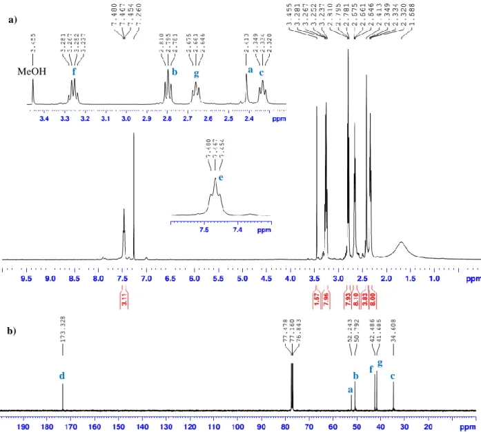
Figure 17 – Cationic, chelating N,O-ruthenium(II)-arene-PTA salicylaldimine metallodendrimers (adapted from ref. ( 181)). 20 Figure 18 - a) 1H-NMR and b) 13C-NMR spectra of G0-PAMAM, in CDCl3. Each signal is marked
with the different type of carbons and protons that are represented with a unique letter
Nitrile based PAMAM dendrimers functionalized with [RuCp(PPh3)2]+ moiety for anticancer applications
Figure 19 - COSY spectrum of G0-PAMAM, in D2O. Each signal is marked with the respective group of protons that are neighbours and linked to each other by a carbon bond. Each type of protons is represented with a unique letter (see scheme 1). 34 Figure 20 - HSQC spectrum of G0-PAMAM, in D2O. Each signal is marked with the respective
type of protons and type of carbon they are directly linked with. Each type is represented
with a unique letter (see scheme 1). 34
Figure 21 - +MS2 spectrum of G0-PAMAM – fragmentation of the molecular ion (517.7 m/z). The molecular structure of the base peak (403.2 m/z) is also represented. 35 Figure 22 - a) 1H-NMR and b) 13C-NMR spectra of G1-PAMAM, in CDCl3. Each signal is marked
with the different type of carbons and protons that are represented with a un ique letter
– see scheme 2). 38
Figure 23 - COSY spectrum of G1-PAMAM, in D2O. Each signal is marked with the respective group of protons that are neighbours and linked to each other by a carbon bond. Each type of protons is represented with a unique letter (see scheme 2). 38 Figure 24 - HSQC spectrum of G1-PAMAM, in D2O. Each signal is marked with the respective
type of protons and type of carbon they are directly linked with. Each type is represented
with a unique letter (see scheme 2). 39
Figure 25 - NMR spectra of purified G0 -(CN)4: a) 1H-NMR in CDCl3; b) 1H-NMR in D2O; c) 13C-NMR in CDCl3. The “e” and “h” signals represents the amide and amine protons, respectively, that are not detected in b) – orange arrows. The red stars represent the
absence of MeOH. 43
Figure 26 - COSY spectrum of purified G0 -(CN)4, in CDCl3. Each signal is marked with the respective group of protons that are neighbours and linked to each other by a carbon bond. Each type of protons is represented with a unique letter (see scheme 1). 45 Figure 27 - HSQC spectrum of purified G0 -(CN)4, in CDCl3. Each signal is marked with the
respective type of protons and type of carbon that are directly l inked with. Each type is represented with a unique letter (see scheme 1). 45
Figure 28 - Molecular structure of G0-(CN)8. 46
Figure 29 - 1H-NMR spectrum of commercial dicyclopentadiene, in CDCl3. Each signal is marked with the respective type of protons that are represented with a unique letter (see scheme
5). 47
Figure 30 - 1H-NMR spectrum of distilled cyclopentadiene, in CDCl3. Each signal is marked with the respective type of protons that are represented with a unique letter (see scheme 5). 48 Figure 31 - 1H-NMR spectrums of the obtained [RuCp(PPh3)2Cl]: a) orange crude crystals; b)
Nitrile based PAMAM dendrimers functionalized with [RuCp(PPh3)2]+ moiety for anticancer applications
marked with the respective type of protons that are represented with a unique letter (see
scheme 4). 49
Figure 32 - 31P-NMR spectrums of the corresponding samples from fig. 14; in CDCl3. The “p” signal corresponds to the phosphorus from the [RuCp(PPh3)2Cl] (see scheme 4). 50 Figure 33 - 1H-NMR spectra of the obtained products from the synthesis reaction of
G0-(CNRuCp(PPh3)2)4(CF3SO3)4: a) the crude fraction after 22h of reaction and b) the
brown powder after filtration; in CDCl3. Each signal is marked with the respective type of protons that are represented with a unique letter (see scheme 1 an d 3). The orange arrows points to the impurities/side -products that are needed to be removed. 54 Figure 34 - 31P-NMR spectrums of the corresponding samples from fig. 18; in CDCl3. The “m”
signal corresponds to the phosphorus from the G0 -(CNRuCp(PPh3)2)4(CF3SO3)4
metallodendrimer (see scheme 1). The orange arrows points to the impurities/side -products that are needed to be removed. 55 Figure 35 - 1HNMR spectra of the obtained products from the synthesis reaction of G0
-(CNRuCp(PPh3)2)4(CF3SO3)4: a) after extracted by DCM and washed by Et2O; b) after
dissolved in DCM and precipitated with Et2O; c) after the second precipitation with Et2O; d) after the third precipitation; in CDCl3. 57 Figure 36 - 3 1P-NMR spectrums of the corresponding samples from fig. 18; in CDCl3. 59 Figure 37 - 13C-NMR spectrum of purified G0-(CNRuCp(PPh3)2)4(CF
3SO3)4, in CDCl3. Each
signal is marked with the respective type of carbons that are represented with a unique
letter (see scheme 1). 60
Figure 38 - HSQC spectrum of purified G0 -(CNRuCp(PPh3)2)4(CF3SO3)4, in CDCl3. Each signal
is marked with the respective type of protons and type of carbon they are directly linked with. Each type is represented with a unique letter (see scheme 1). 60 Figure 39 - NMR spectra of G1-(CN)8: a) 1H-NMR of the crude and b) the puri fied product; c)
13C-NMR after purified; in CDCl3. The red star represents the absence of MeOH. 63 Figure 40 - COSY spectrum of purified G1 -(CN)8, in CDCl3. Each signal is marked with the
respective group of protons that are neighbours and linked to each o ther by a carbon bond. Each type of protons is represented with a unique letter (see scheme 2). 64 Figure 41 - HSQC spectrum of purified G1 -(CN)8, in CDCl3. Each signal is marked with the
respective type of protons and type of carbon they are directly link ed with. Each type is represented with a unique letter (see scheme 2). 65 Figure 42 - 1H-NMR spectra of the a) crude, b) extracted by DCM and washed by Et2O and c)
purified G1-(CNRuCp(PPh3)2)8(CF3SO3)8, in CDCl3. Each signal is marked with the
respective type of protons that are represented with a unique letter (see scheme 2 and 3). The orange arrow represents the impurity/sub -product that was removed after the
Nitrile based PAMAM dendrimers functionalized with [RuCp(PPh3)2]+ moiety for anticancer applications
Figure 43 - 3 1P-NMR spectra of the corresponding samples from fig. 25; in CDCl3. 68 Figure 44 - 13C-NMR spectrum of G1-(CNRuCp)8, in CDCl3. Each signal is marked with the
respective type of carbons that are represented wi th a unique letter (see scheme 2). 70 Figure 45 - HSQC spectrum of purified G1 -(CNRuCp)8, in CDCl3. Each signal is marked with
the respective type of protons and type of carbon they are directly linked with. Each type is represented with a unique letter (see scheme 2). 70 Figure 46 - 1H-NMR spectrum of G0-(CO
2tBu)8: a) crude, b) purified by the L -L extraction and c) washed by dH2O and lyophilized, in CDCl3. Each signal is marked with the respective type of protons that are represented with a unique letter (see scheme 6). The red box point to the vestigial amounts of unreacted tert -butyl acrylate and the red stars represents the impurities of CDCl3 (1H-NMR in fig. 7A in attachment). 77 Figure 47- 13C-NMR spectrum of purified G0-(CO2tBu)8, in CDCl3. Each signal is marked with
the respective type of carbons that are represented with a unique letter (see scheme 6). 78 Figure 48 - +MS (total fragmentation) spectrum of purified G0 -(CO2tBu)8. The molecular ion
(M+·) is represented in blue in the graphic area. The structure of the base peak (643.4
m/z) is also shown. 79
Figure 49 - COSY spectrum of purified G0 -(CO2tBu)8, in D2O. Each signal is marked with the respective group of protons that are neighbours and linked to each other by a carbon bond. Each type of protons is represented with a unique letter (see scheme 6). 80 Figure 50 - HSQC spectrum of purified G0 -(CO2tBu)8, in D2O. Each signal is marked with the
respective type of protons and type of carbon they are directly linked with. Each type is represented with a unique letter (see scheme 6). 80 Figure 51 - Synthesis reaction of G0 -(OH)8: a) Grey suspension of the reaction mixture before
neutralization of LiAlH4; b) Settled white suspension of neutralized LiAlH4. 82 Figure 52 - NMR characterization of G0 -(OH)8: 1H-NMR spectra of the a) impure fraction and
b) pure fraction, and c) 13C-NMR spectrum of the pure fraction, in D2O. Each signal is marked with the respective t ype of protons that are linked to the corresponding carbon – they are represented with a unique letter (see scheme 6). The red stars represent the
impurities of the impure fraction. 84
Figure 53 - COSY spectrum of the pure fraction of G0 -(OH)8, in D2O. Each signal is marked with the respective group of protons that are neighbours and linked to each other by a carbon bond. Each type of protons is represented with a unique letter (see scheme 6). 85 Figure 54 - HSQC spectrum of the pure fraction of G0 -(OH)8, in D2O. Each signal is marked
with the respective type of protons and type of carbon they are directly linked with. Each type is represented with a unique letter (see scheme 6). 85
Nitrile based PAMAM dendrimers functionalized with [RuCp(PPh3)2]+ moiety for anticancer applications
Figure 55 - +MS (total fragmentation) spectrum of the pure fraction of G0 -(OH)8. The molecular ion (M+·) is represented in blue in the graphic area. The structure of the base peak (751.5
m/z) is also shown. 86
List of Schemes
Scheme 1 - Synthetic routes for the synthesis of t he metallodendrimer G0
-(CNRuCp(PPh3)2)4(CF3SO3)4. 32
Scheme 2 - Synthetic routes for the synthesis of the metallodendrimer G1
-(CNRuCp(PPh3)2)8(CF3SO3)8. 37
Scheme 3 Mechanism of reaction of the aza Michael addition for the preparation of the G0/G1
-CN dendrimers (adapted from (202)). 41
Scheme 4 - Cracking of dicyclopentadiene. 47
Scheme 5 - Synthesis reaction of [RuCp(PPh3)2Cl]. 47 Scheme 6 - Synthetic routes for the preparation of G0 -(OH)8 dendrimer. 76 Scheme 7 - Mechanism of reaction of the reduction of the ester groups of G0/G1-CO2tBu with
LiAlH4 to give the G0/G1-OH dendrimers (adapted from ref. (220)). 81 Scheme 8 - Synthetic routes for the preparation of G1 -(OH)16 dendrimer. 88
List of Acronyms
ACN – Acrylonitrile
AgCF3SO3 – Silver trifluoromethanesulfonate AgCl – Silver chloride
ASR – Age-standardized cancer incidence and mortality rates CAS No. – Chemical Abstracts Service number
CDCl3 – Deuterated chloroform CHCl3 – Chloroform
COSY – Correlation spectroscopy Cp – Cyclopentadiene
Nitrile based PAMAM dendrimers functionalized with [RuCp(PPh3)2]+ moiety for anticancer applications
DMSO – Dimethyl sulfoxide D2O – Deuterated water EtOH – Ethanol Et2O – Diethyl ether
FDA – Food and Drug Administration
FTIR – Fourier Transform Infrared Spectroscopy G0 – Generation 0
G1 – Generation 1
G0/G1-CN – Nitrile dendrimers from generations 0 and 1
G0/G1-CNRuCp(PPh3)2)𝑥(CF3SO3)𝑥 – Nitrile Ruthenium metallodendrimers with cyclopentadienyl and
diphenylphosphine ligands from generation 0 and 1
G0/G1-CO2tBu – tert-Butyl ester dendrimers from generations 0 and 1
G0/G1-OH – Hydroxyl dendrimers from generation 0 and 1 HSQC – Heteronuclear single-quantum correlation
ICR – Imidazole-containing complex IR – Infrared
KBr – Potassium bromide
KP46 – tris(8-quinolinolate)gallium(III)
LC-ESI-MS/MS – Liquid Chromatography-Electrospray Ionization-Mass Spectrometry LiAlH4 – Lithium Aluminium hydride
L-L – Liquid-liquid MeOH – Methanol MS – Mass Spectrometry Me4Si or TMS – Tetramethylsilane m/z – Mass-to-charge ratio m – Multiplet
NaCl – Sodium chloride NaH – Sodium hydride NaOH – Sodium hydroxide
Nitrile based PAMAM dendrimers functionalized with [RuCp(PPh3)2]+ moiety for anticancer applications
NMR – Nuclear Magnetic Resonance N2 – Nitrogen
n.d. – not defined
PAMAM – Polyamidoamine PPh3 – Triphenylphosphine ppm – Parts per million q – Quartet
RDC – Ruthenium derived compounds
RuCl3.𝑥H2O – Ruthenium (III) chloride hydrate
[RuCp(η5-C5H5)] – cyclopentadienyl ruthenium (II) complex with a piano stool structure s – Singlet
t – Triplet
THF – Tetrahydrofuran
TlPF6 – Thalium (I) hexafluorophosphate 1D – One-dimensional
1H-NMR – Proton Nuclear Magnetic Resonance 13C-NMR – Carbon-13 Nuclear Magnetic Resonance 2D – Two-dimensional
31P-NMR – Phosphorous-31 Nuclear Magnetic Resonance +MS – Total fragmentation
+MS2 – Second fragmentation +MS3 – Third fragmentation
Nitrile based PAMAM dendrimers functionalized with [RuCp(PPh3)2]+ moiety for anticancer applications
Nitrile based PAMAM dendrimers functionalized with [RuCp(PPh3)2]+ moiety for anticancer applications
1.
METAL-BASED ANTICANCER DRUGS
Metals, specifically transition metals, have a list of properties that offer several advantages in comparison with the more common organic-based drugs, which lead to the design of numerous therapeutic agents: a) a wide range of coordination numbers and geometries, b) “tune-ability” of the thermodynamics and kinetics of ligand substitution, c) accessible redox states and d) a wide structural diversity. These properties of metals or more appropriately of the metal complexes can be exploited for oncology treatment (3-5).
Cancer is a leading cause of death worldwide and was responsible for 8.8 million deaths in 2015. Nearly 1 in 6 deaths is due to cancer and the three deathliest types of cancer are: lung (1.69 million deaths), liver (788 000 deaths) and colorectal (774 000 deaths) (6). In a study from the World Health Organization published in 2014 (7), breast cancer was the most predominant among women while among men, the lung and prostate cancer had the highest incidences (fig. 1).
Figure 1 – Estimated world age-standardized cancer incidence and mortality rates (ASR), from the year of 2012, per 100 000, in men and women (7).
Nitrile based PAMAM dendrimers functionalized with [RuCp(PPh3)2]+ moiety for anticancer applications
Due to the progresses that were made in cancer therapy and diagnosis in the last 27 years, 23% of the cancer death rate has dropped since 1991 but, despite this progress, for the pancreatic, liver and uterine corpus types of cancer, death rates are increasing (8, 9). In the next two decades, about 20 million cancer cases are expected to occur, which leads to the quest for new and improved anticancer agents (10).
Medicinal inorganic chemistry is a prosperous area for cancer research and, just about 50-40 years ago, its potential for designing new anticancer agents has only been elucidated and explored after a) the pioneering work of Köpf H. and Köpf-Maier P. (11) (in the late 1970´s) that have tested transition metal cyclopentadienyl complexes for antitumor activity, and b) the discovery of the biological activity of cisplatin (cis-diamminedichloroplatinum (II), fig. 2) (12, 13).
This metallic coordination compound was firstly synthesized by Peyrone M., in 1844, and Werner A., in 1893, have discovered its chemical structure (14). Nevertheless, just in the mid 1960´s, the findings of Rosenberg B. et al. (15) initiated several preclinical and clinical studies that have revealed its cytotoxic effects and its safety and therapeutic profile. Thus, in 1978,
Figure 2 – Chemical structure (with optimised geometries) of three recognised platinum antic ancer drugs: a) cisplatin; b) carboplatin and c) oxaliplatin (10).
Nitrile based PAMAM dendrimers functionalized with [RuCp(PPh3)2]+ moiety for anticancer applications
cisplatin was approved by FDA (US Food and Drug Administration) to treat patients with testicular and bladder cancer types. Since then, the treatment was extended to head, neck, lung, colorectal and ovarian cancers (16, 17). In the early 1980´s, two additional platinum derivatives were discovered – cis-diamine(cyclobutene-1,1-dicarboxylate-O,O’)platinum (II) (carboplatin) and [(1R,2R)-cyclohexane-1,2-diamine](ethanedioato-O,O’)platinum (II) (oxaliplatin) (both structures are represented in fig. 2) – and nowadays are also approved by FDA for cancer therapy (18, 19). However, the use of cisplatin (the most used anticancer metallodrug) has been associated with several toxic side effects as neurotoxic, cardiotoxic, nephrotoxic (20), ototoxic and hepatoxic (21, 22). Additionally, the resistance to this drug revealed to be the major drawback (23, 24). During the last 40 years, an investigational effort was done to overcome this problem, including the use of cisplatin in combination with targeted anticancer agents but, truly, most of these solutions failed to improve the therapeutic profile of cisplatin in the clinical trials (25-27). Approximately ten other platinum compounds are currently in clinical trials (4) and more recently, it was published some studies that suggests the potential of novel platinum (II) complexes that were tested for anticancer activity in vitro (28) and in vivo (29) but there´s still a long way to go. For this reason, besides the platinum compounds, the development of other inorganic anticancer agents has increased, encompassing a large variety of metal ions and ligands that have been tailored according to the specific biological target.
The main classes of metal-based anticancer drugs include: platinum (II and IV), gold (I and III), palladium (II), ruthenium (II and III), copper (II), bismuth (III), gallium (III), rhenium (I) and tin (IV) derivatives. It has been showed that, some of them, have higher in vitro anticancer activity when compared with cisplatin. Their therapeutic effect in malignant formations is, specially, based in their ability to interact with the DNA (30-36) (examples in fig. 3), leading to its damage and cell death (10, 37). Though, they can exhibit additional interactions with proteins and enzymes (38-43) (fig. 4) and even suffer a transformation in vivo that activate and/or improve their physiochemical, biopharmaceutical and pharmacokinetic properties (prodrug strategy (44-49); examples of these compounds in fig. 5) (4).
Nitrile based PAMAM dendrimers functionalized with [RuCp(PPh3)2]+ moiety for anticancer applications a) b) c) d) a) b) c) d)
Figure 3 - Examples of metal-based compounds that target DNA: a) Cytotoxic Ru (II) arene complex (36); b) Intercalation of a Ru (II) complex into DNA (34); c) [γ-Ru(azpy)2Cl2] (31); d) A cytotoxic Os (II) arene complex (30) (adapted from ref. (4)).
Figure 4 - Examples of metal-based anticancer drugs that target proteins and enzymes: a) Gold(III) meso-tetraarylporphyrins complex (43); b) tris(8-quinolinolate)gallium(III) (KP46) (40); c) DW1 – a ruthenium staurosporin bioconjugate (41); d) Hexacarbonyl dicobalt complex containing a nucleoside ligand (39) (adapted from ref. (4)).
Nitrile based PAMAM dendrimers functionalized with [RuCp(PPh3)2]+ moiety for anticancer applications
1.1.
Ruthenium complexes
The search for new compounds to treat cancer diseases have been relentless and ruthenium complexes are a potential target because of their unique properties: a) their expanded set of octahedral coordination geometry provides them the possibility to occupy a high number of spatial positions (with approximately 30 stereo isomers) which offers them unique possibilities to interact with DNA b) their ability of exist in the biological fluids in different oxidation states from II to IV and d) the facility to exchange with oxygen and nitrogen donor molecules in a similar way to platinum compounds (41, 50, 51). The idea of studying these compounds as anticancer agents started in 1975 with the observation that they, preferentially, localize in tumour tissue. The findings of this pioneering work begun with the hypothesis proposed by Clarke M.J. et al (52-55) that emphasized the mechanism of “activation by reduction” with the
fac-[RuCl3(NH3)3] (fig. 6): Ru (III) (low reactive prodrug) is reduced in Ru (II) in a way that
only the tumour cells get this activated or toxic form of the drug: the low oxygen content in the solid tumour masses creates a reducing environment and the lowering of the pH in the surrounding tissues, as a result of the lactic acid production from glycolysis (51, 56, 57).
Som P. et al (58) studied the transportation of this metal to the cancer cells by transferrin, a molecule present in plasma and other fluids that have specific receptors overexpressed i n the
a) b)
c) d)
R1: ρ-cymene, biphenyl
R2: NMe2, OH, H
Figure 5 – Examples of metal-based anticancer prodrugs: a) Rh (III) complex photoactivatable (48); b) Ferrocifen (49); c) Cobalt–marimastat bioconjugate (47); d) Ru (II) arene complex with iodo and a phenylplazopyridine ligand (46) (adapted from ref. (4)).
Nitrile based PAMAM dendrimers functionalized with [RuCp(PPh3)2]+ moiety for anticancer applications
tumour cells, presumably because of the higher need of iron of these rapidly dividing cells (59). Its strong affinity to transferrin launch the hypotheses that Ru (III) substitute Fe (III) , leading the cancer cells to cellular damage and induced apoptosis. Ruthenium not only have the ability to bind to transferrin but also to albumin (54). Brabec V. (60, 61) discovered the ability of ruthenium to interact with DNA through a different mechanism than cisplatin. This fact is extremely important to overcome the challenging resistance of tumour cells to platinum compounds (55, 62, 63). With the knowledge of this basic but imperative properties of ruthenium, the work of Keppler B. K. et al (64) and Sava G. et al (65, 66) press forward the progress for the ruthenium anticancer drugs. Firstly, a lead structure was found in the imidazole-containing complex (ICR) named KP418 (fig. 6) – imidazolium trans-[tetrachloridobis(1H-imidazole)ruthenate(III)]– that have showed in vivo anticancer activity against autochthonous colorectal cancer (67). This study lead to the discovery of several analogues of the KP418 compound. The first to show promising results was the KP1019 (indazolium trans-[tetrachloridobis(1H-indazole)ruthenate(III)], fig. 6) that have showed to be more active against the studied in vivo model of colon cancer and even more than 5-fluorouacil (5-FU), the standard anticancer drug for the colorectal cancer (68). Thus, this compound was selected for (pre)clinical evaluations and clinical phase I (69-71). In the other hand, Sava G. et al (65, 66) have synthesized DMSO-containing ruthenium complexes that was also inspired by the KP418
compound: the imidazolium trans-[tetrachlorido(1H-imidazole)(S-dimethyl
sulfoxide)ruthenate(III)] or NAMI-A ((72), fig. 6). It was not only active against primary tumours but also against metastases and the process of metastasising. Both compounds, KP1019 and NAMI-A (42, 73, 74) have been clinically developed. In fact, NAMI-A was the first ruthenium compound to enter clinical trials and has completed the phase I study for dose finding, toxicity evaluation and PK determination (51). Another compound, the NKP-1339 (sodium
trans-[tetrachloridobis(1H-indazole)ruthenate(III)], fig. 6), that is the sodium salt equivalent of
KP1019, due to its higher solubility in water, has been selected as a lead candidate for clinical trials. It should be noted that both NAMI-A and NKP-1339 are currently under clinical evaluation (75-79).
Besides this promising ruthenium complexes, in the last two decades, it was published a large number of papers that reported the preparation of novel compounds with biological activity against cancer cells in vitro and in vivo. Complexes such as RM175 (fig. 7a)) represented a breakthrough in the ruthenium mechanism of action since they are based of Ru
Nitrile based PAMAM dendrimers functionalized with [RuCp(PPh3)2]+ moiety for anticancer applications
(II) that is already an active molecule and don´t need to be reduced. The expectations for this type of compounds are high because of the type of interactions with DNA that are conferred by
the “piano-stool” shape which could overcome the mechanism of resistance. In the RM175, the “piano-stool” structure is given by an arene (η6-C
6H5) and the amine ligands represent the “legs”
with one chloride as leaving group (80, 81). Nevertheless, a series of different ligands could be incorporated, such as in the complexes of the RAPTA family (fig. 7b)). In this case, the ligand is a PTA (1,3,5-triaza-7-phosphoadamantane) molecule that enable this complexes to be selectively activated in the hypoxic environment of solid tumours showing antimetastatic (82, 83), antiangiogenic (84, 85) and anticancer (86) properties in vivo (51, 87). Recently it was reported several studies with O,O- and O,N- chelating ligands linked to the arene-Ru (II), showing a potent in vitro cytotoxic activity (88-90). RDC11 (fig. 7c)) (RDC: ruthenium derived compounds) is a complex that showed excellent preclinical data and represents a good candidate for clinical trials. This compound does not cause severe side effects to the liver, neuronal sensory system, kidneys, or blood cells and are free of resistance in several cancer cell lines (91).
Figure 6 – Genealogy of antitumour ruthenium complexes. The represented references refer to the first published studies that have suggested their therapeutic activity in an in vivo tumor model(75).
Nitrile based PAMAM dendrimers functionalized with [RuCp(PPh3)2]+ moiety for anticancer applications
1.1.1.
Cyclopentadienyl Ru (II) complexes
The cyclopentadienyl ruthenium (II) complexes (with the general formula [(η5 -C5H5)Ru(P-P)L]+) (e.g. P-P: 1,2-bis(diphenylphosphine)ethane (dppe) or triphenylphosphine
(PPh3); L: monodentate or bidentate nitrogen donors) have a piano-stool structure and were
being prepared with different type of ligands in the past years. However, the anticancer activity of these compounds has been insufficiently studied, fact that is emphasized with the few number of papers that have been published. Nevertheless, promising results were observed (92, 93). Along this point, some examples of them will be mentioned.
One of the explored aspects were the preparation of ruthenium compounds with the aim to mimic the activity of staurosporine, a biomolecule that has a high affinity to the ATP binding site of the protein kinases, thus inhibiting their biological activity(94-96). This approach is very promising, since kinases are important molecular targets for the cancer treatment (their mutation or deregulation of their activity can induce cancer) (97). Additionally, the syntheses of such biomolecules, as the staurosporin, are often quite difficult because of their complex three-dimensional structure and the need to maintain a specific conformation and special ori entation (98). In the other hand, the organometallic moieties are relatively easy to synthesize in a specific spatial conformation and their specificity to target kinases, in some cases, is even higher than staurosporin (99, 100). An example of this type of ruthenium complexes is represented in fig.
a) b)
c)
Figure 7 – Chemical structures of three ruthenium drug candidates: a) RM175 ([(η6-C6H5C6H5)RuCl (H2NCH2CH2NH2-N,N′)]PF6); b) RAPTA-T (Ru(η6-C6H5Me)(PTA)Cl2) and c) RDC11 ([Ru(phenanthroline)(κ-C,N-(2-phenyl-pyridine)(NCMe)2]PF6 (adapted from ref. (51)).
Nitrile based PAMAM dendrimers functionalized with [RuCp(PPh3)2]+ moiety for anticancer applications
3c) and other more have showed auspicious results. Some examples are the prepared compounds in the studies of Atilla-Gokcumen G.E. et al (101), Pagano N. et al (102), Smalley K.S. et al (103) and Bregmann H., Meggers E. (104).
Ruthenium (II) cyclopentadienyl complexes with the general formula of [(η5
-C5H5)Ru(P-P)L]+ (P-P: 1,2-bis(diphenylphosphine)ethane (dppe) or triphenylphosphine (PPh3);
L: monodentate or bidentate nitrogen donors) has been prepared and studied for anticancer applications by the group of Professor Helena Garcia at Lisbon University (ULisboa) during for, at least, 16 years (105-112). They exhibit anticancer activity against several cancer lines in the nanomolar range with better results when compared with cisplatin (105-107, 111). Among one of the most effective is the TM34 ([(η5-C
5H5)Ru(2,2’- bipy)(PPh3)][CF3SO3], fig. 8a)) (92).
Not only its anticancer activity was studied but also its mechanism of action. More recently, they have prepared quite a few carbohydrate Ru(II) complexes with the same general formula (the ligand is a carbohydrate-derived molecule) (109, 110, 112), which some of them have showed leading cytotoxic results (one of the most promising is presented in fig. 8b)) against the colorectal cancer when compared with the oxaliplatin, the chemotherapeutic metallo-drug used in the treatment of this type of cancer (112).
a) b)
Figure 8 – Molecular structures of two ruthenium complexes with the general formula [(η5-C5H5)Ru(P-P)L]+ (P-P: 1,2-bis(diphenylphosphine)ethane (dppe) or triphenylphosphine (PPh3); L: monodentate or bidentate nitrogen donors): a) TM34 ([(η5-C5H5)Ru(2,2’- bipy)(PPh3)][CF3SO3]) and b) a Ru (II) cyclopentadienyl complex with a carbohydrate-derived ligand (adapted from ref. (112)).
Nitrile based PAMAM dendrimers functionalized with [RuCp(PPh3)2]+ moiety for anticancer applications
2.
METALLODENDRIMERS AS ANTICANCER DRUGS
Metal-based drugs, as it was already mentioned in the previous section, have a high number of attractive properties for anticancer treatment but there are also some limitations: dose-limiting water solubility, lack of cell specificity, toxicity and the primary structure of the metallocomplexes is affected which may alter its biological activity (3). A promising drug delivery strategy, the use of nanocarriers, can potentially overcome a great part of these limitations. In the past decades, nanotechnology had a great progress in chemotherapy (113-116) and different types of nanocarriers, such as dendrimers, have been designed to enhance the pharmacokinetic properties and increase the water solubility of the metal-based drugs. These molecules are created in a specific way in order to transport the drug(s) safely to the target, protecting them from the elimination mechanisms and in vivo degradation and, at the same time, minimizing the toxic effects to the healthy cells (3).
The metal-based anticancer drugs and the concept of multinuclearity is a growing area of research. Combining more than one metal in the same molecule can result in enhanced therapeutic activity (117-121). Furthermore, one way of introducing this concept is by conjugating these therapeutic agents onto dendrimers (122-126).
2.1.
Dendrimers in drug delivery applications
Dendrimers are the result of the advances and innovations carried out in the 20th century in the field of polymer science. These molecules were, for the first time, studied during 1970– 1990 mainly by two different groups: Vögtle F. et al. (127) and Tomalia D.A. et al.(128). Since then, numerous papers related to the synthesis of different families of dendrimers have been published.The expression “dendrimer” was built up from two Greek words “dendrons” meaning “tree” or “branch” and “meros” meaning “part” and, although having a well-defined finite molecular structure, they should be considered a sub-group of hyperbranched polymers (fig. 9) (129, 130). These synthetic polymeric macromolecules are characterized by having high branching points, three dimensional globular shape (131, 132), narrow polydispersity index (133) and a precisely controlled nanoarchitecture with a large number of tailor-made terminal groups which could be finely tuned. There are three distinct domains in a dendrimer´s architecture: a) a core composed by an atom or a molecule (with, at least, two identical chemical
Nitrile based PAMAM dendrimers functionalized with [RuCp(PPh3)2]+ moiety for anticancer applications
functions), at the center; b) branches that arise from the core, represented by repeat units with, at least, one branch junction that are organized in a geometrical progression to originate radially concentric layers called “generations” (represented by the letter “G”); and c) many terminal functional groups (or end groups), generally located at the surface, that are vital in determining the properties of dendrimers (fig. 10) (130).
.
Dendritic architecture is, most frequently, synthetized either by divergent or convergent approaches (fig. 11). In the divergent method (firstly introduced by Tomalia D.A. et al (134)), dendrimer is grown away from a central focal point (i.e.: core extending radially to the periphery); whereas in the convergent method (discovered by Frechet J.M. et al (135)), synthesis starts from the surface and proceeds towards the interior prior to the attachment of pre -synthetized dendrons to the core (136).
Low-generation dendrimers (G1 to G4 dendrimers), when compared with high-generation dendrimers (G5 to G10) have significant advantages: a) are easy to synthesize, purify and characterize using well-known techniques; b) their yield is substantially better; c)
Figure 10 - Schematic representation of the general structure of a dendrimer. G0, G1, G2, G3, and G4 are the generation number (adapted from ref. (130)).
Nitrile based PAMAM dendrimers functionalized with [RuCp(PPh3)2]+ moiety for anticancer applications
demonstrate a low level of branching defects but still high degree of functionalities; d) show absence or reduced backfolding; e) may play an important role in controlling steric effects, the degree of electrophile/nucleophile interaction process and solubility; f) exhibit shorter blood
circulation time; g) show an enhanced permeability and retention (EPR) effect – they accumulate at higher concentrations in the tumor vs. normal tissues; h) suffer easily cell endocytosis and renal excretion (137).
Dendrimers, specifically as nanocarriers, have a number of peculiar properties that make them apposite for that end: a) the capability to transport higher loads of drug with the encapsulation of them in their internal cavities or with their conjugation at the “surface” (138, 139); b) the possibilility of designing the complex with a specific number of monomers, polymeric branches and functional groups (138) and c) their multifunctional surface that enhances the selectivity of the drug (122, 140, 141).
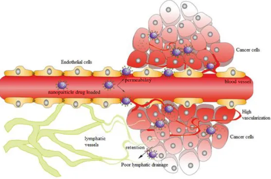
The mechanism of drug delivery is based on two different strategies. The enhanced permeability and retention (EPR) effect (fig. 12) justifies the accumulation of dendrimers preferably in the cancer cells due to their high vascularization and poor lymphatic drainage, which increase the bioavailability of the transported drug and reduce the side effects. However, the degree of vascularization in different tumours is not homogeneous which creates an important limitation for this strategy (142-145). In contrast, the dendritic complex (dendrimer + drug ) could be synthesized with specific moieties at the surface that will interact specifically
Figure 11 - The two most commonly used methods for dendrimers synthesis: divergent and convergent strategies (130).
Nitrile based PAMAM dendrimers functionalized with [RuCp(PPh3)2]+ moiety for anticancer applications
with overexpressed cancer cells receptors. This approach will lead to the internalization of the complex, being more effective (146).
2.1.1.
PAMAM dendrimers
There are many types of dendrimers having different functionalities that were synthetized in the last decades. Some of these include the PAMAM (Polyamidoamine) (147), PPI (PolyPropylene Imine) (148), liquid crystalline (LC) (149), core-shell (tecto) (150), chiral (151), peptide (152), hybrid (153), glycodendrimers (154), metallodendrimers (122, 155) (or metal-containing dendrimers), triazine based (156) and dendritics polyesters (157), polyglycerols (158), phosphorous dendrimers (159-161) and hybrid families of dendrimers.
PAMAM dendrimers were firstly synthetized by Tomalia and co-workers(128) in the mid-1980s and were the first dendritic structures that have been exhaustively investigated and have received widespread attention. These dendrimers have polyamide branches with tertiary amines as branching points and their synthesis is possible by using the divergent method with ethylenediamine or ammonia as initiator core reagents. They can be obtained having surface amino groups or carboxylic acid groups, being commercially available, usually as methanol solutions, up to generation 10 (129, 130).
Figure 12 – Schematic representation of the EPR effect in drug delivery (adapted from ref. (145)).
permeability nanoparticle drug loaded
Nitrile based PAMAM dendrimers functionalized with [RuCp(PPh3)2]+ moiety for anticancer applications
2.2.
Metallodendrimers
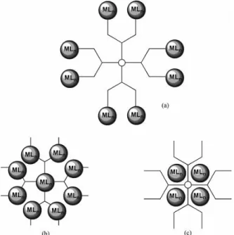
As it was introduced in point 2., the concept of multinuclearity can be applied to improve the effectiveness of the anticancer drugs. Dendrimers can be used as organic scaffolds for the incorporation of metal-based drugs, hence metallodendrimers. Thus, the attractive properties of dendrimers as nanocarriers and the anticancer activity of some metal-based drugs came together to develop an emerging area of interest. These interesting molecules could be prepared with different frameworks (fig. 13): a) the metal could be on the periphery of the metallodendrimer;
b) scattered all over the framework or c) encapsulated in the internal cavities of the dendrimer
(117).
Since the metallodendrimers field is still in expansion (when compared to the other classes of dendrimers for biomedical applications), there are only a few examples of them reported in the literature. The first compounds to show promising cytotoxicity in some resistant and sensitive cancer cell lines was prepared through the combination of an anionic G3.5-PAMAM dendrimer with cisplatin (162, 163).
Figure 13 – Schematic representation of the metallodendrimers with the metallic moieties in several positions: a) on the periphery; b) scattered throughout the framework and c) encapsulated between the structure of the dendrimer (111).
Nitrile based PAMAM dendrimers functionalized with [RuCp(PPh3)2]+ moiety for anticancer applications
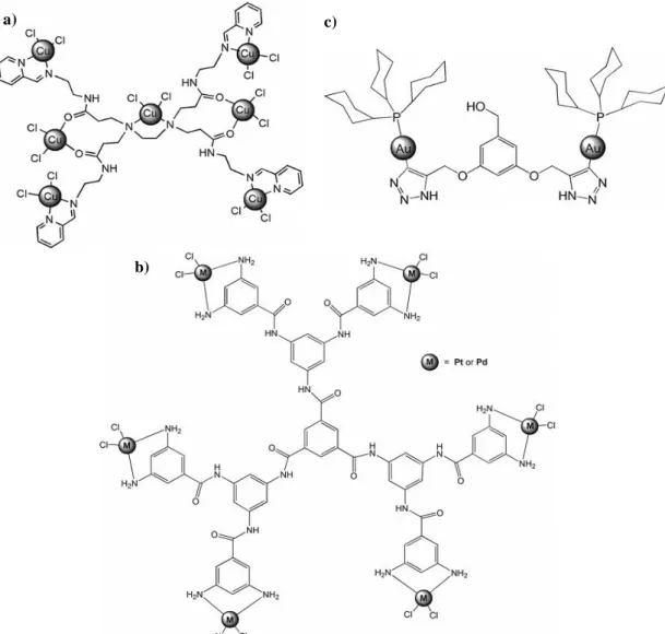
Meanwhile, Zhao C. et al (141) have modified the G1-PAMAM dendrimer to prepare a multinuclear copper complex that also have showed good in vitro results against several cancer cell lines (fig. 14a)). Other two examples of prepared metallodendrimers are the multinuclear Pt (II) and Pd (II) G2-polyamide metallodendrimers (164) (fig. 14b)) and the G1-dinuclear gold(I) metallodendrimers prepared from alkyne-terminated precursors (165) (fig. 14c)).
2.2.1.
Polynitriles compounds as core ligands for the
preparation of metallodendrimers
Nitriles compounds are an important family of molecules due to several reasons, for example: they can be the substrates of the nitrile biocatalysis reaction for industrial production of carboxylic acids, waste treatment and surface modification (166, 167) and represent an essential starting material in organic and inorganic processes (168, 169) with applications on the field of materials (170)and other polymers (171, 172) among others. In fact, they are able to form addition compounds both with nucleophilic and electrophilic reagents. This important property derives from the electronic structure of the triple carbon-nitrogen bond. In terms of nucleophilic addition, they have the ability to stabilize a wide variety of transition metals in different oxidation states (173) in order to form new stable complexes with different properties, namely magnetic (174) and second- and third order non-linear optical properties (169, 170, 175, 176). Thus, they act as ligands strongly activated in the presence of metal ions or electron withdrawing R moieties, as the CF3 (in contrast to their inertness in the free form), which
enhances the rate of reaction in the 106 – 1010 range (177). Their coordination with low-valent and electron rich metal centres, also improve their reactivity towards electrophilic attack. They are also good σ-donors and isoelectronic with other unsaturated ligands – acetylide anions, carbon monoxide, dinitrogen, and isocyanides – which allow them to be involved in substitution or ligand conversion processes (137, 168, 178). In fact, the coordination of nitriles with transition metals, in respect to the metal centre, is described not only in terms of acceptor and electron donor, but also with a contribution of electrostatic interactions (179, 180). These interesting properties of nitriles make them, currently, a target of interest (137, 168).
Nitrile based PAMAM dendrimers functionalized with [RuCp(PPh3)2]+ moiety for anticancer applications
2.2.2.
Ruthenium metallodendrimers
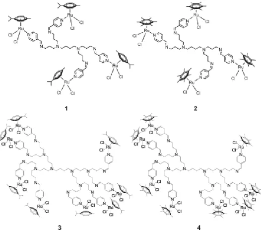
Regarding the ruthenium metallodendrimers as potential anticancer agents there are still few reports available. Some examples are the compounds that were prepared and studied by Govender P. et al (122, 140, 181). The first to be prepared was the multinuclear arene ruthenium complexes coordinated to dendritic polypyridyl scaffolds (fig. 15). These compounds have demonstrated a moderate anticancer activity when compared with cisplatin, against ovarian cancer cell lines. Additionally, they have discovered a correlation between the size of the complex and the obtained cytotoxicity: the octa-coordinated metallodendrimers have showed the best results, when compared with the tetra-coordinated (140).
a) c)
b)
Figure 14 – Molecular structures of three different metallodendrimers: a) multinuclear copper -functionalized, b) a multinuclear Pt (II) and Pd (II) G2-polyamide, and c) G1-dinuclear gold(I) (adapted from ref. (111)).
Nitrile based PAMAM dendrimers functionalized with [RuCp(PPh3)2]+ moiety for anticancer applications
Two years later, they have also prepared a family of chelating, neutral, and cationic first and second generation ruthenium(II) arene complexes based on poly(propyleneimine) dendrimer scaffolds (fig. 16). The in vitro results were similar to their previous studies corroborating the achieved conclusion. They also performed DNA-binding experiments and have found that the observed increased anticancer activity was correlated with their increase of DNA binding activity (181).
Figure 15 - Tetra- and octanuclear arene ruthenium complexes coordinated to dendritic polypyridyl scaffolds (134).
Figure 16 - Chelating N,O- and N,N-ruthenium(II) arene metallodendrimers with poly(propyleneimine) dendrimer as scaffolds. They are coordinated with 4, 8, 16 or 32 n groups (adapted from ref. (181)).
Nitrile based PAMAM dendrimers functionalized with [RuCp(PPh3)2]+ moiety for anticancer applications
The last family of the prepared ruthenium metallodendrimers was a series of neutral, chelating N,O-ruthenium(II)-arene and cationic, chelating N,O-ruthenium(II)-arene-PTA salicylaldimine metallodendrimers (fig. 17). A similar study was performed and the metallodendrimers with 32 end groups have shown the best proliferative activity (122, 182).
As it was already demonstrated, metallodendrimers represents an exciting family of compounds with promising anticancer applications when compared with cisplatin. However, there are several crucial properties that must be extensively studied, such as the pharmacokinetics, biocompatibility, and stability under physiological conditions, toxicity and biodistribution. Thus, in vivo studies are urgent in other to a better understanding of the mechanism of action of these promising therapeutic molecules. Consequently, it is predicted for the next years a continuous increase of interest of the metallodendrimers in the field of anticancer therapy (117).
3.
SCOPE AND OBJECTIVES
The theme of this work was inspired in the attractive and promising anticancer properties of the ruthenium complexes combined with the application of dendrimers as nanocarriers. Additionally, the versatility of dendrimers as building blocks for the preparation of other dendrimers with surface groups of interest, including poly-nitrile groups that will act as core ligands for the preparation of ruthenium metallodendrimers was also considered. Previous studies of our group (Molecular Materials Research Group of CQM – Madeira Chemistry Research Centre) (137, 183-185) as explored the ability of the nitrile groups from functionalized poly(alkylidenamine) dendrimers to coordinate and bridge with a ruthenium cyclopentadienyl
Figure 17 – Cationic, chelating N,O-ruthenium(II)-arene-PTA salicylaldimine metallodendrimers (adapted from ref. (181)).
Nitrile based PAMAM dendrimers functionalized with [RuCp(PPh3)2]+ moiety for anticancer applications
complex [Ru(η5-C5H5(PPh3)2Cl] for the preparation of different cationic metallodendrimers.
Their findings were crucial for the development of this work and, in this way, the main goal was to optimize a synthetic strategy to functionalize nitrile-based PAMAM dendrimers, from generations 0 and 1, with the organometallic moiety [Ru(η5-C
5H5)(PPh3)2Cl]. Other goal was to
fully characterize them with 1D/2D-NMR (one-dimensional/two-dimensional-Nuclear Magnetic Resonance), FTIR (Fourier Transform Infrared Spectroscopy) and MS (Mass Spectrometry) in order to evaluate their structure and determine the exact mass.
Nitrile based PAMAM dendrimers functionalized with [RuCp(PPh3)2]+ moiety for anticancer applications


![Figure 8 – Molecular structures of two ruthenium complexes with the general formula [(η 5 -C 5 H 5 )Ru(P-P)L] + (P-P: 1,2-bis(diphenylphosphine)ethane (dppe) or triphenylphosphine (PPh 3 ); L: monodentate or bidentate nitrogen donors): a) TM34](https://thumb-eu.123doks.com/thumbv2/123dok_br/19274036.984445/31.892.220.682.663.864/molecular-structures-ruthenium-complexes-diphenylphosphine-triphenylphosphine-monodentate-bidentate.webp)