Article
J. Braz. Chem. Soc., Vol. 22, No. 7, 1339-1345, 2011. Printed in Brazil - ©2011 Sociedade Brasileira de Química
0103 - 5053 $6.00+0.00
A
*e-mail: [email protected]
Spectroscopic Characterization of the Exopolysaccharide of
Xanthomonas
axonopodis
pv.
citri
in Cu
2+Resistance Mechanism
Denise Osiro,a,b Roberto W. Assis Francoa and Luiz Alberto Colnago*,a
aEmbrapa Instrumentação Agropecuária, 13560-970 São Carlos-SP, Brazil
bInstituto de Química de São Carlos, Universidade de São Paulo, 13560-970 São Carlos-SP, Brazil
Analizou-se o papel do exopolissacarídeo (EPS) no mecanismo de resistência ao Cu2+ da
Xanthomonas axonopodis pv. citri (Xac) com as espectroscopias de infravermelho com transformada de Fourier (FTIR), ressonância paramagnética eletrônica (RPE) e ressonância magnética nuclear (RMN). Os dados de FTIR demonstraram que as células cultivadas na presença de 0.2 mmol L-1
de CuSO4 produziram uma maior quantidade de exopolissacarídeo piruvatado do que as células
cultivadas na sua ausência. Os dados de RPE indicaram que a quantidade de íons Cu2+ diminuiu
com o tempo de cultura. Os dados de RMN de 13C mostraram a complexação dos íons de Cu2+
com o EPS. Esses resultados mostraram que o EPS tem um papel importante no mecanismo de proteção da Xac ao Cu2+ e este age no mecanismo inicial de proteção, ligando-se aos íons de Cu2+
livres, reduzindo sua difusão e o transporte ativo ao citoplasma . O Cu2+ também induz a produção
de EPS piruvatado, que aumenta a capacidade de coleta e complexação desses íons.
We analyzed the role of exopolysaccharide in the Xanthomonas axonopodis pv. citri (Xac) Cu2+
resistance mechanism by Fourier transform infrared (FTIR), electron paramagnetic resonance (EPR) and nuclear magnetic resonance (NMR) spectroscopies. The FTIR data show that cells cultivated in the presence of 0.2 mmol L-1 of CuSO
4 produce larger amounts of pyruvated exopolissacharide
(EPS) than the ones cultivated in its absence. The EPR data indicate that the amount of Cu2+
decreases with cultivation time. The 13C-CPMAS NMR data also show the complexation of Cu2+
ions to the EPS. The results demonstrate that EPS plays an important role in Xac Cu2+ protection.
Both capsular and slime EPS act as an initial protection mechanism, binding free Cu2+ ions, reducing
their diffusion and their active transport to the cytoplasm. Cu2+ also induces the production of a
highly pyruvated negative EPS, increasing its capture and binding capacity.
Keywords: citrus canker, X. axonopodis pv. citri, exopolysaccharide, EPR, NMR, FTIR
Introduction
Xanthomonas axonopodis pv. citri (Xac) is the causal agent of citrus canker, a major problem in citriculture.1,2 One
of the methods to control the disease is the application of Cu2+ based bactericides, which reduce bacterial populations
on plant surfaces.1,3,4
Copper is an essential element for all living organisms, but its excess is toxic to cells, inducing the formation of free radicals. As it is more toxic to lower organisms such as bacteria and fungi,3 Cu2+ pesticides have been used in
agriculture for over a century, to control plant diseases. This practice has selected copper resistant strains, reducing the eficacy of Cu2+ bactericides.2
Bacterial resistance has been reported in several phytopathogenic genera such as Xanthomonas and
Pseudomonas, which infect hundreds of economically
important plants.3,5-7 Copper resistance has been reported
in Xanthomonas campestris pv. vesicatoria, which infects pepper and tomato; Xanthomonas campestris pv. juglandis, a walnut pathogen; Xanthomonas axonopodis pv. citri,a citrus pathogen and Pseudomonas syringae pv. tomato,a tomato pathogen.3,5,6
The genetic basis for Cu2+ protectionin gram-negative
bacteria such as Xac is usually determined by the cop
operon present in plasmid or in bacterial chromosome.3,5,6
This operon contains four open reading frames (ORFs),
mutations in P. syringae pv. tomato suggest that copA and
copB are essential for resistance and copC and copD are required for full resistance.3,5,6
A characteristic of the Xanthomonas genus is the copious production of exopolysaccharide (EPS), xanthan gum, released by the cells as slime in medium. The EPS is involved in cell protection, but its role in the protection mechanism against heavy metals is not well established. The xanthan gum is a 1,4 linked β-D-glucose polymer. Every alternated glucose contains a negative charged trisaccharide side chain with an internal mannose, glucuronic acid and a terminal mannose. The internal mannose can be acetylated in the O(6) position and the terminal one can be derived by a pyruvic acid moiety, joined by a ketal linkage in the O(4) and O(6) positions, leaving a free carboxylic acid group. The derivation of mannoses is dependent on species, environment (nutrients, pH and temperature) and age.8
In this paper we analyzed role of EPS in Xac Cu2+
resistance mechanism by Fourier transform infrared (FTIR), electron paramagnetic resonance (EPR) and nuclear magnetic resonance (NMR) spectroscopies. Our results show that both capsular and slime EPS play an important role in Xac Cu2+ protection mechanism. They
act as an initial protection mechanism, binding free Cu2+
ions, reducing their diffusion and their active transport to the cytoplasm.
Experimental
Preparation of bacterial samples
The Xanthomonas axonopodis pv. citri (Xac) 306 strain isolated from citrus groves in São Paulo state, Brazil,9
was used to study the EPS role in the Cu2+ resistance
mechanism. The cells were cultivated in predeined M9 medium, at 28 °C in a rotary shaker. The Xac were also cultivated in M9 medium with 0.2 mmol L-1 of CuSO
4.
The optical density of the cultures at 600 nm, 1 cm quartz cells, was monitored in a Shimadzu UV-Visible 1601PC spectrophotometer. The samples with optical density (OD) higher than 1 were diluted before the measurements.
After the interruption of the cultures, the cells were collected by centrifugation (4000 ×g for 20 min). The pellets were suspended in 0.5% NaCl solution and centrifuged twice. The inal pellets were lyophilized and used for the FTIR, EPR and NMR analyses.
Fourier transform infrared spectroscopy (FTIR)
The FTIR spectra were acquired on a Paragon 1000 Perkin Elmer spectrometer, from 4000 to 400 cm-1, 4 cm-1 of
spectral resolution and 64 scans. The samples were prepared in the form of KBr pellets, 1 mg of bacteria and 100 mg of KBr. The spectra were baseline corrected from 3000 to 2800 cm-1 and from 1800 to 900 cm-1 and normalized by the
intensity of the amide II peak (intensity = 1) at 1550 cm-1,
which is related to biomass.10,11
Electron paramagnetic resonance spectroscopy (EPR)
The solid samples were analyzed by EPR spectroscopy using a Bruker EMX spectrometer (Bruker Biospin Corporation, Rheinstetten, Germany) operating in the X-band (9.8 GHz) with a spherical cavity, at 300 K. The relative intensity measurements were referenced by a ruby (Al2O3:Cr3+) located in a ixed position inside the cavity
below the sample. The g-factors were referenced by a MgO:Cr3+ (g = 1.9797) sample attached to the sample to be
analyzed. The following experimental settings were used: microwave power 1 mW; modulation frequency 100 kHz; and modulation amplitude 0.2 mT. The WinEPR SimFonia12
program was used to simulate EPR spectra.
Nuclear magnetic resonance spectroscopy (NMR)
Solid-state 13C NMR spectra were acquired using a
9.4 T Varian Inova 400 spectrometer. The samples were compacted in a 5 mm zircon rotor. The CP/MAS (cross polarization/magic angle spinning) spectra of the samples spinning rate at 9 kHz were recorded with 4 µs pulse width, 13.4 ms acquisition time, 2.5 s of recycle time, and a 60 kHz decoupling ield strength. All spectra were iltered by an exponentially decaying function (line broadening factor of 20 Hz).
Results and Discussion
Results
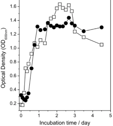
Figure 1 shows the optical density (OD) curves for the
Xac cultures with and without Cu2+ as a function of the
incubation time. The data are shown only up to the 3rd day
to demonstrate the effect of Cu2+ at the beginning of the
culture. Thereafter, the OD remained almost constant up to the end of the experiment (13 days).
The cells in M9 medium without Cu2+ showed a
continuous growth from the beginning (log phase) up to the stationary phase in about 48 h. The cells cultivated in M9 medium with Cu2+ showed a reduction of OD values
was observed in the X. campestris pv. vesicatoria copper
resistant strain.7
The role of EPS in the Xac Cu2+ resistance mechanism
was analyzed by FTIR, EPR and NMR spectroscopies.
FTIR analysis
Figure 2 shows the FTIR spectra of the Xac cells (2a) with its exopolisacharide and the exopolysaccharide, xanthan gum (2b), which is released into the medium as slime.
Figure 2a is the Xac FTIR spectrum showing the major bands due to proteins, xanthan gum and lipids. The signals between 3600 and 3100cm-1 are due to proteins and
xanthan gum, N–H and O–H stretching; between 3000 and 2800 cm-1 due to C–H stretching of methyl and methylene
groups; at 1730 cm-1,dueto C=O stretching of the xanthan
acetate group (Figure 2b) and lipids; from 1700 and 1600 cm-1, due tothe proteins amide I band and to xanthan
O–H angular deformation and to symmetric stretching
of carboxylate group of pyruvate and of glucuronic acid (Figure 2b); from 1700 to 1600 cm-1 due to protein amide
II band, which is used as a internal standard for biomass content10,11 and from 1100 and 900 cm-1 to xanthan C–O
stretching (Figure 2b).
The xanthan gum FTIR spectrum (Figure 2b) also shows weak signals at 1400 cm-1 and at 1250 cm-1 due to
carboxylate asymmetric stretching and to C=O acetate deformation, respectively.
Figure 3 shows some of the FTIR spectra (from 1800 to 900 cm-1) of Xac incubated in M9 medium (3a) and in
M9 medium with Cu2+ (3b). Figure 4 shows the variation
of band intensity at 1730 cm-1, 1640 cm-1, and 1050 cm-1
for the spectra of Figure 3, due to acetate and pyruvate groups in xanthan and to the total xanthan, as a function of incubation time.
Figures 3a and 4a show small reduction in total xanthan content with incubation time. Figures 3b and 4b show a strong signal at 1640 cm-1 in 24 h. The intensities of signals
at 1050 cm-1 and 1730 cm-1 were similar in both media
and were constant with cultivation time, showing a slight increase in the cells cultivated in the presence of Cu2+ (4b);
signal was constant with the cultivation time.
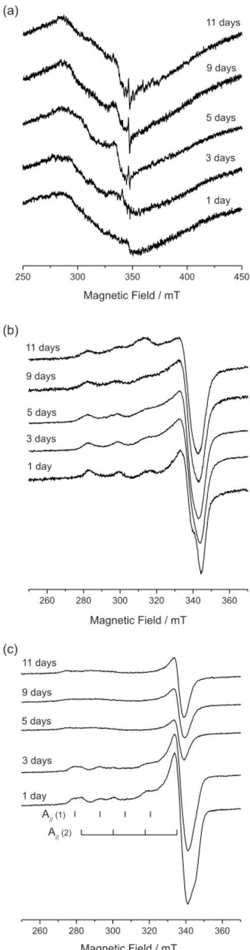
EPR analysis
The interaction of Cu2+ with Xac and xanthan gum
was also analyzed by electron paramagnetic resonance spectroscopy (EPR). The EPR spectra of lyophilized Xac
cultivated in M9 medium without Cu2+ (Figure 5a) show a
broad line (∆H ca. 600 G) that can be associated to Fe+3 in
a ligand ield with tetragonal symmetry.13 The spectra after Figure 1. Optical density of Xac culture without () and with ()
0.2 mmol L-1 of cupric sulfate.
5 days show a sharp line (∆H ca. 10 G) at g = 2.00054, which is attributable to an unknown free radicals.3
Figure 5b shows some of the EPR spectra of lyophilized
Xac cells obtained from the M9 medium with cupric sulfate. The spectra show only the typical signal of Cu2+ in an axial
symmetric environment, with a 3d9 electronic coniguration
and electronic spin S=1/2. The nuclear spin for both 63Cu
(natural abundance 69%) and 65Cu (natural abundance 31%)
isotopes is I=3/2. Therefore (2I+1), i.e., four perpendicular and four parallel hyperine components can be expected, resulting from the dipole-dipole interaction between the magnetic moment of the nucleus and the electronic moment of the paramagnetic ion. The spectra are axial type, having g//, g⊥ > 2, indicating the occupation of Cu2+ in an axial
symmetry of a tetragonally distorted octahedral site.13,14
All the Xac EPR spectra (Figure 5b) were similar, showing a slight reduction in resolution with the incubation time. This suggests an increase of Cu2+ bound to cells,
increasing the Cu2+-Cu2+ dipolar interaction. Nakajima14
showed that the Cu2+ spectral hyperine structure was not
observed only when the concentration in the cells exceed
Figure 4. Normalized FTIR absorbance of the signals at 1730 (), 1640 (), and 1050 cm-1 () from Xac cells for different incubation times in
M9 medium (a) and in medium with 0.2 mmol L-1 of cupric sulfate (b).
400 µg g-1. This suggests that the Cu2+ concentrations in Xac samples were below this value. The EPR spectra in Figure 5b also show that the Cu2+ is bound in a similar
environment, indicating no changes in the binding site in function of incubation time.
The EPR spectra of lyophilized supernatant, with the same mass (Figure 5c), show a continuous reduction of Cu2+
signals as a function of incubation time. The spectra of day 1 and 3 also show an overlap of Cu2+ signals in two distinct
sites. Table 1 lists the g// and g⊥ and A// values. In one site,
Cu2+ spectra show similar parameters to the puriied xanthan
(Table 1) and the other to a Cu2+ complexed to the medium
amino acids. Jung et al.15 obtained values of g
x = 2.038,
gy = 2.068, gz = 2.390 and A// = 13.5 mT, which are similar
to one of the measured supernatant values.
The supernatant spectra after day 3 showed only a broad line for g// signals. This type of reduction in resolution has
also been observed for other bacteria and was also related to the pH below 4.14 However, that was not the case here,
since the media pH was always above 6. The spectra also show the reduction of Cu2+ in the medium, indicating its
reduction or absorption by the bacteria.
The values obtained for Xac and xanthan (Table 1) were similar to those obtained by Nakajima14 for both
gram-negative and gram-positive bacteria. Because the gram-negative and gram-positive bacteria have different cell wall components, he suggested that Cu2+ was sorbed
by the surface proteins.
As the EPR spectra of Xac and xanthan are similar (Figure 5b and c, Table 1), showing identical environment, the Cu2+ have to be complexed preferentially with the OH
and COO− groups of xanthan, which is the major component
of the Xac wall.
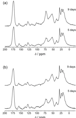
13C solid state NMR spectroscopy analysis
Figure 6 shows the high resolution solid state 13C NMR
spectra of Xac cells cultivated in M9 medium (6a) and M9 medium with Cu2+ (6b). The major signals in the NMR
spectra can also be attributed to proteins and EPS as for FTIR (Figure 2). The strong signal at 175 ppm is due mainly to carbonyl carbons from proteins, fatty acids and EPS. The signals from 140 to 100 ppm were attributed to the aromatic amino acid side chains. The weak signal at 105 ppm is assigned to anomeric carbons of the glucose in xanthan and to C2 of the pyruvic acid ketal group. The strong signal at 75 ppm is assigned to the C2, C3 and C5 of the glucose polymer chain. The signals from 45 to 70 ppm were assigned to the α-carbon proteins and the signals from 15 to 45 ppm to aliphatic amino acid side chains, acetate and pyruvate methyl groups. The fatty acids signals were also detected by the stronger peak at 30 and 130 ppm, attributed to methylene groups and double-bound carbons, respectively. These signals overlapped with protein signals, but are well characterized in single pulse spectra (data not shown), similar to the ones observed in Xylella fastidiosa.16
The carbonyl peak at 175 and the EPS peak at 73 ppm for the cells cultivated without Cu2+ (Figure 6a) were
stronger in 9 days than in 5 days, revealing a high capsular EPS in older cells. The spectra of cells cultivated with Cu2+
(Figure 6b) showed the opposite effect. The signals at 175 and 73 ppm were stronger in 5 days than in 9 days. These differences can be explained by the higher concentration of EPS and pyruvate groups in younger cultures and also by the effect of Cu2+ in longitudinal relaxation time of Table 1. EPR spectra parameters of Cu(II) in Xac, xanthan gum and
supernatant extracted from Xac cultures with cupric sulfate
Samples with Cu(II) ion g// g⊥
Xac 2.269 2.069
Xanthan gum 2.255 2.055
Supernatant - 3 days 2.260 2.325
2.055 2.055
*Cu(Asn)+ 2.390 g
x = 2.038
gy = 2.068 *Reference 15.
Figure 6.13C-CPMAS NMR spectra of Xac cell cultivated in M9 medium:
the EPS carbons. As shown by EPR spectra, the Cu2+ are
bound to capsular EPS in the carboxylate and OH groups. The EPS carbons that are closer to Cu2+ have a very short
relaxation time, giving broad lines, and are not observed in the 13C spectra.
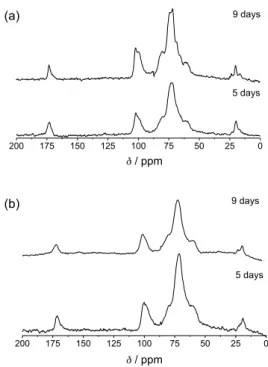
A similar effect can be seen in the solid state 13C NMR
spectra of the xanthan gum extracted from the same cultures (Figure 7), where the 13C signals for pyruvate and
acetate C=O group are at 175 ppm and the CH3 signals
at 24 and 20 ppm, respectively. The signals from 85 and 55 ppm are attributed to carbons 2-6 of glucose, mannose and glucuronic acid. The signals from 105 to 95 ppm are due to quaternary C2 of the pyruvate and the C1-carbon of glucose, mannose and glucuronic acid. All the signals from the xanthan samples cultivated with Cu2+ show broader lines
than the ones cultivated without it.
Conclusions
The FTIR, EPR and NMR spectroscopies show the variation of chemical composition in exopolysaccharide xanthan gum, in lyophilized Xac cultivated with and without of Cu+2 ions, in the media. The spectra show the
toxic effect of Cu+2 ions, which has been used to control the
citrus cancer disease in ield, demonstrated by the increase of the EPS synthesis in the irst stage of the culture.
As indicated in Figures 3 and 4, the cells cultivated in the presence of Cu2+ had a higher EPS content in all the
fermentation times and displayed a very strong FTIR peak at 1640 cm-1, indicating the presence of high carboxylate
content. The extra carboxyl groups in the cells as due to overproduction of pyruvated EPS in response to Cu2+ ions.
It is known that Xanthomonas can modulate the amount of acetate and pyruvate in its EPS as a function of nutrients, pH, temperature etc.8
The presence of more negative pyruvated EPS has a higher afinity to cations in the culture, increasing the rate of active cation transport as well as its binding capacity, as demonstrated by EPR and NMR data (Figures 5 to 7). The more negative pyruvated EPS produced by the cells in the irst days of culture has stronger ability to harvest and bind the lethal Cu2+ ions in culture, keeping them outside
the cells and not reaching the cytoplasm.
The xanthan gum produced as slime by the cells cultivated with Cu2+ was also more pyruvated in the
initial days of culture (Figures 3 and 4). In addition to its harvesting and binding capacity, pyruvated xanthan also increased the culture viscosity, reducing cation diffusion.8
The lower diffusion rate reduced the Cu2+ contact with cells,
thus reducing its level in the cytoplasm.
A similar production of highly pyruvated xanthan has been observed when Xanthomonas campestry was treated with hypochlorous acid, which is known to induce free radicals such as Cu2+ ions. It has been demonstrated that the
production of highly pyruvate xanthan is activated by the free radical inducible regulatory protein, SoxS, which binds DNA SoxS consensus sequence in the gum B promoter region, increasing the transcription of gum mRNAs.17
High expression of EPS has been reported as a response to Cu2+ ions in Xylella fastidiosa, which also causes Citrus
disease and is related to Xac. The results also suggest a synergistic effect between diffusion barriers and other mechanisms associated with bacterial resistance in this phytopathogen.18
Xac cells seem to have a similar response to different lethal compounds, indicating that EPS is involved in a general and non-speciic cell protection mechanism. Since EPS is continuously produced by the cells, Xac cells can be used in a fast protection mechanism, reducing the initial death rate and allowing for activation of speciic protection mechanisms,19 like cop operon, against Cu2+ ions. After
this speciic mechanism becomes effective, the production of highly pyruvated EPS is reduced to normal levels, as illustrated in Figures 3 and 4.
These data also indicate that Cu2+ based bactericides
used in citrus canker control programs must be effective in the irst application, as the cells with normal (low) pyruvated EPS, is not effective in protecting them against Cu2+. If some cells survive the initial spraying, they are
ready to face future applications with highly pyruvated EPS and with the copper operon activated, reducing the
Figure 7.13C-CPMAS NMR spectra of xanthan gum extracted from Xac
effectiveness of Cu2+ bactericides or requiring application
of higher Cu2+ doses.
Acknowledgments
We thank FAPESP and CNPq (Brazilian agencies) for inancial support and Dr. Marcos Antônio Machado for donating the Xac 306 strain.
References
1. del Campo, R.; Russi P.; Mara, P.; Mara, H.; Peyrou, M.; Ponce de Leon I.; Gaggero, C.; FEMS Microbiol. Lett.2009, 298, 143. 2. Mendes, B. M. J.; Cardoso, S. C.; Boscariol-Camargo, R. L.;
Cruz, R. B.; Mourão Filho, F. A. A.; Bergamin Filho, A.; Plant Pathol.2010, 59, 68.
3. Cooksey, D. A.; FEMS Microbiol. Rev. 1994, 14, 381. 4. Graham, J. H.; Gottwald, T. R.; Cubero, J; Achor, D. S.; Mol.
Plant Pathol. 1995, 5, 1.
5. Cooksey, D. A.; Mol. Microbiol. 1993, 7, 1.
6. Lee, Y. A.; Hendson, M.; Panopoulus, N. J.; Schroth, M. N.;
J. Bacteriol. 1994, 176, 173.
7. Ramos, G. B. A; Rosato, Y. B.; Braz. J. Genet. 1996, 19, 551. 8. Casas, J. A.; Santos, V. E.; García-Ochoa, F.; Enzyme Microb.
Technol. 2000, 26, 282.
9. da Silva, A. C. R.; Ferro, J. A.; Reinach, F. C.; Farah, C. S.; Furlan, L. R.; Quaggio, R. B.; Monteiro-Vitorello, C. B.; Van Sluys, M. A.; Almeida, N. F.; Alves, L. M. C.; do Amaral, A. M.; Bertolini, M. C.; Camargo, L. E. A.; Camarotte, G.; Cannavan, F.; Cardozo, J.; Chambergo, F.; Clapina, L. P.; Cicarelli, R. M. B.; Coutinho, L. L.; Cursino-Santos, J. R.; El-Dorry, H.; Faria, J. B.; Ferreira, A. J. S.; Ferreira, R. C. C.; Ferro, M. I. T.; Formighieri, E. F.; Franco, M. C.; Greggio, C. C.; Gruber, A.; Katsuyama, A. M.; Kishi, L. T.; Leite, R. P.; Lemos, E. G. M.; Lemos, M. V. F.; Locali, E. C.; Machado, M. A.; Madeira, A.
M. B. N.; Martinez-Rossi, N. M.; Martins, E. C.; Meidanis, J.; Menck, C. F. M.; Miyaki, C. Y.; Moon, D. H.; Moreira, L. M.; Novo, M. T. M.; Okura, V. K.; Oliveira, M. C.; Oliveira, V. R.; Pereira, H. A.; Ross,i A; Sena, J. A. D.; Silva, C; de Souza, R. F.; Spinola, L .A. F.; Takita, M. A.; Tamura, R. E.; Teixeira, E. C.; Tezza, R. I. D.; dos Santos, M. T.; Trufi, D.; Tsai, S. M.; White, F. F.; Setubal, J. C.; Kitajima, J. P.; Nature2002, 417, 459.
10. Naumann, D. In Encyclopedia of Analytical Chemistry; Meyers, R. A., ed., John Wiley & Sons: Chichester, UK, 2000, pp. 102-131.
11. Osiro, D.; Colnago, L .A.; Otoboni, A. M. M. B.; Lemos, E. G. M.; de Souza, A. A.; Coletta Filho, H. D.; Machado, M. A.;
FEMS Microbiol. Lett. 2004, 236, 313.
12. Bruker Analytische Messtechnik GmbH; WINEPR SimFonia shareware, Version 1.25; Rheinstetten, Germany, 1996. 13. Griscom, D. L.; J. Non-Cryst. Solids 2004, 40, 211. 14. Nakajima, A.; Water Res.2002, 36, 2091.
15. Jung, K.; Ristori, S.; Martini, G.; Spectrochim. Acta, Part A
2000, 56, 341.
16. Osiro, D.; Muniz, J. R. C.; Coletta Filho, H. D.; de Souza, A. A.; Machado, M. A.; Garratt, R. C.; Colnago, L.A.; Biochem. Biophys. Res. Commun. 2004, 323, 987.
17. Rao, Y. M.; Sureshkumar, G. K.; Biotechnol. Bioeng. 2001, 72, 62.
18. Rodrigues, C. M.; Takita, M. A.; Coletta-Filho, H. D.; Olivato, J. C.; Caserta, R.; Machado, M. A.; Souza, A. A.; Appl. Microbiol. Biotechnol.2008, 77, 1145.
19. Chan, J. W. Y. F.; Goodwin, P. H.; Biotechnol. Adv.1999, 17, 489.
Submitted: March 1, 2010
Published online: March 22, 2011