Nº de ordem:06/D/2009
TESE DE DOUTORAMENTO
Apresentada à UNIVERSIDADE DA MADEIRA
Para obtenção do grau de Doutor José Luís da Silva Santos
FUNCTIONALIZATION OF DENDRIMERS
FOR IMPROVED GENE DELIVERY TO
MESENCHYMAL STEM CELLS
Júri:
Reitor da Universidade da Madeira Doutor Abhay Pandit
Doutora Ana Paula Gomes Moreira Pêgo Doutor David Kelham Smith
Doutora Helena Maria Pires Gaspar Tomás Doutor João Manuel Cunha Rodrigues Doutor Pedro Lopes Granja
FUNCTIONALIZATION OF DENDRIMERS
FOR IMPROVED GENE DELIVERY TO
MESENCHYMAL STEM CELLS
José Luís da Silva Santos
Tese submetida à Universidade da Madeira
com vista à obtenção do grau de Doutor em Química na Especialidade de Química de Materiais
Trabalho efectuado sob a Orientação de: Professora Doutora Helena Maria Pires Gaspar Tomás
Doutor Pedro Lopes Granja
i
ACKNOWLEDGEMENTS
This thesis was only possible to accomplish due to the support of several persons and institutions. To them, I would like to express a deep sense of gratitude.
Foremost, I would like to acknowledge my supervisor, Prof. Helena Tomás, without whom this thesis, and what I have accomplished, would not be possible. Her support, enthusiasm, and input were of invaluable significance for the contents of this thesis. Her warm encouragement and constant guidance have contributed greatly to my professional development.
I would like to express my gratitude to my co-supervisor, Dr. Pedro Granja for his precious help during the last 4 years. He is a good friend and mentor. I greatly appreciate the discussions about the various ideas, hypotheses and manuscripts and I thank him for his encouragement and the creation of an exceptional working atmosphere.
I owe a special word of thanks to Dr. Ana Paula Pêgo, who has provided me numerous valuable advices and helpful discussions. She was always nearby to meet and talk about my work, to proofread abstracts and papers and improve their contents.
I am very grateful to all members of the Molecular Materials Research Group who have directly or indirectly contributed for this thesis. In particular, I would like to thank Prof. João Rodrigues that introduced the field of dendrimers as a topic of research in Centro de Química da Madeira (CQM) and with whom I have learnt the beauty of synthetic chemistry. A special thanks to my dear friends Deepti Pandita (thanks for teaching me the importance of viewing all situations with a carpe diem attitude), João Figueira and Manuel Jardim (many thanks for the help in NMR experiments), Susana Sargo, Hewerson Tavares, Rita Castro and Elena Oramas for their priceless support, enthusiasm, motivation and for sharing so many good moments.
ii I would also like to thank the non-teaching staff (Ana Paula Tentem and Ana Paula Andrade) of the Department of Chemistry of the University of Madeira (UMa) for their timely help and assistance whenever required.
I am also sincerely grateful to Hugo Oliveira, Sílvia Bidarra, Ana Patrícia Cardoso, Ana Paula Filipe and Eliane Vale (from Instituto de Engenharia Biomédica, INEB) and Susana Oliveira (from Instituto de Biologia Molecular e Celular, IBMC) for their friendship, attention, support and enthusiasm throughout the good and less good moments especially during my stay in Porto. I would also like to take this opportunity to thank the staff of INEB for their warmth and responsiveness as their administrative work certainly facilitated my research work.
This thesis will be incomplete without a special thank to the institutions or entities and projects that provided the laboratory facilities and the financial support to conduct the research. To the Fundação para a Ciência e Tecnologia (FCT) for the financial support through the Ph.D. Grant (Ref. SFRH/BD/19450/2004, co-financed by the programs POCI 2010 and FEDER) and the Project DENDRALGENE (Ref. PTDC/SAU-BEB/71161/2006). To the Portuguese National Nuclear Magnetic Resonance Network (PTNMR-REDE/1517/RMN/2005-POCI2010/FEDER).
To the University of Madeira, CQM (UMa) and INEB/IBMC (University of Porto) for the laboratory and other physical resources that allowed the execution of this work. The financial support of UMa through the “research funds program” is also acknowledged (less is better than nothing!).
I am also very grateful to Hospital Central do Funchal for providing me the access to the Flow Activated Cell Sorting Equipment. I want to express my deeply-thank to Dr. Elisabete Santos for the friendship and technical guidance during the analysis.
The support of CS Madeira Hotel is also acknowledged.
iii Tatiana Segura (UCLA, USA), respectively, for kindly providing the hBMP-2 and the Luciferase/Green Fluorescent Protein plasmid DNA.
Lastly, I would like to express my gratitude to the most important persons in my life, to whom this thesis is also dedicated.
I express my true and special thanks to my father and my mother for the endless support, unconditional love, concern and enthusiasm.
Special mention to my brothers Sergio and Roberto, my sister and my brother-in-law Juan who have been always supportive at whatever moments… a special thank!
To my nephew Tiago for his special energy and joy which gave strength to keep me ongoing in an “easier way”.
I am forever indebted to my ultimate inspiration and unending source of love, Rosa.
v
ABSTRACT
Disease, injury, and age problems compromise human quality of life and continuously motivate the search for new and more efficacious therapeutic approaches. The field of Tissue Regeneration and Engineering has greatly evolved over the last years, mainly due to the combination of the important advances verified in Biomaterials Science and Engineering with those of Cell and Molecular Biology. In particular, a new and promising area arose – Nanomedicine – that takes advantage of the extremely small size and especial chemical and physical properties of Nanomaterials, offering powerful tools for health improvement. Research on Stem Cells, the self-renewing progenitors of body tissues, is also challenging to the medical and scientific communities, being expectable the appearance of new and exciting stem cell-based therapies in the next years.
The control of cell behavior (namely, of cell proliferation and differentiation) is of key importance in devising strategies for Tissue Regeneration and Engineering. Cytokines, growth factors, transcription factors and other signaling molecules, most of them proteins, have been identified and found to regulate and support tissue development and regeneration. However, the application of these molecules in long-term regenerative processes requires their continuous presence at high concentrations as they usually present short half-lives at physiological conditions and may be rapidly cleared from the body. Alternatively, genes encoding such proteins can be introduced inside cells and be expressed using cell’s machinery, allowing an extended and more sustained production of the protein of interest (gene therapy). Genetic engineering of stem cells is particularly attractive because of their self-renewal capability and differentiation potential. For Tissue Regeneration and Engineering purposes, the patient’s own stem cells can be genetically engineered in vitro and, after, introduced in the body (with or without a scaffold) where they will not only modulate the behavior of native cells (stem cell-mediated gene therapy), but also directly participate in tissue repair.
vi and being investigated use cationic molecules as carriers for DNA. In this case, gene delivery and gene expression level remain relatively low when primary cells are used.
The main goal of this thesis was to develop and assess the in vitro potential of polyamidoamine (PAMAM) dendrimers based carriers to deliver genes to mesenchymal stem cells (MSCs). PAMAM dendrimers are monodispersive, hyperbranched and nanospherical molecules presenting unique characteristics that make them very attractive vehicles for both drug and gene delivery. Although they have been explored for gene delivery in a wide range of cell lines, the interaction and the usefulness of these molecules in the delivery of genes to MSCs remains a field to be explored. Adult MSCs were chosen for the studies due to their potential biomedical applications (they are considered multipotent cells) and because they present several advantages over embryonic stem cells, such as easy accessibility and the inexistence of ethical restrictions to their use.
This thesis is divided in 5 interconnected chapters.
Chapter I provides an overview of the current literature concerning the various non-viral systems investigated for gene delivery in MSCs. Attention is devoted to physical methods, as well as to chemical methods that make use of polymers (natural and synthetic), liposomes, and inorganic nanoparticles as gene delivery vectors. Also, it summarizes the current applications of genetically engineered mesenchymal stem cells using non-viral systems in regenerative medicine, with special focus on bone tissue regeneration.
vii assays including alkaline phosphatase activity (an early marker of osteogenesis), osteocalcin production, calcium deposition and mineralized nodules formation (late osteogenesis markers). Results show that a low transfection level is enough to induce in vitro osteogenic differentiation in MSCs.
Next, from Chapter III to Chapter V, studies are shown where several strategies are adopted to change the interaction of PAMAM dendrimers with MSCs cell membrane and, as a consequence, to enhance the levels of gene delivery. In Chapter III, generations 5 and 6 of PAMAM dendrimers are surface functionalized with arginine-glycine-aspartic acid (RGD) containing peptides – experiments with dendrimers conjugated to 4, 8 and 16 RGD units were performed. The underlying concept is that by including the RGD integrin-binding motif in the design of the vectors and by forming RGD clusters, the level of transfection will increase as MSCs highly express integrins at their surface. Results show that cellular uptake of functionalized dendrimers and gene expression is enhanced in comparison with the native dendrimers. Furthermore, gene expression is dependent on both the electrostatic interaction established between the dendrimer moiety and the cell surface and the nanocluster RGD density.
In Chapter IV, a new family of gene delivery vectors is synthesized consisting of a PAMAM dendrimer (generation 5) core randomly linked at the periphery to alkyl hydrophobic chains that vary in length and number. Herein, the idea is to take advantage of both the cationic nature of the dendrimer and the capacity of lipids to interact with biological membranes. These new vectors show a remarkable capacity for internalizing pDNA, being this effect positively correlated with the –CH2– content present in the hydrophobic corona. Gene expression is also greatly enhanced
using the new vectors but, in this case, the higher efficiency is shown by the vectors containing the smallest hydrophobic chains.
Finally, chapter V reports the synthesis, characterization and evaluation of novel gene delivery vectors based on PAMAM dendrimers (generation 5) conjugated to peptides with high affinity for MSCs membrane binding - for comparison, experiments are also done with a peptide with low affinity binding properties. These systems present low cytotoxicity and transfection efficiencies superior to those of native dendrimers and partially degraded dendrimers (Superfect®,
viii is shown to be cell surface receptor-mediated. Overall, results show the potential of PAMAM dendrimers to be used, as such or modified, in Tissue Regeneration and Engineering. To our knowledge, this is the first time that PAMAM dendrimers are studied as gene delivery vehicles in this context and using, as target, a cell type with clinical relevancy.
ix
RESUMO
As doenças, acidentes e problemas relacionados com a velhice comprometem a qualidade de vida humana, motivando a contínua procura de novas e mais eficientes abordagens terapêuticas. Ao longo dos últimos anos, o domínio da Engenharia e Regeneração de Tecidos conheceu uma enorme evolução, nomeadamente, devido à combinação dos importantes avanços ocorridos na Ciência e Engenharia de Biomateriais com os da Biologia Molecular e Celular. Em particular, surgiu uma nova e promissora área científica – a Nanomedicina – que tira vantagem do tamanho extremamente pequeno e das propriedades químicas e físicas únicas dos nanomateriais, oferecendo ferramentas poderosas para a melhoria da saúde humana. A pesquisa em células estaminais (CE), células com capacidade de se auto-renovar e de dar origem aos vários tecidos do corpo, constitui também um desafio para as comunidades médicas e científicas, sendo expectável que, nos próximos anos, surjam novas e estimulantes terapias baseadas em CE.
x As células podem ser modificadas geneticamente recorrendo a métodos virais ou não-virais. Os vírus, como resultado da sua evolução ao longo de milhões de anos, são extremamente eficientes na entrega de genes em vários tipos de células, incluindo em células de origem primária. Contudo, os riscos associados ao seu uso (tais como infecções e reacções imunológicas) estão a direccionar a investigação para sistemas não-virais capazes de transferir eficientemente o material genético para dentro das células. Entre eles, estão a ser investigados métodos químicos que fazem uso de moléculas cationicas como transportadores de DNA. Neste caso, a transferência e expressão de genes continua a ser relativamente baixa aquando do uso de células de origem primária.
O principal objectivo desta tese foi desenvolver e avaliar o potencial dos dendrímeros à base de poliamidoamina (PAMAM) como veículos para a entrega/transferência de genes, in vitro, em células estaminais mesenquimatosas (CEM). Os dendrimeros PAMAM são moléculas com um baixo índice de polidispersão, altamente ramificadas e nano-esféricas, apresentando características únicas que os tornam veículos atractivos para a libertação de drogas e genes. Apesar de terem sido investigados, ao longo dos últimos anos, como veículos para a entrega de genes numa grande variedade de linhas celulares, a sua interacção e aplicabilidade na entrega de genes em CEM continua a ser um campo por explorar. Neste trabalho, foram utilizadas CEM de origem não-embrionária (células adultas) devido ao seu elevado potencial em aplicações biomédicas (são consideradas multipotentes) e às variadas vantagens que apresentam em relação às células estaminais embrionárias, tais como o seu fácil acesso e a não existência de restrições éticas ao seu uso.
Esta tese encontra-se dividida em 5 capítulos interligados:
xi No capítulo II, é avaliada a capacidade dos dendrimeros PAMAM com grupos amino à superfície para transfectar CEM. Em primeiro lugar, foi avaliado o nível de transfecção obtido com estas moléculas usando DNA plasmídico (pDNA) que codifica o gene da β-Galactosidase. O efeito da geração do dendrímero, do número de passagens celulares, e da razão N:P (onde N representa o número de aminas primárias no dendrímero e P o número de grupos fosfato na cadeia de DNA) no nível de transfecção é avaliado, tendo-se observado que os níveis de transfecção foram sempre relativamente baixos. Seguidamente, utilizou-se um plasmídeo que codifica a proteína morfogenética do osso 2 (BMP-2), uma proteína conhecida pelo seu importante papel na proliferação e diferenciação das CEM. O teor de BMP-2 produzido pelas células transfectadas foi avaliado através de um ensaio ELISA e o seu efeito nos marcadores osteogénicos foi avaliado através de métodos clássicos que incluíram a actividade da fosfatase alcalina (um marcador precoce da osteogénese), a produção de osteocalcina, a deposição de cálcio e a formação de nódulos de mineralização (marcadores tardios da osteogénese). Os resultados obtidos demonstram que um baixo nível de transfecção é suficiente para induzir a diferenciação osteogénica in vitro de células estaminais mesenquimatosas.
xii O capítulo IV reporta a síntese de uma nova família de vectores para transferência génica baseada num cerne constituído por um dendrímero PAMAM (geração 5), o qual foi aleatoriamente funcionalizado à superfície com cadeias alquilo hidrofóbicas que variam em comprimento e número. Neste caso, a ideia é tirar partido da natureza catiónica dos dendrímeros e da capacidade de interacção dos lípidos com as membranas biológicas. Estes novos vectores demonstram uma capacidade notável para internalizar pDNA, sendo este efeito positivamente correlacionado com o teor de -CH2- presente na coroa hidrofóbica. A utilização dos novos
vectores também resulta num aumento da transfecção, mas, neste caso, esta é superior para os vectores que apresentam as cadeias hidrofóbicas mais curtas.
Por último, o capítulo V reporta a síntese, caracterização e avaliação de uma nova família de vectores para transferência génica baseado em dendrímeros PAMAM (geração 5) funcionalizados com péptidos que exibem uma elevada afinidade pela membrana das CEM – para efeitos comparativos, todos os estudos foram também efectuados com dendrímeros funcionalizados com péptidos de baixa afinidade para as CEM. Os sistemas sintetizados apresentam baixa citotoxicidade e eficiências de transfecção superiores às do dendrímeros não modificados e, ainda, dos dendrímeros parcialmente degradados (Superfect®, um produto
comercial). Ademais, comprova-se que, com esta abordagem biomimética, o processo de transferência génica é mediado pelos receptores à superfície.
xv
TABLE OF CONTENTS
Acknowledgements ... i
Abstract ... v
Resumo ... ix
Table of contents ... xv
List of figures ... xxi
List of tables ... xxvii
Short curriculum vitae ... xxix
List of publications (SCI) ... xxxi
SECTION 1. ... 1
CHAPTER I.
Non-Viral Gene Delivery to Mesenchymal Stem Cells: Strategies and Applications... 3
Abstract ... 5
1. General introduction ... 6
2. Mesenchymal stem cells ... 9
3. Non-viral gene delivery to mesenchymal stem cells ... 10
3.1. Chemical Methods ... 11
3.1.1. Liposome-based vectors ... 11
3.1.2. Synthetic polymer-based vectors ... 12
3.1.3. Natural polymer-based vectors... 15
3.1.4. Dendrimer-based vectors ... 16
3.1.5. Inorganic nanoparticles ... 18
3.2. Physical Methods ... 19
3.2.1. Electroporation and Nucleofection ... 19
xvi 3.2.3. Molecular vibration ... 21
3.2.4. Nanoinjection ... 21
4. Therapeutic applications ... 22
4.1. Bone tissue ... 22
4.2. Cartilage tissue ... 26
4.3. Other tissues ... 28
5. Key issues and problems to be solved in non-viral gene delivery to MSCs ... 29
5.1. Type of non-viral gene delivery system ... 29
5.2. MSCs vs the recruitment of body cells ... 30
5.3. Source of MSCs ... 30
5.4. 2D (static culture) vs 3D environment (perfusion culture) ... 31
5.5. Genes to be delivered ... 31
6. Conclusion ... 31
References ... 32
SECTION 2. ... 43
CHAPTER II.
Osteogenic Differentiation of Mesenchymal Stem Cells using PAMAM dendrimers as Gene Delivery Vectors ... 45
Abstract ... 47
1. Introduction ... 48
2. Materials and methods ... 49
2.1. Materials and reagents ... 49
2.2. Isolation and culture of bone marrow-derived mesenchymal stem cells ... 49
xvii 2.4. Plasmid propagation and isolation ... 50
2.5. Cytotoxicity studies ... 51
2.6. Transfection assays using a β-galactosidase reporter gene ... 52
2.7. Transfection assays using a plasmid carrying the hBMP-2 gene ... 53
2.8. Expression of the BMP-2 protein ... 53
2.9. Alkaline phosphatase activity assay ... 54
2.10. Osteocalcin (OC) secretion assay ... 54
2.11. Calcium deposition assay ... 54
2.12. Von Kossa staining ... 55
2.13. Statistical analysis ... 55
3. Results ... 55 3.1. Cytotoxicity studies ... 55
3.2. Transfection assays using a β-galactosidase reporter gene ... 56
3.3. Transfection assays using a plasmid carrying the hBMP-2 gene ... 59
4. Discussion ... 63
References ... 65
CHAPTER III.
Delivery of the BMP-2 Gene into MSCs: a Biomimetic approach using RGD Nanoclusters based on Poly(amidoamine) Dendrimers ... 69
Abstract ... 71
1. Introduction ... 72
2. Materials and methods ... 74
2.1. Materials and reagents ... 74
2.2. Experimental determination of primary amine group content of dendrimers ... 74
xviii 2.4. Plasmid DNA amplification... 76
2.5. Complex assembly ... 77
2.6. Agarose gel electrophoresis retardation assays ... 77
2.7. Pico green assay for evaluation of pDNA condensation ... 77
2.8. Pico green assay for evaluation of salt-induced complex dissociation ... 78
2.9. Isolation and culture of rat bone marrow-derived mesenchymal stem cells ... 78
2.10. Cellular uptake studies by fluorescence-activated cell sorting (FACS) ... 79
2.11. Gene delivery studies ... 79
2.12. Cytotoxicity studies ... 80
2.13. Fluorescence microscopy studies ... 81
2.14. Statistics ... 82
3. Results and discussion... 82
3.1. Synthesis and characterization of PAMAM dendrimers/RGD conjugates ... 82
3.2. Characterization of the complexes formed by PAMAM dendrimers/RGD conjugates and pDNA . 84
3.3. Cellular uptake of the complexes formed by pDNA and PAMAM dendrimers/RGD conjugates ... 86
3.4. Gene delivery using the PAMAM dendrimers/RGD conjugates ... 90
4. Conclusions ... 94
References ... 95
CHAPTER IV.
Functionalization of Poly(amidoamine) Dendrimers with Hydrophobic Chains for Improved Gene Delivery in Mesenchymal Stem Cells ... 99
Abstract ... 101
1. Introduction ... 102
2. Materials and methods ... 105
xix 2.2. Experimental determination of primary amine group content of dendrimers ... 105
2.3. Synthesis and characterization of the functionalized dendrimers ... 106
2.4. Functionalized dendrimers/pDNA complexes assembly ... 107
2.5. Agarose gel electrophoresis shift assay ... 107
2.6. Pico green intercalation assay ... 108
2.7. Serum nucleases protection assay ... 108
2.8. Dynamic light scattering and zeta potential measurements ... 108
2.9. Isolation and culture of rat bone marrow-derived mesenchymal stem cells ... 109
2.10. Cellular uptake studies using fluorescence-activated cell sorting (FACS) ... 109
2.11. Gene delivery studies- expression of the Luc gene ... 110
2.12. Gene delivery studies - expression of the EGFP gene ... 110
2.13. Cytotoxicity of the functionalized dendrimers ... 111
2.14. Fluorescence microscopy studies ... 111
2.15. Statistics ... 112
3. Results and discussion... 112
3.1. Synthesis and characterization of the functionalized dendrimers ... 112
3.2. Characterization of the complexes formed by the functionalized dendrimers and pDNA ... 113
3.3. Cellular uptake of the complexes formed by the functionalized dendrimers and pDNA ... 117
3.4. Gene expression studies ... 119
4. Conclusions ... 124
References ... 125
CHAPTER V.
xx Abstract ... 127
1.Introduction ... 132
2. Materials and methods ... 133
2.1. Materials and reagents ... 133
2.2. Experimental determination of primary amine group content of dendrimers ... 133
2.3. Synthesis and characterization of peptide-functionalized G5 PAMAM dendrimers ... 134
2.4. Polyplex preparation ... 135
2.5. Agarose gel electrophoresis retardation assay ... 136
2.6. Pico green intercalation assay ... 136
2.7. Dynamic light scattering and zeta potential measurements ... 136
2.8. Isolation and culture of rat bone marrow-derived mesenchymal stem cells ... 137
2.9. Cellular uptake studies by fluorescence-activated cell sorting (FACS) ... 137
2.10. Intracellular trafficking of pDNA ... 138
2.11. Gene delivery studies ... 138
2.12. Cytotoxicity studies ... 139
2.13. Statistics ... 140
3. Results and discussion... 140
References ... 150
SECTION 3. ... 153
CHAPTER VI.
Conclusions and Perspectives ... 155
1. Conclusions ... 157
xxi
LIST OF FIGURES
SECTION 1
CHAPTER I
Non-Viral Gene Delivery to Mesenchymal Stem Cells: Strategies and Applications
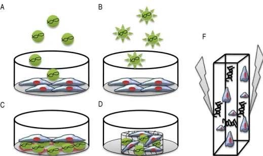
Figure 1. Schematic of DNA delivery to MSCs by chemical (A, B, C and D) and physical methods (F). Non-viral
gene delivery vectors are based on a number of different materials that support the packing of DNA into nanometer sized particles. (A) Conventional non-viral gene delivery (bolus delivery). (B) Ligand-receptor-mediated gene delivery. (C) Substrate-Ligand-receptor-mediated gene delivery (reverse transfection). (D) Scaffold-Ligand-receptor-mediated gene delivery (3D environment) ... 8
Figure 2. Gene delivery strategies. Therapeutic transgenes may be delivered to bone defects by (A) direct
application of gene carrier to the defect or loading the gene carriers onto a biomaterial support (the biomaterial support can deliver the DNA to cells inside the surrounding tissue or can target those cells infiltrating the scaffols or gels), (B) carriers incorporated into the biomaterial support together with cells (hybrid system), or (C) genetic engineering of cells in culture and subsequent implantation into the defect ... 22
SECTION 2
CHAPTER II
Osteogenic Differentiation of Mesenchymal Stem Cells using PAMAM Dendrimers as Gene Delivery Vectors
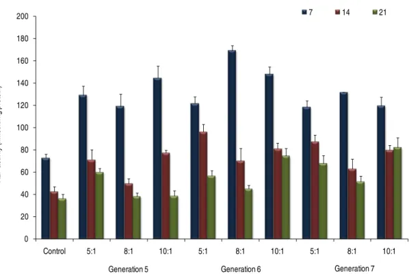
Figure 1. Effect of dendrimer concentration on the percentage of metabolic activity of MSCs in relation to the
control (0 nM): (A) for dendrimers and (B) for polyplexes - this range includes dendrimer concentrations used in later transfection assays (table 2). Values represent means from six replicates ± s.e.m. ... 56
Figure 2.In vitroβ-Galactosidase activity in MSCs 2 days after transfection using G5-7 PAMAM dendrimers at (A) different N:P ratios, using cells from the second passage, and (B) at a fixed N:P ratio of 10, using cells from different passages. For comparison, the β-Galactosidase activity in MSCs and HEK 293T cells, 2 days after transfection and using G6 PAMAM dendrimers, is also presented (C). Results for non-transfected cells (control) and naked DNA (DNA) are also shown. Values represent means from 6 replicates ± s.e.m. ... 57
Figure 3. X-Gal Staining for (A) non-transfected MSCs, (B) transfected MSCs and (C) transfected HEK 293T
cells, using G6 dendrimers at a N:P ratio of 10 ... 58
Figure 4. In vitro BMP-2 expression by MSCs 3 days after transfection. Values represent means from three replicates ± s.e.m. ... 58
xxii Figure 6. Time course of osteocalcin content secreted by MSCs transfected with a plasmid containing the
hBMP-2 gene using generations 5, 6 and 7 of PAMAM dendrimers at various N:P charge ratios.The values represent means from three replicates ± s.e.m. ... 61
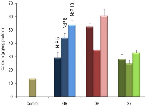
Figure 7. Calcium deposition on extracellular matrix. The cells were transfected with a plasmid DNA containing
the hBMP-2 gene using generations 5, 6 and 7 of PAMAM dendrimers, and cultured for 21 days. The values represent means from three replicates ± s.e.m. ... 62
Figure 8. Von Kossa staining showing matrix mineralization. (A) Non transfected MSCs were compared with
MSCs transfected with a plasmid DNA containing the hBMP-2 gene using PAMAM dendrimers of generations (B) 5, (C) 6 and (D) 7 at a N:P charge ratio of 10. Cells were cultured for 21 days ... 62
CHAPTER III
Delivery of the BMP-2 Gene into MSCs: a Biomimetic approach using RGD Nanoclusters based on
Poly(amidoamine) Dendrimers
Figure 1. Schematic illustration of the synthesis of PAMAM dendrimers/RGD conjugates (pathway A+B), and of the indirect estimation of the number of peptide units by spectrophotometry (pathway A+B+C). Note: The amine content of G5 PAMAM dendrimers (115 amines/dendrimer) was previously determined experimentally ... 76
Figure 2.1H NMR spectra (400MHz, in D2O) of (A) G5 PAMAM/RGD conjugate (sixteen peptides per dendrimer)
and native G5 PAMAM dendrimer, inset is a magnified image of part of the spectrum from 3.4-4.0 ppm; (B) G6 PAMAM/RGD conjugate (sixteen peptides per dendrimer) and native G6 PAMAM dendrimer, inset is a magnified image of part of the spectrum from 3.4-4.0 ppm ... 83
Figure 3. Electrophoretic pattern of pDNA complexes with G5 (A), RGD4-G5 (B), RGD8-G5 (C) and RGD16-G5
(D) vectors. Lane 1, pDNA only; Lane 2-8, for an N:P ratio of 0.25, 0.5, 1, 2, 5, 8 and 10, respectively ... 84
Figure 4. PicoGreen (PG) assay results at various N:P ratios for (A) G5 and G5 PAMAM dendrimers/RGD
conjugates and (B) G6 and G6 PAMAM dendrimers/RGD conjugates. The results are reported as the relative percentage of PG fluorescence, where 100% intensity was observed for a N:P of 0 (pDNA only). Data represents the mean ± s.e.m., n=3 ... 85
Figure 5. Salt induced dissociation of the complexes formed by pDNA and (A) G5 and G5 PAMAM
dendrimer/RGD conjugates and (B) G6 and G6 PAMAM dendrimers/RGD conjugates. The PicoGreen assay was used to estimate the amount of pDNA released. Results are expressed as the mean ± s.e.m., n=3 ... 85
Figure 6. MSCs uptake of the complexes formed by pDNA and (A) G5 and G5 PAMAM dendrimer/RGD
xxiii Figure 7. Microscopy RITC-labeled pDNA visualization in culture, 4 h post-transfection. Live cell images were
captured. (A), (B), (C) and (D) are, respectively, the merged images of bright field mode (gray) and fluorescence mode using G5, RGD4-G5, RGD8-G5 and RGD16-G5 vectors. (E), (F), (G) and (H) are, respectively, the merged images of bright field mode (gray) and fluorescence mode using G6, RGD4-G6, RGD8-G6 and RGD16-G6 vectors. The micrographs were obtained at an original magnification of 200× ... 88
Figure 8. Cellular distribution of (RITC)-labeled pDNA in MSCs at 2 h, 4 h and 24 h post transfection. (A), (B),
(C), and (D) represents cells transfected using G5, RGD8-G5, G6 and RGD8G6 vectors, respectively. The cell nuclei were stained with DAPI (blue), and the acidic late endosome and lysosome compartments were stained with LysoSensor Green DND-189 (green). The bar represents 10 µ m ... 90
Figure 9. (A) Luc gene expression achieved with G5 dendrimer based vectors. (B) Luc gene expression
achieved with G6 dendrimer based vectors. (C) Cytotoxicity evaluation of the complexes formed by pDNA and G5 dendrimer based vectors, 48 h post-transfection. (D) Cytotoxicity evaluation of the complexes formed by pDNA and G6 dendrimer based vectors, 48 h post-transfection. Results are expressed as the mean ± s.e.m., n=6 ... 91
Figure 10. Fluorescence microscopy images showing Enhanced Green Fluorescent Protein expression 24 h
post-transfection. The micrographs (A), (B), (C) and (D) are respectively the transfected MSCs with G5, RGD4-G5, RGD8-G5 and RGD16-G5. The micrographs (E), (F), (G) and (H) are respectively the transfected MSCs with G6, RGD4-G6, RGD8-G6 and RGD16-G6.
The micrographs were obtained at an original magnification of 100×... 92
Figure 11 Cytotoxicity evaluation of dendrimers/RGD conjugates without being complexed with pDNA . Data
represents the mean ± s.e.m., n=6 ... 93
Figure 12. BMP-2 gene expression achieved with (A) G5 dendrimer based vectors and (B) G6 dendrimer based
vectors. Results are expressed as the mean ± s.e.m., n=3... 94
CHAPTER IV
Functionalization of Poly(amidoamine) Dendrimers with Hydrophobic Chains for Improved Gene Delivery in
Mesenchymal Stem Cells
Figure 1. Strategy of synthesis followed in dendrimer surface functionalization with alkyl chains (activation of the
fatty acid + conjugation reaction) ... 107
Figure 2.1H NMR (400MHz) spectra of functionalized dendrimers in D2O ... 113
Figure 3. Agarose gel electrophoresis shift assay results for N:P ratios ranging from 0 (pDNA only) to 8: (A) G5,
xxiv Figure 4. PicoGreen assay. The results are reported as the relative percentage of PG fluorescence, where 100%
intensity was observed for a N:P of 0 (pDNA only). Results are expressed as the mean ± s.e.m obtained from three independent experiments ... 115
Figure 5.(A) Complex size (mean diameter) and polydispersity indices assessed by DLS, and (B) complexes ζ
-potential. Results were obtained for a N:P ratio of 5. Results are expressed as the mean ± s.e.m obtained from three independent experiments ... 116
Figure 6. pDNA protection from serum nucleases achieved with G5 (lanes 9-11), La1-G5 (lanes 13-15), La2-G5
(lanes 17-19), My1-G5 (lanes 21-23), My2-G5 (lanes 25-27), Pa1-G5 (lanes 29-31), and Pa2-G5 (lanes (33-35). Results were obtained for a N:P ratio of 5. Serum only (lanes 1-3) and naked pDNA added of serum (lanes 5-7) were used as controls. Control samples without addition of serum and SDS correspondent to pDNA (lane 4) and complexes formed with G5 (lane 8), La1-G5 (lane 12), La2-G5 (lane 16), My1-G5 (lane 20), My2-G5 (lane 24), Pa1-G5 (lane 28) and Pa2-G5 (lane 32) were also analyzed. Band 1 is the position of sample loading (complexes without serum and SDS). Bands 2 and 3 are relaxed and supercoiled pDNA, respectively. Band 4 results from the complex between serum and SDS ... 117
Figure 7. (A) Complex uptake by MSCs after 1h of contact. The line shows the number of cells positive for
PG-labeled pDNA whereas the bars reveal the amount of pDNA delivered per cell (displayed as the mean average fluorescence intensity). Results are expressed as the mean ± s.e.m obtained from three independent experiments. (B) Correlation between the total number of –CH2– groups contained in dendrimer hydrophobic corona and both the number of cells positive for PG-labeled pDNA (squares) and the amount of pDNA delivered per cell (circles). (C) RITC-labeled pDNA visualization in live cell cultures by fluorescence microscopy, 4 h post-transfection. Original magnifiation: 200x. (D) Distribution of RITC-labeled pDNA inside cells, 2, 4 and 24 h after transfection, when using G5 and La2-G5. Cell nuclei were stained with DAPI (blue).
Original magnification: 1000x ... 118
Figure 8. (A) Luc gene expression achieved with the functionalized dendrimers, 48, 72 and 120 h
post-transfection. SF was used for comparison. (B) Complex cytotoxicity evaluation 24 h post-post-transfection. Results are expressed as the mean ± s.e.m. and were obtained from two independent experiments ... 120
Figure 9. Fluorescence microscopy images showing Enhanced Green Fluorescent Protein expression 24 h
post-transfection. Original magnification: 100x ... 121
Figure 10. Cytotoxicity evaluation of the functionalized dendrimers. Each data point represents the mean ±
xxv CHAPTER V
Receptor-Mediated Gene Delivery using PAMAM Dendrimers Conjugated with Peptides Recognized by
Mesenchymal Stem Cells
Figure 1. The two-step reaction for the synthesis of peptide-functionalized G5 PAMAM dendrimers (pathway
A+B), and for indirect estimation of the number of peptide units by spectrophotometry (pathway A+C). Note: The amine content of G5 PAMAM dendrimers (115 amines/dendrimer)
was previously determined experimentally ... 135
Figure 2.1H NMR of (A) G5-(HAB)8 and of (B) G5 native dendrimer. The inset shows the imidazole proton peaks
from the amino acid histidine ... 142
Figure 3. (A) Agarose gel retardation assay results for N:P ratios ranging from 0 (pDNA only) to 8: (a) G5; (b)
G5-(HAB)2; (c) G5-(HAB)4; (d) G5-(HAB)8; (e) G5-(LAB)2; (f) G5-(LAB)4. Binding is shown by the inhibition of
pDNA electrophoretic mobility (band 1). Bands 2 and 3 show the relaxed and supercoiled forms of pDNA, respectively. (B) PicoGreen assay. The results are reported as the relative percentage of PG fluorescence, where 100% intensity was observed for a N:P of 0 (pDNA only). Results are expressed as the mean ± s.e.m obtained from three independent experiments ... 143
Figure 4. (A) Polyplex size (mean diameter) and polydispersity indices assessed by DLS, and (B) polyplexes ζ
-potential. Results were obtained for a N:P ratio of 5. Results are expressed as the mean ± s.e.m obtained from three independent experiments ... 144
Figure 5. Polyplex uptake by MSCs after 2 h of contact. The line shows the number of cells positive for
PG-labeled pDNA, whereas the bars reveal the amount of pDNA delivered per cell (displayed as the mean average fluorescence intensity). Results are expressed as the mean ± s.e.m obtained from three independent experiments ... 145
Figure 6. Cellular distribution of (RITC)-labeled pDNA (2 and 4 h post-transfection) using native dendrimers and
G5-(HAB)4 and G5-(LAB)4 as vectors. The acidic late endosome and lysosome compartments were stained with
LysoSensor Green DND-189 (green), and the nucleus with DAPI (blue). Original magnification: 1000x ... 146
Figure 7. (A) Luc gene expression achieved with HAB peptide-functionalized dendrimers, (B) Luc gene
expression achieved with LAB peptide-functionalized dendrimers, and (C) comparison of Luc gene expression 48 h post-transfection achieved with HAB peptide-functionalized dendrimers for different pDNA concentrations (1 µ g.cm-2 and 2 µ g.cm-2). (D) Cytotoxicity evaluation 24 h post-transfection. Results are expressed as the mean ±
s.e.m. and were obtained from two independent experiments ... 147
Figure 8. Fluorescence microscopy images showing Enhanced Green Fluorescent Protein expression 24 h
post-transfection using: (A) naked DNA, (B) G5 PAMAM dendrimers, (C) Superfect, (D) G5-(HAB)2, (E) G5-(HAB)4, (F)
xxvi Figure 9. Cytotoxicity evaluation of peptide-functionalized dendrimers. Each data point represents the mean ±
s.e.m. of two independent measurements ... 149
Figure 10. (A) Luc gene expression achieved with HAB peptide-functionalized dendrimers with and without
xxvii
LIST OF TABLES
SECTION 2
CHAPTER II
Osteogenic Differentiation Ff Mesenchymal Stem Cells using PAMAM Dendrimers as Gene Delivery Vectors
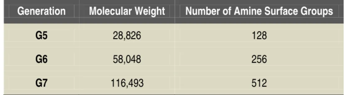
Table 1.Molecular weight and number of amine surface groups for the three generations of PAMAM dendrimers
used ... 50
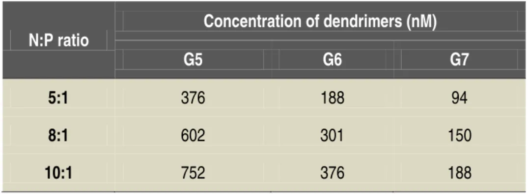
Table 2. Concentration of dendrimers in solution for the N:P charge ratios used in the
transfection experiments ... 53
CHAPTER III
Delivery of The BMP-2 Gene into MSCs: A Biomimetic approach using RGD Nanoclusters based on
Poly(Amidoamine) Dendrimers
Table 1. Average number of PDP and of peptides per dendrimer, respectively, after reactions A and B.. ... 83
CHAPTER IV
Functionalization of Poly(amidoamine) Dendrimers with Hydrophobic Chains for Improved Gene Delivery in
Mesenchymal Stem Cells
Table 1. Identification of the developed gene delivery vectors and characterization
of their hydrophobic moieties. ... 114
CHAPTER V
Receptor-Mediated Gene Delivery using PAMAM Dendrimers Conjugated with Peptides Recognized by
Mesenchymal Stem Cells
xxix
SHORT
CURRICULUM VITAE
José Luis Santos was born in 1982, in Funchal, Portugal. He graduated in Chemistry (4 years course) in the University of Madeira and, in 2005, received the Best Chemistry Student Award from the University of Madeira.
In 2005, he was awarded a Ph.D. grant from the Fundação para a Ciência e Tecnologia (FCT) and, since then, he is a Ph.D. Student at the University of Madeira, developing his experimental work integrated in the Molecular Materials Research Group at Centro de Química da Madeira (CQM), Department of Chemistry, University of Madeira. His Ph.D. work was done under the supervision of Prof. Helena Tomás (CQM and University of Madeira) and co-supervision of Dr. Pedro L. Granja (INEB-Instituto de Engenharia Biomédica, University of Porto).
In the period covered by the Ph.D. grant, he participated in 12 Advanced Courses/Summer Schools covering diverse topics related with the theme of his Ph.D. thesis. He has also contributed for several research projects and activities developed at CQM and UMa that are described in more detail below.
As a result of his research efforts, he is author or co-author of 6 publications in international peer-reviewed journals (2 published and 4 submitted for publication). His work also resulted in 5 oral presentations and 13 poster presentations mostly in international scientific meetings.
PROFESSIONAL FORMATION
2009- International Symposium “20 years of Biomagical Engineering in Porto” (Porto, Portugal).
2009- 10th Advanced Course in Cell-Materials Interactions – Self assembly: from nature to clinics
(Porto, Portugal).
2008- 5th Marie Curie Cutting Edge Conference on Synthesis and applications of self-assembling
materials at nano-scale (Funchal, Portugal).
2007- 8th Advanced Course in Cell-Materials Interactions – Inflamation in Tissue Repair and
Regeneration
2007-
(Porto, Portugal).
1st TERMIS-EU Summer School on Key Elements of Tissue Engineering (Funchal,
xxx
2007- 3rd Joint Summer School on Hybrid Materials, organized in the scope of FAME european
research network (Funchal, Portugal).
2007- Gordon Research Conference on Biomaterials: Biocompatibility / Tissue Engineering (Plymouth, New Hampshire, USA).
2007- 3rd Marie Curie Cutting Edge Conference on Biomineralisation of polymeric materials,
bioactive biomaterials and biomimetic methodologies (Funchal, Portugal).
2007- Microscopy course at the Faculty of Pharmacy/University of Porto (Porto, Portugal).
2006- 7th Advanced Course in Cell-Materials Interactions – Regenerative Medicine (Porto,
Portugal).
2006- 1st Marie Curie Cutting Edge Conference on New developments on polymers for tissue
engineering: replacement and regeneration (Funchal, Portugal).
2005- 6th Advanced Course in Cell-Materials Interactions at Molecular level
DENDRALGENE - Design of new gene delivery vectors based on dendrimers, alginate and the RGD sequence for bone tissue engineering. Funding through Fundação para a Ciência e Tecnologia, Ref. PTDC/SAU-BEB/71161/2006 (175000 euros). Researcher of the project since September 2006.
(Porto, Portugal).
PARTICIPATION IN PROJECTS
DENDRIFAT - Functionalization of dendrimers using fatty acids for improved efficiency as gene delivery vectors. Funding through UMa (458 euros). Researcher of the project during 2008.
Novel drug/gene delivery platforms based on the self-assembly of dendrimer-single strand DNA conjugates (Post-Doc project of Deepti Pandita, PhD). Collaborator since
2009.
In Vitro study of Human Stem Cell behavior on Yb:YAG laser irradiated Titanium in presence or absence of Hydroxyapatite (part of the PhD project of Dr. Hewerson Tavares, visiting researcher in CQM in 2008) . Collaborator during 2008.
xxxi
LIST OF PUBLICATIONS (SCI)
1. Santos JL
2. Alves NM, Shi J, Oramas E,
, Oramas E, Pego AP, Granja PL, Tomás H. Osteogenic differentiation of mesenchymal stem cells using PAMAM dendrimers as gene delivery vectors. J. Controlled Release 2009; 134: 141-148 (2008 Impact Factor: 5.690).
Santos JL
3.
, Tomás H, Mano JF. Bioinspired superhydrophobic poly(L-lactic acid) surfaces prevent the adhesion and proliferation of bone marrow derived cells. J. Biomed. Mater. Res. Part A. 2009 (in press). DOI: 10.1002/jbm.a.32210 (2008 Impact Factor: 2.706).
Santos JL
4.
, Pandita D, Rodrigues J, Pêgo AP, Granja PL, Balian G, Tomás H. Receptor-mediated gene delivery using PAMAM dendrimers conjugated with peptides recognized by mesenchymal stem cells (submitted).
Santos JL
5.
, Oliveira H, Pandita D, Rodrigues J, Pêgo AP, Granja PL, Tomás H. Functionalization of Poly(amidoamine) Dendrimers with hydrophobic chains for improved gene delivery in mesenchymal stem cells (submitted).
Santos JL
6. Pandita D,
, Pandita D, Rodrigues J, Pêgo AP, Granja PL, Tomás H. Non-viral gene delivery to mesenchymal stem cells: strategies and applications (submitted).
Santos JL
ABSTRACTS IN REFEREED JOURNALS
, Rodrigues J, Pêgo AP, Granja PL, Tomás H. Delivery of the BMP-2 gene into MSCs: a biomimetic approach using RGD nanoclusters based on poly(amidoamine) dendrimers (submitted).
1. Santos JL, Rodrigues J, Pêgo AP, Granja PL, Tomás H. Novel conjugates of dendrimers
and fatty acids for gene delivery applications. Tissue Eng. A 2008; 14:S888-S889.
xxxii ORAL COMMUNICATIONS
1. Santos JL
2. Song W, Veiga DD, Shi J, Oramas E,
, Pandita D, Rodrigues J, Granja PL, Pêgo AP, Tomás H. Dendrimers surface engineering for improved cellular uptake and gene delivery to Mesenchymal Stem Cells. 4th
International Meeting of the Portuguese Society for Stem Cells and Cellular Therapy - Novel Frontiers in Stem Cell Research. Lisbon, Portugal: April 29-30, 2009.
Santos JL
3.
, Tomás H, Alves NM, Mano JF. Bioinspired surfaces with extreme wettability ranges to control cell attachment and protein adsorption. Tissue Engineering & Regenerative Medicine International Society - Asia-Pacific Chapter Meeting 2008 (TERMIS-AP 2008 Meeting). Taipei, Taiwan: November 6-8, 2008.
Santos JL
4.
, Oramas E, Pêgo AP, Granja PL, Tomás H. In vitro osteoblastic differentiation of MSCs using PAMAM Dendrimers as delivery vectors for the hBMP-2 gene. 8th World
Biomaterials Congress. Amsterdam, The Netherlands: May 28-June 01, 2008.
Santos JL
5.
, Oramas E, Pego AP, Granja PL, Tomás H. Osteogenic differentiation of mesenchymal stem cells through gene delivery. 3rd International Meeting of the Portuguese
Society for Stem Cells and Cellular Therapy - From Molecular Mechanisms to Therapeutical applications. Faro, Portugal: April 23-24, 2008.
Santos JL
POSTER COMMUNICATIONS
, Oramas E, Pego AP, Granja PL, Tomás H. In vitro osteogenesis via gene delivery. 5th Marie Curie Cutting-Edge Conference - Synthesis and applications of
self-assembling materials at nano-scale. Funchal, Portugal: April 14-18, 2008.
1. Santos JL
2.
, Pandita D, Rodrigues J, Granja P, Pêgo AP, Balian G, Tomás H. Receptor-Mediated gene delivery in Mesenchymal Stem Cells by PAMAM dendrimers conjugated with Osteotropic peptides. 10th Advanced Course in Cell-Materials Interactions. Porto, Portugal:
June 22-25, 2009.
Santos JL, Pandita D, Rodrigues J, Granja PL, Pêgo AP, Tomás H. Dendrimers surface
engineering for improved cellular uptake and gene delivery to Mesenchymal Stem Cells. 4th
xxxiii
3. Santos JL
4.
, Oramas E, Pego AP, Granja PL, Tomás H. PAMAM dendrimers used as vectors for gene delivery into Mesenchymal Stem Cells. NanoSpain 2008. Braga, Portugal: April 14-18, 2008.
Santos JL
5.
, Oliveira H, Oramas EG, Pego AP, Granja PL, Tomás H. Gene therapy platform for bone regeneration using dendrimers and osteogenic plasmids systems. Gordon research Conference on Biomaterials: Biocompatibility / Tissue Engineering. Plymouth, New Hampshire, USA: July 22-27, 2007.
Santos JL
6.
, Oramas EG, Pego AP, Granja PL, Tomás H. Polyamidoamine dendrimer vectors for gene delivery: Physicochemical characterization and transfection efficiency evaluation. 8th
Advanced Summer Course in Cell-Materials Interactions. Porto, Portugal: June 18-22, 2007.
Santos JL
7.
, Oramas EG, Pego AP, Granja PL, Tomás H. In vitro gene delivery studies using polyamidoamine dendrimers: physicochemical characterization of dendriplexes and transfection efficiency evaluation. 3rd Marie Curie Cutting Edge Conference - Biomineralisation of polymeric materials, bioactive biomaterials and biomimetic methodologies. Funchal, Portugal: June 4-8, 2007.
Santos JL
8. Rocha-Sousa L,
, Granja PL, Tomás H. In vitro evaluation of gene delivery in bone marrow stromal cells by polyamidoamine dendrimers. 14th Annual Congress of the European Society of Gene Therapy (ESGT). Athens, Greece: November 9-12, 2006.
Santos JL
9.
, Pelláez-Abellán E, Guastaldi AC, Tomás H. Cytocompatibility modified by spark-anodization, 4º Congresso Latino-Americano de Órgãos Artificiais e Biomateriais. Caxambú, Brasil: August 8-11, 2006.
Santos JL
10.
, Pêgo AP, Oliveira H, Granja PL, Tomás H. PAMAM dendrimers as gene delivery vectors for bone tissue engineering. 7th Advanced Course in Cell-Material Interactions. Porto,
Portugal: June 19–23, 2006.
Santos JL, Pêgo AP, Oliveira H, Granja PL, Tomás H. Use of polyamidoamine Dendrimers
as gene delivery vectors for bone tissue engineering. 1st Marie Curie Cutting Edge
xxxiv
11. Santos JL
12.
, Granja PL, Tomás H. PAMAM dendrimers as vectors for gene delivery into human primary osteoblastic cells – preliminary cytotoxicity studies. XIII Annual Congress of the European Society for Gene Therapy (ESGT). Prague, Czech Republic: October 29 – November 1, 2005.
Santos JL
13.
, Rodrigues J. Degradation of organic dyes by TiO2 nanoparticules under visible
artificial light or natural solar irradiation. 6th Conference on Inorganic Chemistry (Portuguese
Society of Chemistry). Funchal, Portugal: March 31 – April 2, 2005.
Santos JL
ORAL COMMUNICATIONS IN
, Rodrigues J. Photocatalytic degradation of Congo Red using WO3-Preliminary
study. XIX Congress of the Portuguese Society of Chemistry. Coimbra, Portugal: April 15-17,
2004.
CQM AND INEB
1.
INTERNAL MEETINGS
Santos JL
2.
, Pandita D, Rodrigues J, Pêgo AP, Granja PL, Tomás H. Functionalization of poly(amidoamine) dendrimers for improved gene delivery . 4th Materials Line Meeting.
Funchal, Portugal: January 30, 2009.
Santos JL
3. Sargo S,
, Oramas E, Pêgo AP, Granja PL, Tomás H. In Vitro osteoblastic differentiation of MSCs using PAMAM dendrimers as delivery vectors for the hBMP-2 gene. 3rd Materials Line
Meeting. Funchal, Portugal: January 25, 2008.
Santos JL
4.
, Rodrigues J, Pêgo AP, Granja PL, Tomás H. PAMAM dendrimers/alginate nanoparticles for gene delivery. 3rd Materials Line Meeting. Funchal,
Portugal: January 25, 2008.
Santos JL
5.
, Granja PL, Tomás H. Gene therapy for bone regeneration. 2nd Materials Line
Meeting. Funchal, Portugal: January 26, 2007.
Santos JL, Granja PL, Tomás H. Bone Tissue engineering via local gene delivery. 19th
xxxv OTHER ACTIVITIES
Collaboration in the organization of several scientific conferences:
• 6th Conference on Inorganic Chemistry (Portuguese Society of Chemistry). Funchal,
Portugal: March 31 – April 2, 2005.
• 1st Marie Curie Cutting Edge Conference - New developments on polymers for tissue
engineering: replacement and regeneration. Funchal, Portugal: June 1–5, 2006.
• 3rd Marie Curie Cutting Edge Conference - Biomineralisation of polymeric materials, bioactive biomaterials and biomimetic methodologies. Funchal, Portugal: June 4-8, 2007.
PhD student representative in the scope of the external evaluation of University of Madeira, Portugal, performed by the European University Association (EUA), 2008.
Collaboration in several events for enhancing public awareness of science regularly organized by Madeira Chemistry Research Centre (CQM)/Department of Chemistry (University of Madeira):
“A Química é Divertida” (Chemistry is Fun) – a project (one week of experimental demonstrations) to promote the scientific culture in Chemistry and Biochemistry among the young people. Collaborator in the period 2004-2008.
“Ocupação científica de jovens nas férias” (Scientific occupation of young people during summer holidays) – activity promoted at the national level by “Agência Nacional para a cultura Científica e Tecnológica – Ciência Viva”. Collaborator in the period 2004-2009. Participation in a TV program for science popularization among children (Panda Doc
series, Panda Channel from Cable TV, 2008).
Collaboration in practical lessons (laboratory works) of the disciplines: Biomaterials and Tissue Engineering (Master in Applied Biochemistry, Univ. of Madeira) and Molecular and Cellular Characterization Techniques (3rd year, Degree in Biochemistry, Univ. of Madeira).
Collaborator in 2007/2008 and 2008/2009.
CHAPTER I.
CHAPTER I. Non-Viral Gene Delivery to Mesenchymal Stem Cells
5
CHAPTER I.
Non-Viral Gene Delivery to Mesenchymal Stem Cells: Strategies and Applications*
ABSTRACT
Mesenchymal stem cells (MSCs) can be isolated from several tissues in the body, have the ability to self-renewal, show immune suppressive properties and are multipotent, being able to generate various cell types. At present, due to their intrinsic characteristics, MSCs are considered very promising in the area of tissue engineering and regeneration. In this context, genetic modification can be a powerful tool to control the behavior and fate of these cells and be used in the design of new cellular therapies. Viral systems are very effective in the introduction of exogenous genes inside MSCs. However, the risks associated with their use are leading to an increasing search for non-viral approaches to attain the same purpose even if MSCs have been shown to be more difficult to transfect by this way. Progress has been made in the last years in the development of chemical and physical methods for non-viral gene delivery. Herein, the application of those methods specifically to MSCs is reviewed and their success in the delivery of therapeutic genes discussed. Key issues in non-viral gene delivery to MSCs are addressed and problems that remain to be solved highlighted.
This chapter is based on the following publication:
Santos JL, Pandita D, Rodrigues J, Pêgo AP, Granja PL, Tomás H, Non-Viral Gene Delivery to Mesenchymal Stem
CHAPTER I. Non-Viral Gene Delivery to Mesenchymal Stem Cells
6
1. GENERAL INTRODUCTION
A huge number of studies have demonstrated the importance of mesenchymal stem cells (MSCs) in regenerative medicine and tissue engineering1-9. Indeed, they can be isolated from
several tissues throughout the body, expanded in culture, and stimulated to differentiate into mesenchymal and non-mesenchymal cell lineages, characteristics that make them attractive in the biomedical area. Furthermore, MSCs are harvested from adult individuals (autologous cells can be used), thus not raising ethical problems in their manipulation and use10. Also MSCs have
been shown to be able to suppress immune responses in various situations like organ transplantation and autoimmune diseases9,11. Indeed, there is now overwhelming evidence that
the mechanisms of action of MSCs in tissue repair include not only cell engraftment and differentiation but also signaling through paracrine secretions and cell-to-cell contacts.
MSCs applications can be potentiated using gene delivery approaches12-18. Processes
such as proliferation, migration, cell-cell and cell-matrix interactions, differentiation, apoptosis, and secretion of soluble signaling molecules can be controlled through the introduction of exogenous genes inside cells that will result in the synthesis of a specific protein (therapeutic protein). In regenerative medicine and tissue engineering, genetic modification is preferred over the exposition of cells to growth factors and cytokines as the short half-life and body clearance of these molecules may imply the use of either high (non physiological) concentrations or repeated administrations to produce the desired biological effect. Also recombinant proteins may not provide the therapeutic benefit of natural proteins13. In fact, the power of cell genetic engineering
was recently highlighted by experiments that showed the possibility of obtaining pluripotent stem-like cells (induced pluripotent stem cells, iPS cells) by transferring genes encoding for certain transcription factors into non-pluripotent cells19,20.
A simple strategy of gene therapy can be followed where genes are directly delivered into the site of injury13,21. In this case, administration to the target tissue/organ can be done by
CHAPTER I. Non-Viral Gene Delivery to Mesenchymal Stem Cells
7 after, implanted in the injury site13,21,22. During the in vitro process, MSCs can be cultured in cell
culture dishes or already in a three-dimensional (3D) environment (scaffold) that, after cell colonization, will be implanted in the patient body. In fact, a number of studies have demonstrated that MSCs can be used for cell-mediated gene therapy and as a tool for understanding the molecular mechanisms leading to repair and regeneration of complex tissues and organs12-18,21,22.
Both simple gene therapy and cell-mediated gene therapy make use of gene delivery systems. An ideal gene delivery system should present various features, like ensure protection of DNA, achieve desired level of gene transfer and expression, exhibit specific targeting, prevent non-specific interactions with blood components, be nontoxic and non-immunogenic, and be cost effective23-25. Broadly, gene delivery systems can be divided in two major categories: viral and
non-viral systems14,23-25. The traditional method to introduce a therapeutic gene into MSCs
involves the use of viral vectors, such as lentivirus, retrovirus, adeno-associated virus and adenovirus derived vectors26-28. Although viral vectors present high efficiencies in gene transfer (a
process called “infection”) and may allow stable gene expression, their clinical applications are currently narrowed due to potential problems, such as oncogenic transformation, pathogenic risk, and induction of immune responses23-25. Furthermore, they present difficulty in large-scale
production and size limits for exogenous DNA23-25. These complications gave rise to substantial
efforts in the development of alternative non-viral systems (gene transfer is, in this case, called
“transfection”) which may be additionally classified in physical and chemical methods (Figure 1). Physical methods comprise the simplest, but ineffective, way of delivering genes to cells that is to use naked DNA (without any carrier) in contact with cells14,29. They also include physical
manipulations of the cells that will result in improved gene transfer such as microinjection, particle bombardment, electroporation, sonoporation and laser irradiaton14,29,30. Chemical methods imply
the use of natural or synthetic materials (the so called “non-viral vectors”) that are able to transfer the genetic material into the cell31-34. Non-viral systems offer several advantages, like any or low
immunogenicity (thus, more than one application to the patient may be applied), no risk of infectious disease, flexibility towards the molecular size of loaded DNA, and also low cost23-25.
When non-viral vectors are used, there is also the possibility of engineering them for targeting specific tissues35-37. Actually, non-viral vectors offer a great structural and chemical versatility,
CHAPTER I. Non-Viral Gene Delivery to Mesenchymal Stem Cells
8 barriers for an improved gene delivery and expression will be overcome31-34. Unfortunately,
non-viral systems present also some drawbacks. On one hand, physical methods are more difficult to be applied in an in vivo situation and can cause tissue damage29,30,38. On the other hand, for
chemical methods: (a) transfection efficiency compared to viral vectors, both in vitro and in vivo, is low25,39; (b) gene expression is transient25,39; (c) toxicity may be high40; (d) the level of
transfection strongly varies with the cell type14,41,42. Indeed, most of non-viral vectors have only
been tested in cell lines, being difficult to extrapolate the in vitro results to a clinical situation. Often, primary cells, as is the case of MSCs, are refractory to transfection using the majority of non-viral vectors42.
Figure 1. Schematic of DNA delivery to MSCs by chemical (A, B, C and D) and physical methods (F). Non-viral gene
delivery vectors are based on a number of different materials that support the packing of DNA into nanometer sized particles. (A) Conventional non-viral gene delivery (bolus delivery). (B) Ligand-receptor-mediated gene delivery. (C) Substrate-mediated gene delivery (reverse transfection). (D) Scaffold-mediated gene delivery (3D environment).
The advantages of exerting genetic control over MSCs behavior and fate associated with the need to move towards non-viral gene delivery represent a current challenge. Research is being done not only focused on the development of better and safer non-viral gene delivery methods but also on the understanding of the mechanisms of the gene delivery process and of its effects on the behavior of MSCs. Herein, we start by brief reviewing the current knowledge on the nature and properties of MSCs, followed by an overview of the use of non-viral gene delivery systems specifically for MSCs transfection. The different strategies used in the delivery of therapeutic genes to those cells for tissue engineering and regeneration applications are
A B
C D
CHAPTER I. Non-Viral Gene Delivery to Mesenchymal Stem Cells
9 exemplified and discussed. Finally, the main aspects and problems to be solved related with non-viral gene delivery to MSCs are highlighted.
2. MESENCHYMAL STEM CELLS
MSCs were first described in the pioneering studies of Fridenstein, Owen and co-workers in the 60s and 70s43-48, followed by intensive work on the study of the properties of these
cells49,50. They were isolated from rat marrow based on their adherence to plastic tissue culture
surfaces and were shown to be able to differentiate towards the osteoblastic, adipogenic and chondrogenic lineages. Early studies demonstrated that these cells proliferated rapidly in vitro and gave rise to distinct colonies from single precursors, being thus called “fibroblast colony forming units” (CFU-F). The term “mesenchymal stem cells” came after, being first applied by Caplan in 1991, reflecting their “stem cell” nature51 – that is, capacity for self-renewal (when
dividing, they give rise to at least one daughter cell identical to the initial cell) and retention of differentiation potential (MSCs are considered multipotent and can differentiate into specific mesenchymal cell types under the control of environmental cues). Meanwhile, several other names have been attributed to these cells, such as “marrow stromal cells”, “mesenchymal stromal cells”, “skeletal stem cells”, “multipotent mesenchymal stromal cells”, and “multipotent adult progenitor cells” This controversy in cell’s nomenclature is certainly related with the reduced knowledge that still exists concerning the nature, functions and differentiation potential of MSCs1,7,52.
For many years MSCs have been isolated from low-density mononuclear cell population of bone marrow based on their selective adherence to plastic surfaces, compared to hematopoietic cells which are non-adherent and can therefore be removed through medium changes53-56. MSCs
reactivity to monoclonal antibodies was also applied to sort a homogeneous population of cells with defined phenotype by flow cytometry (FACS)57-59. Indeed, along the years, several
CHAPTER I. Non-Viral Gene Delivery to Mesenchymal Stem Cells
10 with hematopoietic cell lineages and readily differentiated into osteoblasts, adipocytes and chondroblasts. However, up to now, all the isolation methods used suffer from lack of absolute specificity due to the lack of specific “markers” that can be employed to select MSCs.
Besides bone marrow, MSCs were also isolated from other adult and foetal tissues – e.g., cord blood, placenta, amniotic fluid, heart, skeletal muscle, adipose tissue, synovial tissue, brain, spleen, liver, kidney, lung, thymus, pancreas, etc.1-9,61-70. The wide distribution of these cells in
the body raised the hypothesis that MSCs exist in perivascular niches5. Furthermore, there are
reports about MSCs differentiation not only into mesenchymal tissue lineages but also into non-mesenchymal tissue lineages (such as epithelial, endothelial, and neural cells)1-9,52,71. Some
discussion exists however around these discoveries and the characterization of these cells and so, in order to distinguish them from MSCs derived from bone marrow (the most studied source of MSCs), it was suggested that they should be called “MSCs-like cells”9. In an interesting review, it
was even suggested that MSCs under certain conditions (i.e. depending on the cell environment), can de-differentiate returning to a stem state and becoming pluripotent52.
As explained before, the potential of delivering genes to MSCs (or MSCs-like cells) has been recognized and a broad range of gene delivery systems are now available for the effect. Non-viral systems, being more versatile and safer, are increasingly attractive for the researchers and clinicians that are working in this field.
3. NON-VIRAL GENE DELIVERY TO MESENCHYMAL STEM CELLS
Non-viral gene transfer is normally accomplished using a plasmid DNA (circular molecules of double stranded DNA, pDNA) which contains the gene encoding the protein of interest under the transcriptional control of several eukaryotic regulatory elements and is amplified in bacteria72.
CHAPTER I. Non-Viral Gene Delivery to Mesenchymal Stem Cells
11 formed endosomes and decomplexation must occur 33,73,74. The all process of gene transfer will
then depend on the preparation, purification and composition of the pDNA, but also on the type of cell used as target, on the non-viral system chosen for the transfection and on the experimental conditions used in the process. Due to all these variables, a strict comparison among the research works published on this subject is difficult to make. However, it is possible to extract general conclusions from a literature survey and to get an idea of the best directions to be followed.
3.1. Chemical Methods
3.1.1. Liposome-based vectors
Cationic lipids have polar heads and non-polar tails, and as such, can self-assemble forming liposomes33,75. The positive charge associated with these structures allows their
interaction with the anionic molecules of pDNA, being possible to use them as gene delivery vehicles. The DNA molecules are either entrapped in the internal aqueous space of liposomes or bounded on their surface, in both cases forming “lipoplexes”. It is believed that, after endocytosis, lipoplexes destabilize the endosomal membrane causing the release of DNA into the cytoplasm. “Lipofection” is a term currently used to mean transfection mediated by liposomes.
Several lipid transfection reagents commercially available, like Lipofectin® and Lipofectamine™, Effectene®, Fugene® 6, and Metafectene®, etc., are now being used for genetic
engineering of cells76-79. Even if liposome-based DNA delivery systems were one of the earliest
strategies used to introduce exogenous genetic material into animal cells, lipofection studies specifically dealing with MSCs are scarce. Hoelters et al. transfected human MSCs (hMSCs) with three different liposome-based transfection reagents and a plasmid containing the sequence for enhanced green fluorescent protein (EGFP)76. The highest fraction of fluorescent cells was
obtained using Lipofectamine™2000 (50%), followed by Metafectene® (22%), and Lipofectamine