Abstract published online: April 12, 2010
Full paper published online: May 30, 2010
Original paper. ISSN 1678-9199.
Effect of Curcuma zedoaria crude extract against tumor progression and immunomodulation
Carvalho FR (1), Vassão RC (2), Nicoletti MA (3), Maria DA (1)
(1) Laboratory of Biochemistry and Biophysics, Butantan Institute, São Paulo, São
Paulo State, Brazil; (2) Laboratory of Cell Biology, Butantan Institute, São Paulo, São
Paulo State, Brazil; (3) Department of Pharmacy, University of São Paulo, São
Paulo, São Paulo State, Brazil.
ABSTRACT: The aim of the present work was to study the effect of the crude extract of Curcuma zedoaria on peripheral blood cells and tumor progression in C57Bl/6J
mice injected with B16F10 murine melanoma cells. The intraperitoneal therapy
showed a significant increase in total white and red blood cell counts, a decrease in
peritoneal cell number and tumor volume reduction, whereas the oral administration
revealed a noteworthy augmentation only in total leukocyte count. These results
contribute to evaluate the importance of alternative treatments that employ
phytotherapic compounds against tumor progression and its possible
immunomodulation.
KEY WORDS: melanoma, tumor, hematology, Curcuma zedoaria.
CONFLICTS OF INTEREST: There is no conflict.
CORRESPONDENCE TO:
DURVANEI AUGUSTO MARIA, Laboratório de Bioquímica e Biofísica, Instituto
Butantan, Av. Vital Brasil, 1500, São Paulo, SP, 05.503-900, Brasil. Phone:+55 11
INTRODUCTION
Curcuma zedoaria Roscoe (Zingiberaceae family) is a phytotherapic agent
traditionally cultivated in Asian countries and largely employed in oriental medicine.
According to the traditional Chinese medicine, C. zedoaria rhizomes contain several
specific sesquiterpenes that are effective against flatulent colic, debility of the
digestive organs, amenorrhea, hepatocirrhosis, cancer, oxidation and human blood
aggregation (1-5). Moreover, its rhizome is generally used as stomachic in Japan
whereas in Chinese medicine it has been prescribed for the treatment of blood
stagnation syndromes and for promoting menstruation (6).
Among the main hepatoprotective sesquiterpenes isolated from the aqueous acetone
extract of C. zedoaria rhizome, furanodiene, germacrone, curdione, neocurdione,
curcumenol, isocurcumenol, aerugidiol, zedoarondiol, curcumenone and curcumin
revealed potent effect on D-galactosamine- (D-Gain) and lipopolysaccharide- (LPS)
induced acute liver injury in mice. Action mechanisms for their hepatoprotective
activity were elucidated based on the inhibitory effect on D-Gain-induced cytotoxicity
in primary cultured rat hepatocytes, LPS-induced nitric oxide (NO) production in
cultured mouse peritoneal macrophages, and D-Gain and tumor necrosis factor-α
(TNF-α) induced liver injury in mice (6).
More than 20 sesquiterpenes, curcuminoids, and ethyl p-methoxycinnamate were
reported as chemical constituents of C. zedoaria (7, 8). In a previous study, a crude
ethanolic extract of C. zedoaria showed inhibitory effect against OVCAR-3 cells
(human ovarian cancer) (7). Furthermore, a fraction of C. zedoaria ethanolic extract
has shown potent antitumoral and enzymatic actions in vitro (7).
In the present study, we evaluated the effects of crude C. zedoaria extract
administered either orally and intraperitonially, as well as the impact of extract
sesquiterpenes on B16F10 melanoma growth.
MATERIALS AND METHODS Fluid Extract Preparation
One thousand grams of dried root were divided into three portions: 500 g, 300 g and
200 g. The 500-g sample was humidified, saturated with the extraction liquid (ethanol
70° GL) and percolated. The first aliquot of 200 cm3 of percolate was stored until use,
cm3 each, so that the more concentrated extract was separated from the last weak
percolates.
The second amount of dried root (300 g) was moistened, humidified and saturated
with the percolates collected during the previous step. The first volume of 300 cm3
was collected and percolation continued up to 1,000 cm3, which was collected in
200-cm3 aliquots. Finally, the third fraction of the root (200 g) was moistened, humidified
and saturated with the remaining percolates and processed similarly to the second
sample. Five hundred cubic centimeters of percolate were collected and mixed with
the two samples (200 and 300 cm3) previously obtained, thus totalizing 1,000 cm3 of
fluid extract.
Animals and Treatment
Groups of five 1.5-month-old C57Bl/6J male mice, weighing between 20 and 25 g,
were intraperitoneally injected with or orally received, once a week, different
Curcuma zedoaria doses diluted in water. The control group was injected with the
same concentrations of the extraction liquid diluted in water. The other groups
received two different doses (orally or intraperitoneally) of Curcuma zedoaria, 0.1 and
0.2%, diluted in water.
The animals were weighed weekly and 10 μL of blood was collected from the venous
retrorbital plexus in the presence of heparin (5,000 UI, Roche). Blood smears from
each sample were spread on glass slides, fixed with methanol for five minutes,
air-dried and stained (1:4) with Giemsa® (Merck, Germany) for differential leukocyte
count. Total saline diluted red blood cell suspension and Turk stained (1:10) total
leukocytes were counted under a microscope Axiovert 40 MAT® (Zeiss, Germany)
with a hemocytometer chamber of the Malassez type.
After three months (oral route treatment) and two months (intraperitoneal treatment),
the animals were killed and underwent necropsy, then, peritoneum cells were
washed with 2 mL of iced PBS. The peritoneal exudate was collected and total
resident cells were counted on Malassez chambers. The different types of leukocytes
were placed on glass slides (Cytospin® Centrifuge, Eppendorf, USA), stained with
DifQuick® (Merck, Germany) and, afterward, analyzed and counted under a
microscope. The results concerning the effects of different doses of Curcuma
statistical analysis (Student's t-test: p < 0.05, 0.01 > p > 0.001 and p < 0.001). Data
were expressed as mean ± SD for each experimental group.
Treatment of Primary Tumor Growth of B16F10 Cells
B16F10 murine melanoma cells were obtained from the Ludwig Institute of Cancer,
Switzerland. Groups of five C57Bl/6J mice were subcutaneously injected with 5 x 10⁴
B16F10 melanoma cells. Subsequently, the animals were treated with Curcuma
zedoaria extract as aforementioned. The tumor growth was observed weekly and its
volume was calculated using the following formula: (length)2 x (width) x (π/6). The
efficacy of the treatment was calculated as the ratio of tumor size in treated animals
to the tumor size in control animals.
The statistical analysis was performed using the Student's t-test (p < 0.05, 0.01 > p >
0.001 and p < 0.001).
RESULTS
Our results indicate that the oral treatment of normal mice with the extract does not
induce significant alterations on red blood cells number; whereas in the
intraperitoneal administration there was an increase on red blood cell count after 15
and 60 days of treatment (Figure 1 – A, B and C).
B
C
Figure 1. Analysis of total erythrocytes in the blood of C57BL/6J mice treated with (A) 0.1%; (B) 0.2% or (C) 1% Curcuma zedoaria crude extract.
Data are expressed as mean ±SD.
There was no weight lost among the different treated groups when they were
compared with control groups (data not shown).
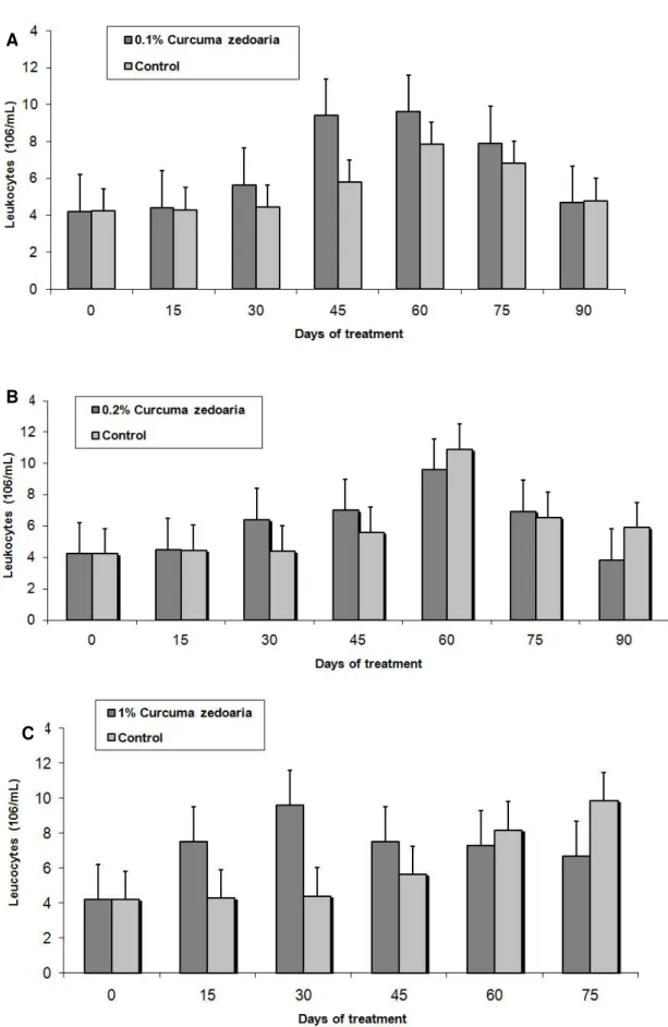
The intraperitoneally treated group showed an increase in total leukocyte count 15,
30 and 45 days after treatment (Figure 2 – C), while the oral administration led to an
augmentation in total leukocyte number 30 and 45 days after treatment at both
A
B
C
Figure 2. Analysis of total leukocytes in the blood of C57BL/6J mice treated with (A) 0.1%; (B) 0.2% or (C) 1% Curcuma zedoaria crude extract.
The differential analysis of orally treated mice showed an increase in the lymphocyte
count 30 and 45 days after treatment with 0.1% Curcuma zedoaria extract; whereas
regarding the 0.2% extract, an elevation was noticed 15 and 30 days after the
administration. Similarly, 0.1% Curcuma zedoaria extract augmented the number of
neutrophils 15 days after treatment, reached a peak within 60 days and, then,
returned to normal levels in 90 days. The same behavior was observed in animals
treated with the 0.2% extract (Figure 3 – A and B).
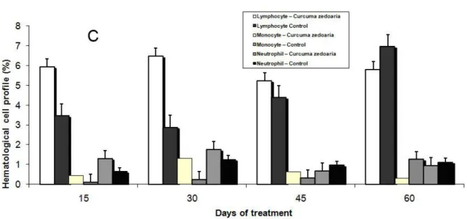
The differential analysis of the intraperitoneal administration revealed an increase in
lymphocyte number after 15 and 30 days, a monocyte increase after 15, 30 and 45
days and an elevation of neutrophils 15 days after treatment. The number of
Figure 3. Differential analysis of leukocytes in the blood of C57BL/6J mice treated with (A) 0.1%; (B) 0.2% or (C) 1% Curcuma zedoaria crude extract.
Data are expressed as mean ±SD.
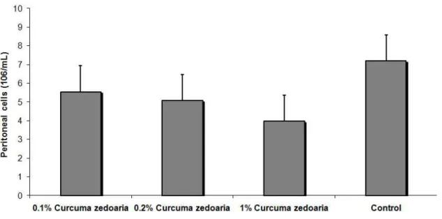
There was no alteration in the number of peritoneal cells concerning orally treated
animals; however, mice intraperitoneally treated had a significant decrease in
peritoneal cell count (Figure 4) The differential analysis of peritoneal cells showed a
reduced number of macrophages in the animal groups that received orally 0.1% C.
zedoaria extract treated and intraperitoneally 1% extract (Figure 5).
After the B16F10 melanoma cell inoculation, animals were daily observed. Data in
Figure 6 (A) display a reduction in the tumor volume during the first couple of weeks
of intraperitoneal treatment. There was no alteration in the tumor area (Figure 6 – B).
After the crude extract treatment, animals were killed and tumors and lymphatic
nodes were analyzed. Photomicrographs of dorsal tumors and draining nodes
showed infiltrated tumor cells in the lymphatic system with mast cells granulations
and also a large number of mast cells in primary tumors (Figure 7). The
Figure 4. Cell migration to the peritoneal cavity. C57BL/6J mice were treated with 0.1, 0.2 and 1% Curcuma zedoaria crude extract.
Data are expressed as mean ±SD.
Figure 5. Differential cell migration to the peritoneal cavity. C57BL/6J mice were treated with 0.2 or 1% Curcuma zedoaria crude extract.
A
B
Figure 6. (A) Analysis of dorsal B16F10 tumor growth in C57BL/6J mice treated with 0.2% Curcuma zedoaria. (B) Analysis of dorsal B16F10 tumor area in C57BL/6J mice treated with 0.2% Curcuma zedoaria.
Figure 7. Light micrograph of infiltrated tumor cells in lymph nodes after dorsal implantation of B16F10 melanoma cells followed by Curcuma zedoaria crude extract
Figure 8. Effects of Curcuma zedoaria on subcutaneous B16F10 tumor growth. B16F10 melanoma cells (5 x 10⁴/mouse) were subcutaneously injected in the dorsal
region of C57BL/6J mice, in the absence (controls) or presence of C. zedoaria
extract (0.2%). The tumor volumes were monitored by periodic caliper measurement.
(A) Group treated with C. zedoaria and (B) control group.
DISCUSSION
To study the regulation of B16F10 melanoma tumour growth by a phytotherapic
agent in vitro and in vivo, firstly, antitumor and inflammatory responses to Curcuma
zedoaria extract treatment were assessed. Recently, considerable attention has been
focused on identifying naturally occurring chemopreventive substances capable of
inhibiting, retarding, or reversing the tumoral growth. Numerous phenolic substances
present in fruits and vegetables or in medicinal plants have been found to retain
potential cancer chemopreventive activities. For most known chemopreventive
phytochemicals, however, their protective effects are based solely on results of
animal tests.
Dehydrocurdione, a major sesquiterpene, has shown an analgesic effect in mice by
decreasing the number of writhes induced by acetic acid and inhibiting
carrageenan-induced paw edema. The curcuma extract has also proven to promote an important
decrease in free radicals production. Moreover, the anti-inflammatory potency of
Several works on epidemiology and animal model studies demonstrated that natural
compounds, which possess antioxidant or anti-inflammatory properties, could inhibit
carcinogenesis(9-12).
In vitro studies have shown that a fraction from the ethanolic extract of C. zedoaria
has a powerful antitumoral action and promotes macrophage activation, causing an
increase in phagositosis and in oxidative burst in vivo. In addition, it was noticed that
high molecular weight compounds have strong macrophage-stimulating activity and
low molecular weight compounds (curcumin, curzerenone and zedoarone) present
antifungal activity (2).
Curcumin (diferuloylmethane) is a yellow odourless pigment isolated from the
rhizome of tumerics (Curcuma longa Linn, Zingiberaceae and Curcuma zedoaria
Roscoe). This substance possesses anti-inflammatory and antioxidant properties, as
well as inhibits lipopolysaccharide and interferon-induced production of nitric oxide or
nitrite in macrophages (13-21). Curcumin is one of the most extensively investigated
and well-defined chemopreventive phytochemicals and has been shown to protect
against skin, oral, intestinal and colon carcinogenesis and also to suppress
angiogenesis and metastasis in a variety of animal tumour models (22-24). It also
inhibits the proliferation of cancer cells by arresting them in various phases of the cell
cycle and by inducing apoptosis (22, 24).
Dehydrocurdione and lactones are the major sesquiterpenes in zedoary. The
anti-inflammatory effect of dehydrocurdione is likely to be different from that of
non-stereoidal anti-inflammatory drugs, while sesquiterpene lactones have been shown to
inhibit activation of a transcriptional factor NFkB, which, in turn, regulates different
gene transcription related to cellular inflammatory response (25).
Modern medicine attributes most cases of cancer to changes in DNA that reduce or
eliminate the normal controls over cellular growth, maturation and programmed cell
death. Herb prescriptions that used to be given to cancer patients as the sole therapy
primarily contained plants that were said to remove or counteract toxins, eliminate or
diminish accumulations, restore normal circulation and promote the functions of the
internal organs. In an effort to remove these accumulations (cell masses, such as a
tumor), an herb that was often utilized is curcuma (26).
Curcuma zedoaria contains volatile oils and when its extract is injected into mice that
have tumors, the tumors shrink. It is believed that the enhanced fibrinolysis and other
and consume or destroy the tumor cells. In Chinese medicine, C. zedoaria is
employed to treat cervical cancer by injecting its oil into the tumor mass. Moreover, it
is believed that this type of herbs have low toxicity (26).
The ethanolic extract of C. Zedoaria contains three identified active curcuminoids,
namely curcumin, demethoxycurcumin and bisdemethoxycurcumin, which were
found to possess inhibitory activity against tumor growth and to be cytotoxic to
murine sarcoma, transformed NIH 3T3 fibroblast cells, human colon cancer cell lines
(HCT-15 and HT-29), human embryonic kidney cell line (HEK293), human
hepatocellular carcinoma and Hep-2 cells (7, 10, 11). Furthermore, Kim et al. (3)
found that the polysaccharide fraction from C. zedoaria effectively inhibited the
growth of implanted sarcoma 180 in mice.
One method of cancer therapy currently under investigation is the inhibition of tumor
angiogenesis. The blood vessels of surrounding normal tissue are eliminated or
displaced by the tumor, thus weakening the surrounding tissue and making it more
susceptible to replacement by tumors mass. A fibrin coating that prevents immune
cells of the blood stream from entering and destroying the tumor often surrounds
tumors. Blood-activating herbs are usually prescribed in Chinese medicine to cancer
patients in order to promote positive effects against cancer. A group of herbs
classified as blood-activating, including C. zedoaria, are used to counter all those
problems, direct cancer-inhibiting actions on tumor cells and promote immune
system attack against cancer cells (26).
The maintenance of homeostasis in normal mammalian tissue reflects a critical
balance between cell proliferation and cell death via apoptosis. Selected
chemopreventive vitamins or phytochemicals have the propensity for suppressing or
retarding the growth of cancer cells (27-29). Moreover, some phenolic substances
derived from Zingiberaceae plants have potent anti-inflammatory activity and induce
apoptosis in human cancer cell lines (11, 30, 31). Additionally, curcumin from C.
zedoaria also protects against chemically induced liver damage in experimental
animals (32, 33). Similarly, another study observed that topical application of
curcumin onto mouse dorsal skin significantly inhibited epidermal cyclooxygenase
and lipooxygenase (34). Seo et al. (35) reported a suppressive effect of zedoary
rhizome on pulmonary metastasis of B16 melanoma cells.
The majority (60%) of the identified and approved antineoplastic drugs in the 1990s
possibilities to find substances of interest in oncology. Some chemotherapy drugs
currently used against diverse tumors are derived from natural plants, such as
alkaloids that have basic chemical properties and usually contain at least one
nitrogen atom in a heterocyclic ring obtained from Vinca rosea, etoposide from
Podophyllum peltatum and taxol isolated from Taxus brevifolia tree. There are other
substances that originate from microorganisms, such as L-asparaginase from
Escherichiacoli and cytarabine from Cryptotethya crypta (36).
Natural products probably represent the most accessible and abundant source of
substances of yet unknown structures, some presenting antitumoral activity and new
specific mechanisms of action, eventually not described until the present date.
REFERENCES
1.Yoshioka T, Fujii E, Endo M, Wada K, Tokunaga Y, Shiba N, et al. Antiinflamatory
potency of dehydrocurdione, a zedoary-derived sesquiterpene. Inflamm Res.
1998;47(12):476-81.
2. Jang MK, Sohn DH, Ryu JH. A curcuminoid and sesquiterpenes as inhibitors of
macrophage TNF-α release from Curcuma zedoaria. Planta Med. 2001;67(6): 550-2.
3. Kim KI, Kim JW, Hong BS, Shin DH, Cho HY, Kim HK, et al. Antitumor,
genotoxicity and anticlastogenic activities of polysaccharide from Curcuma zedoaria.
Mol Cells. 2000;10(4):392-8.
4. Kim KI, Shin KS, Jun WJ, Hong BS, Shin DH, Cho HY, et al. Effects of
polysaccharides from rhizomes of Curcuma zedoaria on macrophage functions.
Biosci Biotechnol Biochem. 2001;65(11):2369-77.
5. Lee H, Lin JY. Antimutagenic activity of extracts from anticancer drugs in Chinese
medicine. Mutat Res. 1988;204(2):229-34.
6. Matsuda H, Ninomiya K, Morikawa T, Yoshikawa M. Inhibitory effect and action
mechanism of sesquiterpenes from Zedoariae rhizome on
D-galactosamine/lipopolysaccharide-induced liver injury. Bioorg Med Chem Lett.
1998;8(4):339-44.
7. Syu WJ, Shen CC, Don MJ, Ou JC, Lee GH, Sun CM. Cytotoxicity of curcuminoids
and some novel compounds from Curcuma zedoaria. J Nat Prod.
8. Anuchapreeda S, Leechanachai P, Smith MM, Ambudkar SV, Limtrakul P.
Modulation of P-glycoprotein expression and function by curcumin in
multidrug-resistant human KB cells. Biochem Pharmacol. 2002;64(4):573-82.
9. Rajakrishnan V, Shiney SJ, Sudhakaran PR, Menon VP. Effect of curcumin on
ethanol-induced stress on mononuclear cells. Phytother Res. 2002;16(2):171-3.
10. Hanif R, Qiao L, Shiff SJ, Rigas B. Curcumin, a natural plant phenolic food
additive, inhibits cell proliferation and induces cell cycle changes in colon
adenocarcinoma cell lines by a prostaglandin-independent pathway. J Lab Clin Med.
1997;130(6):576-84.
11. Jiang MC, Yang-Yen HF, Yen JJ, Lin JK. Curcumin induces apoptosis in
immortalized NIH 3T3 and malignant cancer cell lines. Nutr Cancer.
1996;26(1):111-20.
12. Limtrakul P, Anuchapreeda S, Lipigorngoson S, Dunn FW. Inhibition of
carcinogen induced c-Ha-ras and c-fos proto-oncogenes expression by dietary
curcumin. BMC Cancer. 2001;1(1):1.
13. Amom HP, Wahl MA. Pharmacology of Curcuma longa. Planta Med. 1991;
57(1):1-7.
14. Sharma OP. Antioxidant activity of curcumin and related compounds. Biochem
Pharmacol. 1976;25(15):1811-2.
15. Rao TS, Basu N, Siddiqui HH. Anti-inflammatory activity of curcumin analogues.
Indian J Med Res 1982;75(1):574-8.
16. Mukhopadhyay A, Basu N, Ghatak N, Gujral PK. Anti-inflammatory and irritant
activities of curcumin analogues in rats. Agents Actions. 1982;12(4):508-15.
17. Toda S, Miyase T, Arichi H, Tanizawa H, Takino Y. Natural antioxidants. III.
Antioxidative components isolated from rhizome of Curcuma longa L. Chem Pharm
Bull (Tokyo). 1985;33(4):1725-8.
18. Srivastava R, Srimal RC. Modification of certain inflammation-induced
biochemical changes by curcumin. Indian J Med Res. 1985;81(1):215-23.
19. Lin JK, Shih Ca. Inhibitory effect of curcumin on xanthine dehydrogenase/oxidase
induced by phorbol-12-myristate-13-acetate in NIH3T3 cells. Carcinogenesis.
1994;15(8):1717-21.
20. Sugiyama Y, Kawakishi S, Osawa T. Involvement of the β-diketone moiety in the
antioxidative mechanism of tetrahydrocurcumin. Biochem Pharmacol.
21. Brouet I, Ohshima H. Curcumin, an anti-tumor promoter and anti-inflammatory
agent, inhibits induction of nitric oxide synthase in activated macrophages. Biochem
Biophys Res Commun. 1995;206(2):533-40.
22. Surh YJ, Chun KS. Cancer chemopreventive effects of curcumin. Adv Exp Med
Biol. 2007;595(1):149-72.
23. Patel BB, Majumdar AP. Synergistic role of curcumin with current therapeutics in
colorectal cancer: minireview. Nutr Cancer. 2009;61(6):842-6.
24. Ravindran J, Prasad S, Aggarwal BB. Curcumin and cancer cells: how many
ways can curry kill tumor cells selectively? AAPS J. 2009;11(3):495-510.
25. Lyss G, Schmidt TJ, Merfort I, Pahl HL. Helenalin, an anti-inflammatory
sesquiterpene lactone from Arnica, selectively inhibits transcription factor
NF-kappaB. Biol Chem. 1997;378(9):951-61.
26. Zhu YP, Woerdenbag HJ. Traditional Chinese herbal medicine. Pharm World Sci.
1995;17(4):103-12.
27. Lotan R. Retinoids and apoptosis: implications for cancer chemoprevention and
therapy. J Natl Cancer Inst. 1995;87(22):1655-7.
28. Yu R, Mandlekar S, Harvey KJ, Ucker DS, Kong AN. Chemopreventive
isothiocyanates induce apoptosis and caspase-3-like protease activity. Cancer Res.
1998;58(3):402-8.
29. Sundaram SG, Milner JA. Diallyl disulfide induces apoptosis of human colon
tumour cells. Carcinogenesis. 1996;17(4):669-73.
30. Lee E, Park KK, Lee JM, Chun KS, Kang JY, Lee SS, et al. Suppression of
mouse skin tumour promotion and induction of apoptosis in HL-60 cells by Apinia
oxyphylla Miquel (Zingiberaceae). Carcinogenesis. 1998;19(8):1377-81.
31. Kuo ML, Huang TS, Lin JK. Curcumin, an anti-oxidant and anti-tumor promoter,
induces apoptosis in human leukemia cells. Biochim Biophys Acta.
1996;1317(2):95-100.
32. Kiso Y, Suzuki Y, Watanabe N, Oshima Y, Hikino H. Antihepatotoxic principles of
curcuma longa Rhizomes 1. Planta Med. 1983;49(11):185-7.
33. Soni KB, Rajan A, Kuttan R. Reversal of aflatoxin induced liver damage by
turmeric and curcumin. Cancer Lett. 1992;66(2):115-21.
34. Huang MT, Lysz T, Ferraro T, Abidi TF, Laskin JD, Conney AH. Inhibitory effects
of curcumin on in vitro lipooxygenase and cyclooxygenase activities in mouse
35. Seo WG, Hwang JC, Kang SK, Jin UH, Suh SJ, Moon SK, et al. Suppressive
effect of Zedoariae rhizome on pulmonary metastasis of B16 melanoma cells. J
Ethnopharmacol. 2005;101(1-3):249-57.
36. Beach M. China opens drug market by revising pharmaceutical law. Lancet.