Loss of 4q21.23-22.1 Is a Prognostic
Marker for Disease Free and Overall
Survival in Non-Small Cell Lung Cancer
Faik G. Uzunoglu1., Ebba Dethlefsen2., Annkathrin Hanssen2
, Michaela Wrage2, Lena Deutsch1, Katharina Harms-Effenberger1,2, Yogesh K. Vashist1,
Matthias Reeh1, Guido Sauter3, Ronald Simon3, Maximillian Bockhorn1, Klaus Pantel2, Jakob R. Izbicki1, Harriet Wikman2*
1.Department of General, Visceral and Thoracic Surgery, University Medical Center of Hamburg-Eppendorf, Hamburg, Germany,2.Institute of Tumour Biology, University Medical Center of Hamburg-Eppendorf, Hamburg, Germany,3.Department of Pathology, University Medical Center of Hamburg-Eppendorf, Hamburg, Germany
*h.wikman@uke.de
.These authors contributed equally to this work.
Abstract
This study was performed to assess the prognostic relevance of genomic aberrations at chromosome 4q in NSCLC patients. We have previously identified copy number changes at 4q12-q32 to be significantly associated with the early hematogenous dissemination of non-small cell lung cancer (NSCLC), and now aim to narrow down potential hot-spots within this 107 Mb spanning region. Using eight microsatellite markers at position 4q12-35, allelic imbalance (AI) analyses were performed on a preliminary study cohort (n586). Positions indicating clinicopathological and
prognostic associations in AI analyses were further validated in a larger study cohort using fluorescencein situhybridization (FISH) in 209 NSCLC patients. Losses at
positions 4q21.23 and 4q22.1 were shown to be associated with advanced
clinicopathological characteristics as well as with shortened disease free (DFS) and overall survival (OS) (DFS:P50.019; OS:P50.002). Multivariate analyses identified
the losses of 4q21.23-22.1 to be an independent prognostic marker for both DFS and OS in NSCLC (HR 1.64–2.20, allP,0.04), and especially in squamous cell lung
cancer (P,0.05). A case report study of a lung cancer patient further revealed a loss
of 4q21.23 in disseminated tumor cells (DTCs). Neither gains at the latter positions, nor genomic aberrations at 4q12, 4q31.2 and 4q35.1, indicated a prognostic relevance. In conclusion, our data indicate that loss at 4q21.23-22.1 in NSCLC is of prognostic relevance in NSCLC patients and thus, includes potential new tumor suppressor genes with clinical relevance.
OPEN ACCESS
Citation:Uzunoglu FG, Dethlefsen E, Hanssen A, Wrage M, Deutsch L, et al. (2014) Loss of 4q21.23-22.1 Is a Prognostic Marker for Disease Free and Overall Survival in Non-Small Cell Lung Cancer. PLoS ONE 9(12): e113315. doi:10.1371/ journal.pone.0113315
Editor:Sai Yendamuri, Roswell Park Cancer Institute, United States of America
Received:March 24, 2014
Accepted:October 26, 2014
Published:December 11, 2014
Copyright:ß2014 Uzunoglu et al. This is an open-access article distributed under the terms of theCreative Commons Attribution License, which permits unrestricted use, distribution, and repro-duction in any medium, provided the original author and source are credited.
Data Availability:The authors confirm that all data underlying the findings are fully available without restriction. All relevant data are within the paper and its Supporting Information files.
Funding:This work was supported by the Hamburger Krebsgesellschaft e.V., Hamburg, Germany. The funders had no role in study design, data collection and analysis, decision to publish, or preparation of the manuscript.
Introduction
Genomic instability is one of the key hallmarks of cancer [1]. Numerous studies on genomic instability in non-small cell lung cancer (NSCLC) have highlighted specific deletion patterns for both metastatic and primary tumors [2–4]. Copy number aberrations in NSCLC can be found at multiple regions, some being specific for the histological subtype, and some being dependent on the severity of histopathological changes (e.g. tumor stage or grading) [5–11] Furthermore, the occurrence of copy number changes has shown to be an early event in lung cancer pathogenesis. This can be found in combination with a sequential pattern of loss of heterozygosity (LOH), which begins with the allelic loss on chromosome 8p, followed by 3p and 9p deletions [7,12,13].
We have previously shown that early hematogenous dissemination of tumor cells is driven by a specific pattern of genomic changes. In addition to this pattern,
a large deletion of chromosome 4q12-q32 (.107 Mbp) indicates a strong
association with the presence of disseminated tumor cells (DTC) in bone marrow, as well as brain metastases of lung cancer patients [14]. However, the prognostic relevance of this region has not yet been investigated. The aim of this study was to narrow down the relevant hotspot regions and to investigate their prognostic impact in NSCLC. In addition, we sought to analyze whether this loss could also be detected in DTCs of a NSCLC patient.
For this purpose, we first performed allelic imbalance (AI/LOH) analyses in a preliminary study cohort (n586). For further verification, a fluorescencein situ hybridization (FISH) analysis of 209 NSCLC patients was performed. A loss within a less than 4 Mb-spanning region at 4q21.23-4q22.1 was identified as a significant negative prognostic marker for disease free as well as overall survival in NSCLC. Furthermore, we performed consecutive immunofluorescence (IF) and FISH analyses on the DTCs of a single NSCLC patient, showing a loss of 4q21.23 in the primary tumor. The same loss could also be detected in the DTCs.
Materials and Methods
Samples
This study was approved by the Ethics Committee of the chamber of physicians, Hamburg, Germany. Written informed consent was obtained from all patients. All clinical investigation has been conducted according to the principles expressed in the Declaration of Helsinki. All tumor samples were obtained during surgical resections at the University Medical Centre Hamburg-Eppendorf or associated surgical departments. Clinicopathological data were extracted from a prospective database, and follow-up data were obtained by interviews with the general practitioner or the patient at the outpatient department.
cancer cell types, 37 patients (43.0%) had a squamous cell carcinoma (SqCC) and 49 (57.0%) an adenocarcinoma (AC). The median follow-up time was 21.4 months (2–60). Further detail is given in S1 Table.
For DNA copy number aberrations (FISH) at 4q, a tissue microarray (TMA), consisting of 209 evaluable primary lung cancer patients, was used, with a median age of 62.3 years at time of surgery. Gender distributions were comparable to the AI study cohort, with a similar predominance of male patients (68.4%). The FISH study cohort encompassed 88 (42.1%) patients with SqCCs, 78 (37.1%) with ACs, 34 (16.3%) with large-cell lung carcinoma, and nine patients (4.5%) with neuroendocrine lung cancer. The median follow up time was 24.7 months (2.5–60). Further details are given in S1 Table.
All patients were reclassified according to the seventh edition of the TNM classification of malignant tumors [15]. In regard to the administration of adjuvant therapy, the following specified criteria have been applied since 2004:$ Stage II patients received adjuvant chemotherapy with Cisplatin and Vinorelbin. Staged Ib patients were evaluated for adjuvant therapy if the tumor was.4 cm or in patients with invasion into vein (V+) or invasion into lymphatic vessel (L+).
Adjuvant chemotherapy followed by radiation (50–60 Gy) was discussed for $
Stage III. Patient characteristics for both study cohorts are shown in S1 Table.
DNA isolation
Genomic DNA of matched carcinoma (fresh-frozen) and pathologically-verified non-malignant lung tissue or peripheral blood leukocyte, taken prior to surgery was extracted and purified according to the manufacturer’s protocol using the QIAamp tissue kit (Qiagen, Hilden, Germany) or InnuPREP DNA Microkit (AnalytikJena, Jena, Germany). If necessary, manual microdissection was performed, in order to obtain a tumor cell content of at least 70%. DNA concentration was determined by NanoDrop ND-1000 Spectrophotometer (Wilmington, DE) and samples were diluted to 10 ng/ml and stored at 220
˚
C until use.Allelic imbalance analysis
Based on our previous study, four hotspot regions represented at positions 4q12, 4q21.23, 4q31.2 and 4q35.1 were chosen for further analysis [14]. For each region, two microsatellite markers were used to assess the frequency and clinical relevance of AI (see S2 Table, for details of all microsatellite markers). Forward primers were labeled with a fluorescent dye (6-FAM) for subsequent capillary electro-phoresis. PCRs were carried out in a 10 ml reaction mix consisting of 10 ng DNA
template, 2.5 mM deoxyribonucleotide triphosphate mix (Invitrogen, Darmstadt, Germany), 2.5 pmol sense and antisense primer (MWG, Ebersberg, Germany),
0.25 U AmpliTaq Gold Polymerase (Applied Biosystems) and 5 ml nuclease-free
Genetic Analyzer (Applied Biosystems, Freiburg, Germany), using a mixture of 40 ml formamide (Hi-Di), 0.2 ml Genescan-500-ROX Standard as well as 0.1 ml of
PCR product and denaturation at 94
˚
C for 2 min was performed and the length of allele fragments and fluorescent intensity was assessed. The alleles were defined as the two highest peaks within the expected size range and a ratio of $1.5 between the peak heights of the tumor and normal alleles were scored as AI. For overall quality assurance, 10% of used samples were randomly used for repeated analysis. The concordance of allelic status was .99%.Fluorescence
in situ
hybridization (FISH) analysis
DNA copy number loss analysis of two hot-spot regions based on the AI analyses was assessed in a larger study population by fluorescence in situ hybridization (FISH). Three different BAC probes targeting the regions 2 (RP11-570L13: 4q21.23 and RP11-1053C2: 4q22.1) and 3 (RP11-634D8: 4q31.2) were hybridized on a TMA. The utilized BAC probes were obtained from Source BioScience
LifeSciences (Nottingham, UK). One mg of each BAC probe was labeled by
random priming (BioPrime Labeling System, Invitrogen) with fluorescently labeled dUTPs (RP11-570L13 and RP11-634D8: Spectrum Orange, RP11-1053C2: Spectrum Green; Enzo Life Sciences, Abbott). As a reference, a centromere probe (CEP 10, SpectrumAqua) was employed (Enzo Life Sciences, Abbott). In order to control the specificity and quality of the fluorescently-labeled BAC probes, the FISH was first tested on metaphase chromosome spreads. Afterwards, protocols for the paraffin slides were established for each BAC probe separately. First, the slides were incubated at 60
˚
C for one hour before they were deparaffinized in xylene and hydrated in an ethanol series. The fixation was achieved by placing the slides into a solution of 2% formaldehyde and methanol at -20˚
C, followed by rinsing them in phosphate-buffered saline (1x PBS) and pre-treating them with a pre-treatment solution (Invitrogen) at 90˚
C for 10 min. After PBS washing, the slides were treated with a pre-warmed enzyme-reagent (Invitrogen) at 37˚
C for 10 min. Again, they were washed in PBS and then dehydrated in an ethanol series. Finally, the slides were denatured at 85˚
C for 5 min, before they were incubated with probe hybridization mixtures at 37˚
C overnight. Post-hybridization washingwas then performed in 2 x SSC, 0.3% NP-40 buffer at 70
˚
C and roomtemperature, followed by a second hydration in an ethanol series. DAPI solution was applied for the detection of morphologically intact non-overlapping tumor cells. On average, fluorescent signals of 32 tumor cells for each probe were counted (range 11–57). In order to evaluate the experimental bias, as well as to define the cut-offs of the signal-to-centromere ratio, the hybridization signals of 10 normal control samples (also located on the TMA) were counted.
Consecutive immunofluorescence staining and FISH analysis
For the isolation of DTCs, mononuclear cells from bone marrow aspirates of NSCLC patients were isolated by FICOLL gradient centrifugation. The cells were then cyto-centrifuged onto glass slides. To detect the DTCs, an immunocy-tochemistry staining was performed according to the APAAP method (alkaline phosphatase anti alkaline phosphatase), using an antibody against cytokeratins(A45-B/B3, MicroMet, Martinsried, Germany) [16]. An isotype-matched, murine
monoclonal antibody (MOPC 21, IgG1; Sigma- Aldrich) served as negative control. DTC positive patients were used for analyzing the loss of 4q22.1 by FISH analysis. FISH was performed on tumor cells that were detected beforehand by IF staining with a Cy3 labeled anti-cytokeratin antibody (A45-Cy3; MicroMet). For cell fixation, solution B (MicroMet) was used. After a PBS washing step,
unspecific binding sites were blocked with 10% AB serum (Biotest AG, Dreieich, Germany). Another PBS washing was followed by 45 min A54-Cy3 antibody treatment (1:300). Cells were again washed and DAPI (CellSearch) was applied for nuclear staining. DTCs were identified in an automated fashion (Ariol Scan, Leica Biosystems, Nussloch, Germany) and localized with the England Finder for re-identification. For consecutive FISH analysis, cells were washed with PBS and
incubated for 7 min in a pre-warmed proteinase K (0.1 mg/ml) solution (20 mM
Tris-HCl pH 7.5, 0.2% CaCl2xH2O ad 50 ml Aqua dest.). Thereafter, cells were dehydrated in an ethanol series and denatured for 5 min at 75
˚
C (70%Formamide, 0.6 x SSC, pH 7.4). Cells were again dehydrated and probe hybridization mixtures were denatured for 5 min at 75
˚
C. Hybridization wasperformed over night at 37
˚
C. Post-hybridization washing was done in 50%Formamide/2x SSC, in 2x SSC and in 0.1x SSC at 45
˚
C. After applying DAPI, the DTCs were relocated and fluorescent signals of centromere 3 and RP11-1053C2 probes were counted. A ratio of ,0.75 was defined as a loss.Statistical analysis
Results
Microsatellite analysis
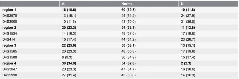
The study cohort used for the microsatellite analysis consisted of a total of 86-matched carcinoma and normal tissue samples of lung cancer patients that underwent surgery. A microsatellite analysis was performed in a total of four regions on chromosome 4q, using two polymorphic markers for each region. Allelic markers close to each other revealed highly similar frequencies of deletion leading to final loss of heterozygosity frequencies of 18.6% (n516) in region 1; 23.3% (n520) in region 2; 25.6% (n522) in region 3 and 34.9% (n530) in region 4. The rate of non-informative cases ranged from 16.3% to 36.0%, depending on the marker. By using two markers for AI detection in each region, the non-informative cases could be reduced to ranges from 2.3% to 15.1%. Regions 1 and 3 had concordant results (normal/loss), whereas region 2 contained three disconcordant results and region 4 contained one of each. All disconcordant results were repeated and the results remained identical, indicating a possible breakpoint between the markers. In total, 45.3% (n539) revealed at least one allelic loss within the analyzed regions. Within these patients, 17.9% (n57) revealed a loss in all regions. Detailed numeric results are given intable 1 andS1 Figure, supporting information.
AI on chromosome 4q and clinicopathological characteristics
There was no evidence of a significant statistical correlation between AI with clinicopathological characteristics in the four investigated regions. However, 83.3% (n515) of patients with AI in region 2 developed a relapse in contrast to 16.7% (n53) without AI (P50.073).AI on chromosome 4q as a prognostic marker for survival
In univariate survival analyses, a trend towards shorter median disease free (DFS) and overall survival (OS) in patients with AI in region 1 and 2 was evident (median survival difference from 11.9 months to 14 months; details are given in
S3 Table). However, differences did not reach statistical significance (P.0.05). Since the study cohort for survival analyses was limited to 68-77 cases, including UICC stage IV patients as well as patients with positive resection margins, confounding effects on survival analysis were to be expected. This was evident for region 2. Therefore, following stratification by age, gender, tumor stage, and resection margins parameters, the presence of AI in region 2 was proven to be an
independent prognostic marker for DFS (HR 3.82, P50.044). A similar
FISH copy number analysis
Based on the reported allelotyping results, we sought to verify these results in a larger study population by FISH analysis. Two different BAC probes, one hybridizing to region 2 and one hybridizing to region 3 were established (S5 Table).
For the region 4q21.23 detected by RP11-570L13, a DNA loss was seen in 24.9% (n552) and a DNA gain in 4.3% (n59) of the analyzed samples. A loss detected at this position revealed a significant histological type-dependent correlation, with 38.0% (n530) loss in SqCC in contrast to 22.7% (n515,P50.049) in AC. The analysis with probe RP11-1053C2 revealed loss in 30.6% and gain in 7.2% (n515), again revealing a significantly more frequent loss rate in SqCC (44.6%, n533) compared to AC (23.3%,n517,P50.022).
Using probe RP11-634D8 (region 3), the rate of successful analysis was 71.6% (n5204). A DNA copy number loss was detected in 36.8% (n577) and a gain in 7.7% (n516) of the analyzable samples.
Copy number loss on chromosome 4q and clinicopathological
characteristics
Copy number loss at position 4q21.23 (RP11-570L13) showed a significant correlation with advanced tumor size (P50.005), but with no other pathological characteristics. Copy number loss at position 4q22.1 (RP11-1053C2) revealed a significant association with the presence of lymph node metastases (loss pN0: 25.2%, n526 versus loss $pN1: 44.0%, n537;P50.005) as well as advanced UICC stage (loss UICC I: 25.3%, n523 versus loss UICC $II: 41.4%,n541;
P50.032). Copy number loss at position 4q31.2 (RP11-634D8) showed a
Table 1.Allelotyping results.
AI Normal NI
region 1 16 (18.6) 60 (69.8) 10 (11.6)
D4S2978 13 (15.1) 44 (51.2) 24 (27.9)
D4S3000 10 (11.6) 43 (50.0) 31 (36.0)
region 2 20 (23.3) 54 (62.8) 11 (12.8)
D4S1534 14 (16.3) 49 (57.0) 17 (19.8)
D4S414 15 (17.4) 44 (51.2) 23 (26.7)
region 3 22 (25.6) 50 (58.1) 13 (15.1)
D4S1565 20 (23.3) 48 (55.8) 17 (19.8)
D4S1588 8 (9.3) 30 (34.9) 15 (17.4)
region 4 30 (34.9) 54 (62.8) 2 (2.3)
D4S3047 20 (23.3) 47 (54.7) 16 (18.6)
D4S2930 27 (31.4) 43 (50.0) 14 (16.3)
AI: allelic imbalance; NI: non-informative; values in parenthesis are percentages.
significant correlation with advanced tumor size (P50.019). However, no further correlations with clinicopathological characteristics were evident (S5 Table).
Copy number loss on chromosome 4q as a prognostic marker for
survival
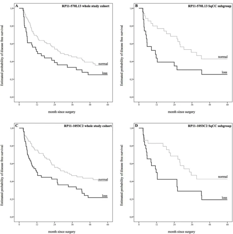
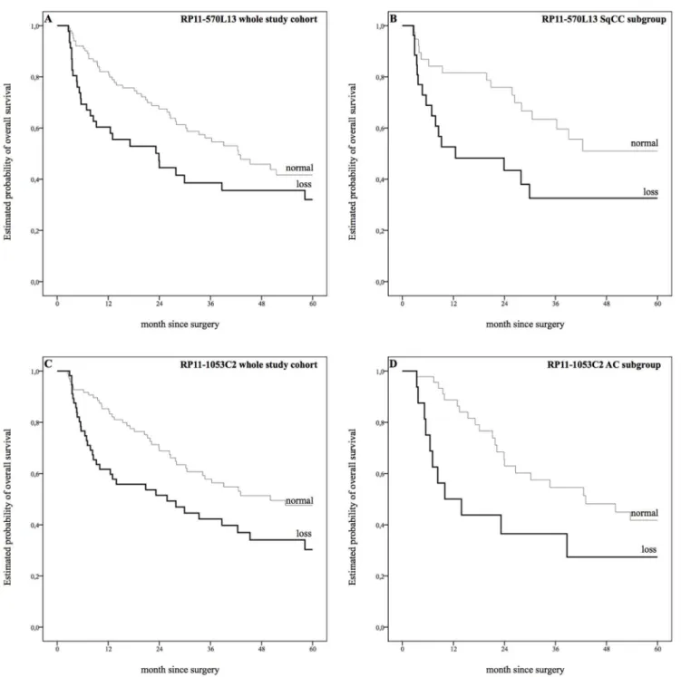
In the context of a univariate analysis, copy number loss at position 4q21.23 (RP11-570L13)revealed a non-significant trend towards shorter median DFS (12.5
months versus 28.0 months, P50.056). Subgroup analyses, however, showed a
strong correlation with DFS in SqCC subgroup (11.2 months versus 39.1 months, P50.031; fig. 1, S6 Table). A similar effect on OS was reported for the whole
study cohort (23.9 months versus 42.6 months, P50.033) and SqCC subgroup
(12.5 months versus 41 months, P50.031, fig. 2, S6 Table).
Copy number loss at position 4q22.1 (RP11-1053C2) showed significant correlations with shorter median DFS within the whole study cohort as well as
within the SqCC subgroup (12.5 months versus 32.7 month, P50.010 and 11.7
months versus 35.5 month, P50.027; fig. 1,S6 Table). Furthermore, a loss of 4q22.1 was associated with shorter OS within the whole study cohort and the AC subgroup, whereas no correlation was seen within the SqCC subgroup (whole
study cohort: 25.8 months versus 40.3 months, P50.012; AC subgroup: 10.0
months versus 43.1 months, P50.035, fig. 2,S6 Table).
In contrast, copy number gains did not reveal a correlation with survival at any of the analyzed positions. In line with the results of the AI study cohort, copy number loss at region 3 (RP11-634D8, position 4q31.2) had no impact on survival (S6 Table).
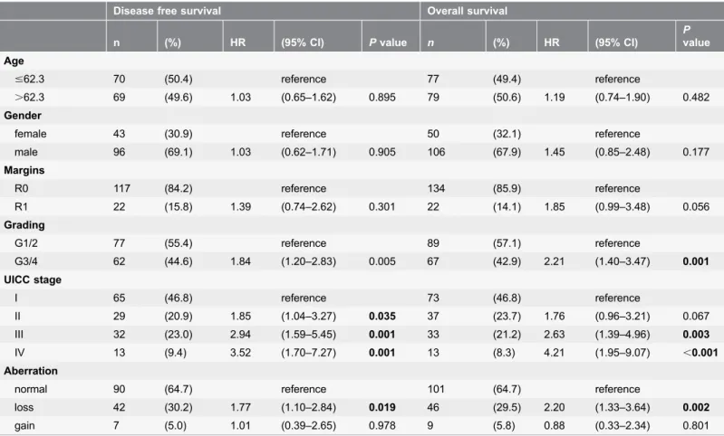
Multivariate analyses identified copy number loss at position 4q21.23 (RP11-570L13) as a negative prognostic marker for DFS and OS for the whole study cohort (cox proportional hazard model, stratified by age, gender, margin
clearance, grading and UICC stage; DFS: HR 1.77 (1.10–2.84 95% CI), P50.019;
OS: HR 2.20 (1.33–3.64 95% CI), P50.002, table 2) as well as for the SqCC
subgroup (DFS: HR 2.85 (1.35–6.04 95% CI), P50.006; OS: HR 2.77 (1.26–6.13
95% CI), P50.012, S7 Table). Similar results were seen for copy number loss at position 4q22.1 concerning the whole study group (RP11-1053C2; DFS: HR 1.64 (1.03–2.61 95% CI), P50.038; OS: 1.84 (1.12–3.01 95% CI), P50.015, table 3), whereas subgroup analysis did not reach statistical significance (S8 Table). A second model of multivariate analyses including the administration of adjuvant chemotherapy was performed (information available in 116 patients). The results stayed significant when the administration of adjuvant chemotherapy was included (data not shown).
In summary, both positions in region 2, namely 4q21.23 (RP11-570L13) and 4q22.1 (RP11-1053C2), were identified as a negative prognostic marker for DFS as well as OS within the whole study cohort. However, only loss at position 4q21.23 reached significance also in the SqCC subgroup. A flow chart summarizing the survival analyses of the study cohort, the study progress and results are given inS2
Fig. 1. Estimated probability of disease free survival.A: Copy number loss at position 4q21.23 (RP11-570L13) revealed a trend towards shorter median disease free survival (DFS) in univariate analysis (12.5 months versus 28.0 months,P50.056). B: Copy number loss at position 4q21.23 (RP11-570L13) in the squamous cell carcinoma (SqCC) subgroup showed strong correlations with shorter DFS (11.2 months versus 39.1 months)P50.031. C: Copy number loss at position 4q22.1 (RP11-1053C2) showed significant correlations with shorter DFS (12.5 months versus 32.7 months),P50.010. D: Copy number loss at position 4q22.1 (RP11-1053C2) showed significant correlations with shorter DFS in SqCC subgroup (11.7 months versus 35.5 months),P50.027. Survival plots for gain aberrations were faded out for the sake of clarity.
Fig. 2. Estimated probability of overall survival.A: Copy number loss at position 4q21.23 (RP11-570L13) revealed strong correlations with shorter median overall survival (OS) in univariate analysis (23.9 months versus 42.6 months),P50.033. B: Copy number loss at position 4q21.23 (RP11-570L13) in the squamous cell carcinoma (SqCC) subgroup showed strong correlations with shorter OS (12.5 months versus 41 months),P50.031 C: Copy number loss at position 4q22.1 (RP11-1053C2) showed significant correlations with shorter DFS (25.8 months versus 40.3 months),P50.012. D: Copy number loss at position 4q22.1 (RP11-1053C2) showed significant correlations with shorter DFS in adenocarcinoma subgroup (10.0 months versus 43.1 months), P50.035. Survival plots for gain aberrations were faded out for the sake of clarity.
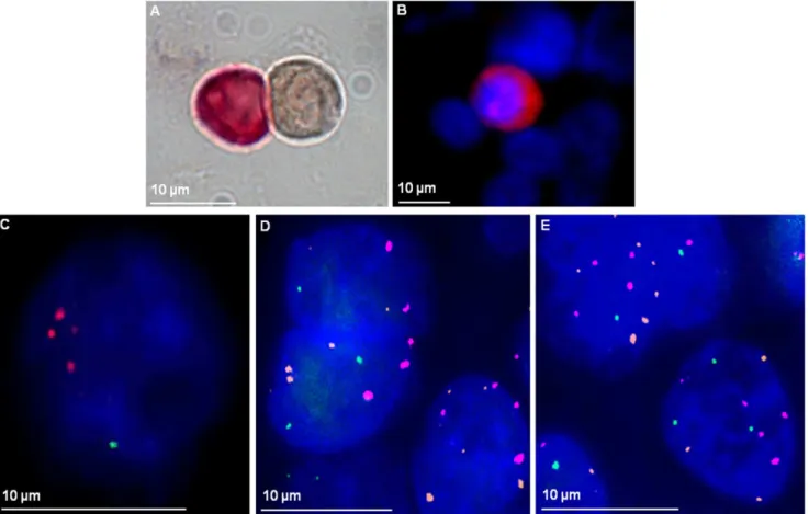
Case report of 4q21.23 loss in DTCs
Based on our earlier finding that 4q12-32 loss correlates with positive DTC status, we wanted to investigate whether a loss of this region is also present in DTCs from a patient with 4q loss in the primary tumor[14]. A loss of 4q22.1 was detected in 77% (17/22) of the patients DTCs. The range of RP11-1053C2 probe and centromere 3 signals were heterogeneous, varying from 1 to 5 signals (mean 1.7 and 2.7 respectively). FISH analysis was also performed on paraffin-embedded primary tumor and lung relapse material from the same patient (fig. 3). These results again revealed a loss of 4q22.1 and high degree of heterogeneity in the individual cells ranging from 0–4 signals in primary tumor and 0–5 in tumor relapse material.
Discussion
In a previous study, we were able to identify five chromosomal regions
differentiating patients with or without early tumor dissemination. Loss of
4q12-Table 2.Multivariate analysis RP11-570L13.
Disease free survival Overall survival
n (%) HR (95% CI) Pvalue n (%) HR (95% CI)
P value
Age
#62.3 70 (50.4) reference 77 (49.4) reference
.62.3 69 (49.6) 1.03 (0.65–1.62) 0.895 79 (50.6) 1.19 (0.74–1.90) 0.482 Gender
female 43 (30.9) reference 50 (32.1) reference
male 96 (69.1) 1.03 (0.62–1.71) 0.905 106 (67.9) 1.45 (0.85–2.48) 0.177 Margins
R0 117 (84.2) reference 134 (85.9) reference
R1 22 (15.8) 1.39 (0.74–2.62) 0.301 22 (14.1) 1.85 (0.99–3.48) 0.056
Grading
G1/2 77 (55.4) reference 89 (57.1) reference
G3/4 62 (44.6) 1.84 (1.20–2.83) 0.005 67 (42.9) 2.21 (1.40–3.47) 0.001 UICC stage
I 65 (46.8) reference 73 (46.8) reference
II 29 (20.9) 1.85 (1.04–3.27) 0.035 37 (23.7) 1.76 (0.96–3.21) 0.067
III 32 (23.0) 2.94 (1.59–5.45) 0.001 33 (21.2) 2.63 (1.39–4.96) 0.003
IV 13 (9.4) 3.52 (1.70–7.27) 0.001 13 (8.3) 4.21 (1.95–9.07) ,0.001
Aberration
normal 90 (64.7) reference 101 (64.7) reference
loss 42 (30.2) 1.77 (1.10–2.84) 0.019 46 (29.5) 2.20 (1.33–3.64) 0.002
gain 7 (5.0) 1.01 (0.39–2.65) 0.978 9 (5.8) 0.88 (0.33–2.34) 0.801
Cox regression hazard model was used for multivariate analysis to assess the prognostic value of aberrations. HR, hazard ratio; CI, confidence interval; UICC, Union for International Cancer Control.
q32 showed the strongest correlation with the positive DTC status. Seventy-three genes within this region were found down regulated among the bone marrow-positive NSCLC patients, indicating a likely presence of tumor suppressor genes within this region [14]. In this study, we were able to narrow down the target region and identify copy number losses within a less than 4 Mb-spanning region on chromosome 4q21.23-22.1 as independent prognostic markers for DFS, as well as OS in NSCLC. The presence of copy number losses within this region was associated with at least 18-month shorter median survival, compared to patients with normal copy numbers. Furthermore, the presence of copy number losses caused an increased risk of disease recurrence and death ranging from 1.6 up to 2.9 within 60 months of follow-up, independent of established prognostic markers for NSCLC (dependent on histological subtype and position of copy number loss). Furthermore, in our case report study, we were able to demonstrate that the loss of chromosomal region 4q21.23 is not only present in both the primary lung tumor and in tumor relapse tissue, but also in a large fraction of very
heterogeneous DTCs. This emphasizes the significance of this chromosomal region for tumor dissemination. Uniparental disomy is one mechanism of how a
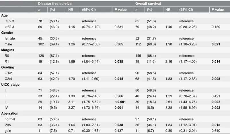
Table 3.Multivariate analysis RP11-1053C2.
Disease free survival Overall survival
n (%) HR (95% CI) Pvalue n (%) HR (95% CI) Pvalue
Age
#62.3 78 (53.1) reference 85 (51.8) reference
.62.3 69 (46.9) 1.15 (0.74–1.79) 0.531 79 (48.2) 1.40 (0.88–2.25) 0.159 Gender
female 45 (30.6) reference 52 (31.7) reference
male 102 (69.4) 1.26 (0.77–2.06) 0.365 112 (68.3) 1.90 (1.10–3.28) 0.021 Margins
R0 128 (87.1) reference 145 (88.4) reference
R1 19 (12.9) 1.89 (1.04–3.44) 0.038 19 (11.6) 2.16 (1.17–4.00) 0.014 Grading
G1/2 84 (57.1) reference 96 (58.5) reference
G3/4 63 (42.9) 1.70 (1.11–2.60) 0.014 68 (41.5) 1.83 (1.17–2.85) 0.008 UICC stage
I 71 (48.3) reference 80 (48.8) reference
II 33 (22.4) 1.39 (0.78–2.48) 0.266 40 (24.4) 1.29 (0.70–2.37) 0.421 III 29 (19.7) 3.11 (1.75–5.52) ,0.001 30 (18.3) 2.61 (1.43–4.76) 0.002
IV 14 (9.5) 3.27 (1.73–6.56) 0.001 14 (8.5) 3.28 (1.55–6.95) 0.002
Aberration
normal 83 (56.5) reference 97 (59.1) reference
loss 53 (36.1) 1.64 (1.03–2.61) 0.038 56 (34.1) 1.84 (1.12–3.01) 0.015 gain 11 (7.5) 0.71 (0.30–1.68) 0.437 11 (6.7) 0.80 (0.31–2.04) 0.640
Cox regression hazard model was used for multivariate analysis to assess the prognostic value of aberrations. HR, hazard ratio; CI, confidence interval; UICC, Union for International Cancer Control.
loss of heterozygosity in cells might originate [17]. In NSCLC we are not aware of studies indicating uniparental disomy at 4q. Unfortunately we could not conduct any specific research on whether the AI in our samples was due to uniparental disomy, as the samples for AI and FISH analyses were not overlapping. We were not able to make any statement whether a 4q loss occurs more frequently in a polysomic case of chromosome 4 as we used centromere 10 as reference. In our original paper describing the association between loss of 4q and positive DTC status we investigated 30 NSCLC patients by array CGH. In this rather small number of cases, we could not find any correlation between loss of 4q and total number of chromosomal aberrations [14], indicating that loss of 4q is specifically associated with metastatic behavior, independent of the level of chromosomal instability in the tumor.
Loss of various sized regions at chromosome 4q has been associated with lower survival rates in many types of epithelial cancer including colorectal cancer, ductal
Fig. 3. Loss of 4q21.23 in DTCs and tumor tissue of a NSCLC patient.Bone-marrow cells of the patient were immunocytochemically stained against cytokeratin using the APAAP method. A positive DTC (red) and a negative leukocyte (brown) are shown.B: Bone-marrow cells of the patient were stained fluorescently against cytokeratin (red signal) followed by FISH analysis inC) with RP11-1053C2 probe and Cen3 probe (RP11-1053C2 probe: 1 green signal; Cen3 probe: 3 spectrum orange signals; nuclear staining in DAPI). Cen7 probe (Cen7 probe: spectrum aqua displayed in the pseudo-color magenta) was in addition used for FISH analysis onD) primary tumor (2–5 orange signals/cell, 2–4 magenta signals/cell and 0–2 green signals/cell) andE) tumor relapse FFPE material (3–6 orange, 4–5 magenta and 1–3 green signals/cell).
pancreatic adenocarcinoma, hepatoblastoma and oral squamous cell carcinoma [3,18–22]. Moreover, in NSCLC, losses of 4q have been associated with metastatic lung adenocarcinomas. However, the size and location of the 4q deletions varies between the different studies [3,18–22].
Besides the identification of the 4q target region in NSCLC and its potential clinical relevance, our data indicates histological subtype-specific deviations in terms of allelic loss frequencies and prognostic relevance. Subgroup analysis revealed a significantly higher rate of allelic loss in SqCC probes in contrast to AC probes. In line with these differences, loss at 4q21.23 (with marker RP11-570L13) within region 2 was identified as a negative prognostic marker for DFS and OS within the whole NSCLC study cohort and especially the SqCC subgroup. This region was not found to be an independent prognostic marker in the AC subgroup. The observed histological subtype-specific allelic loss frequencies with variable influence on survival are in line with former studies reporting several chromosomal regions differentiating SqCC from AC, including the more common loss of 4q in SqCC [5,8–10,23,24]. Moreover, numerous studies have indicated that these different genomic alterations of AC and SqCC might provide auspicious opportunities for prospective targeted therapies of lung cancer patients [11]. As the LCLC and neuroendocrine lung cancer subgroups were too small for subgroup analyses, further studies are required in order to assess the potentially deviating impact of allelic imbalances on 4q on these histological subtypes.
Our study cohort did not include precancerous lesions. As a consequence, it is still unclear when these crucial allelic imbalances occur within the timeline of lung cancer pathogenesis. Further studies are needed, in order to evaluate whether the detected 4q hotspot region may serve as a potential diagnostic marker for precancerous lesions or early-stage lung cancer detection, e.g. based on sputum diagnostics as it has been proposed for other genetic aberrations in NSCLC [25].
In conclusion, our data indicates that genomic profiling of NSCLC patients, may be a complementary tool to determine NSCLC patients with aggressive
tumor biology and corresponding poor prognosis [26]. Whereas genomic
aberrations on the entire chromosome 4q seem to be very frequent, only the loss at 4q21.23-22.1 was shown to be of prognostic relevance in our NSCLC study cohort, especially in SqCC of the lung. Further studies for stratification of the reported results are warranted as well as studies evaluating the presence of potential new tumor suppressor genes within region 4q21.23-22.1.
Supporting Information
S1 Figure. Allelotyping results.
doi:10.1371/journal.pone.0113315.s001 (TIFF)
S2 Figure. Flow chart study cohort survival analyses.
doi:10.1371/journal.pone.0113315.s002 (TIFF)
S3 Figure. Flow chart of the study progress.
S1 Table. Cohort characteristics.
doi:10.1371/journal.pone.0113315.s004 (DOC)
S2 Table. Primers used for allelotyping of each region.
doi:10.1371/journal.pone.0113315.s005 (DOC)
S3 Table. Univariate survival analysis microsatellite study cohort.
doi:10.1371/journal.pone.0113315.s006 (DOC)
S4 Table. Multivariate analyses AI study cohort.
doi:10.1371/journal.pone.0113315.s007 (DOC)
S5 Table. Copy number loss on chromosome 4q and clinicopathological characteristics.
doi:10.1371/journal.pone.0113315.s008 (DOCX)
S6 Table. Univariate FISH survival analyses.
doi:10.1371/journal.pone.0113315.s009 (DOC)
S7 Table. Multivariate analyses for 570L13 SqCC subgroup.
doi:10.1371/journal.pone.0113315.s010 (DOC)
S8 Table. Multivariate subgroup analyses for 1053C2.
doi:10.1371/journal.pone.0113315.s011 (DOC)
Acknowledgments
The authors thank Jolanthe Kropidlowski, Ute Eicke-Kohlmorgen and Petra Merkert for their excellent technical assistance.
Author Contributions
Conceived and designed the experiments: FGU AH MW KP JRI HW. Performed the experiments: FGU ED AH MW HW. Analyzed the data: FGU ED AH MW LD KHE YKV MR GS RS MB KP JRI HW. Contributed reagents/materials/analysis tools: FGU ED AH MW LD KHE YKV MR GS RS MB KP JRI HW. Wrote the paper: FGU ED AH HW. Revising for important content: MW LD KHE YKV MR GS RS MB KP JRI Final approval and agreement to be accountable for all aspects of the work in ensuring that questions related to the accuracy or integrity of any part of the work are appropriately investigated and resolved: FGU ED AH MW LD KHE YKV MR GS RS MB KP JRI HW.
References
1. Hanahan D, Weinberg RA(2011) Hallmarks of cancer: the next generation. Cell 144: 646–674.
2. Petersen S, Aninat-Meyer M, Schluns K, Gellert K, Dietel M, et al.(2000) Chromosomal alterations in the clonal evolution to the metastatic stage of squamous cell carcinomas of the lung. British journal of cancer 82: 65–73.
4. Boelens MC, Kok K, van der Vlies P, van der Vries G, Sietsma H, et al.(2009) Genomic aberrations in squamous cell lung carcinoma related to lymph node or distant metastasis. Lung Cancer. Ireland. pp. 372–378.
5. Petersen I, Bujard M, Petersen S, Wolf G, Goeze A, et al.(1997) Patterns of chromosomal imbalances in adenocarcinoma and squamous cell carcinoma of the lung. Cancer research 57: 2331–2335.
6. Virmani AK, Fong KM, Kodagoda D, McIntire D, Hung J, et al.(1998) Allelotyping demonstrates common and distinct patterns of chromosomal loss in human lung cancer types. Genes, chromosomes & cancer. United States. pp. 308–319.
7. Wistuba, II, Behrens C, Milchgrub S, Bryant D, Hung J, et al. (1999) Sequential molecular abnormalities are involved in the multistage development of squamous cell lung carcinoma. Oncogene 18: 643–650.
8. Pei J, Balsara BR, Li W, Litwin S, Gabrielson E, et al.(2001) Genomic imbalances in human lung adenocarcinomas and squamous cell carcinomas. Genes, chromosomes & cancer 31: 282–287.
9. Tai AL, Yan WS, Fang Y, Xie D, Sham JS, et al.(2004) Recurrent chromosomal imbalances in nonsmall cell lung carcinoma: the association between 1q amplification and tumor recurrence. Cancer 100: 1918– 1927.
10. Danner BC, Hellms T, Jung K, Gunawan B, Didilis V, et al.(2011) Prognostic value of chromosomal imbalances in squamous cell carcinoma and adenocarcinoma of the lung. The Annals of thoracic surgery 92: 1038–1043.
11. Pikor LA, Ramnarine VR, Lam S, Lam WL(2013) Genetic alterations defining NSCLC subtypes and their therapeutic implications. Lung Cancer 82: 179–189.
12. Wistuba, II, Behrens C, Virmani AK, Milchgrub S, Syed S, et al.(1999) Allelic losses at chromosome 8p21-23 are early and frequent events in the pathogenesis of lung cancer. Cancer research 59: 1973– 1979.
13. Pan H, Califano J, Ponte JF, Russo AL, Cheng KH, et al. (2005) Loss of heterozygosity patterns provide fingerprints for genetic heterogeneity in multistep cancer progression of tobacco smoke-induced non-small cell lung cancer. Cancer research 65: 1664–1669.
14. Wrage M, Ruosaari S, Eijk PP, Kaifi JT, Hollmen J, et al.(2009) Genomic profiles associated with early micrometastasis in lung cancer: relevance of 4q deletion. Clinical cancer research: an official journal of the American Association for Cancer Research 15: 1566–1574.
15. Sobin LH, Gospodarowicz MK, Wittekind C (2010) International Union against Cancer. TNM classification of malignant tumours. Chichester, West Sussex, UK; Hoboken, NJ: Wiley-Blackwell. 309 p. p.
16. Pantel K, Schlimok G, Angstwurm M, Weckermann D, Schmaus W, et al.(1994) Methodological analysis of immunocytochemical screening for disseminated epithelial tumor cells in bone marrow. Journal of hematotherapy 3: 165–173.
17. Tuna M, Knuutila S, Mills GB(2009) Uniparental disomy in cancer. Trends in molecular medicine 15: 120–128.
18. Brosens RP, Belt EJ, Haan JC, Buffart TE, Carvalho B, et al.(2011) Deletion of chromosome 4q predicts outcome in stage II colon cancer patients. Cell Oncol (Dordr) 34: 215–223.
19. Danner BC, Gerdes JS, Jung K, Sander B, Enders C, et al.(2011) Comparison of chromosomal aberrations in primary colorectal carcinomas to their pulmonary metastases. Cancer genetics 204: 122– 128.
20. Arai Y, Honda S, Haruta M, Kasai F, Fujiwara Y, et al. (2010) Genome-wide analysis of allelic imbalances reveals 4q deletions as a poor prognostic factor and MDM4 amplification at 1q32.1 in hepatoblastoma. Genes, chromosomes & cancer 49: 596–609.
21. Luebke AM, Baudis M, Matthaei H, Vashist YK, Verde PE, et al.(2012) Losses at chromosome 4q are associated with poor survival in operable ductal pancreatic adenocarcinoma. Pancreatology 12: 16–22.
22. Lin SC, Chen YJ, Kao SY, Hsu MT, Lin CH, Yang SC, et al.Chromosomal changes in betel-associated oral squamous cell carcinomas and their relationship to clinical parameters.
24. Fong KM, Zimmerman PV, Smith PJ(1995) Tumor progression and loss of heterozygosity at 5q and 18q in non-small cell lung cancer. Cancer research 55: 220–223.
25. Li R, Todd NW, Qiu Q, Fan T, Zhao RY, et al.(2007) Genetic deletions in sputum as diagnostic markers for early detection of stage I non-small cell lung cancer. Clinical cancer research: an official journal of the American Association for Cancer Research F13: 482–487.