Cortisol: the villain in Metabolic Syndrome?
SÍLVIA PAREDES1, LAURA RIBEIRO21- Faculty of Medicine, University of Porto
2- Department of Biochemistry and Center for Medical Education, Faculty of Medicine, University of Porto
S
UMMARYStudy conducted at Department of Biochemistry, Faculty of Medicine, University of Porto, Porto, Portugal
Article submitted: 10/04/13 Accepted for publication: 05/08/13
Correspondence Laura Ribeiro Department of Biochemistry, Faculty of
Medicine, 4200-319 Porto, Portugal Phone: 351 22 55 13654 Fax: 351 22 55 13655 [email protected]
http://dx.doi.org/10.1590/1806-9282.60.01.017
Conflict of interest: none
Objective: This article reviews the state of the art regarding the association
between glucocorticoid actions and both obesity and insulin resistance, two main features of the metabolic syndrome.
Methods: A methodological assessment of the literature on PubMed and
SciE-LO databases was conducted by using the following terms: stress, metabolic syn-drome, glucocorticoids, obesity, insulin resistance, hypothalamic-pituitary-adre-nal-axis and 11β-hydroxysteroid dehydrogenase.
Results: Chronic stress, mainly through hypothalamic-pituitary-adrenal axis
dysregulation, promotes the accumulation of visceral fat. Reciprocally, obesity promotes a systemic low-grade inflammation state, mediated by increased adi-pokine secretion, which can chronically stimulate and disturb stress system. This vicious cycle, probably initiated by visceral adipose tissue dysfunction, might be the trigger for the development of metabolic syndrome.
Conclusion: Given the strong evidences linking glucocorticoid release,
obe-sity and type 2 diabetes, better understanding of the mechanisms underlying this connection might be useful for prevention and treatment of the meta-bolic syndrome.
Key words: stress; glucocorticoids; obesity; insulin resistance; metabolic
Syn-drome; 11βHydroxysteroid; dehydrogenase.
I
NTRODUCTIONStress is defined as a state of threatened homeostasis1,
and comprises a complex repertoire of physiologic and behavioral responses that aim to restore the challenged body equilibrium2. The hypothalamic-pituitary-adrenal
(HPA) axis and the central and peripheral components of the autonomic nervous system are responsible for crucial functions of the stress system3. Acute activation
of stress reaction leads to a cluster of time-limited, be-havioral and physical changes that are normally adap-tive and aim to improve the chances of individuals to survive3. Inadequate, excessive, and/or prolonged
reac-tions to stress may lead to disease4. Chronic stress, a
pro-longed threat to homeostasis by persistent or frequen-tly repeated stressors, is an important aspect of daily life leading to the development of a wide range of diseases and syndromes3. In fact, long exposure to stress can
dis-rupt the pathways involved in metabolism, growth, re-production, immunity, personality and behavior deve-lopment1.
The metabolic syndrome (MS) can be described as a cluster of metabolic abnormalities, in which fat accumula-tion appears to play a central role and has a thigh relaaccumula-tion- relation-ship with type 2 diabetes5. Obesity has become recognized
as one of the major health problems nowadays with a strong and deleterious impact on health status and health-care costs6. In order to cope with periods of starvation, humans
were genetically programmed to accumulate high amounts of energy. However, modern lifestyles offer open access to food and promote sedentary habits, leading to a progressive cycle of overeating and weight gain. The prevalence of type 2 diabetes mellitus, an important cause of mortality and morbidity worldwide, is expected to worsen in the next de-cades7. The hypothesis that an adverse psychosocial
envi-ronment contributes to the development of obesity and type 2 diabetes8 has been raised. In fact, both intense stress
reac-tivity and an abnormal recovery predict MS development9.
disrupt homeostasis10, causing adverse metabolic effects.
Several studies have shown a significant association between glucocorticoids (GC), visceral fat, type 2 diabetes and MS2,11.
M
ETHODSIn this article we review recent data suggesting that GC ex-cess is implicated in the development of the metabolic com-plications that characterize central obesity and MS. A meth-odological assessment of the literature on PubMed and SciELO databases was conducted without setting limits for year publication but selecting both English and Portu-guese papers with full text availability. The following terms were used: stress, metabolic syndrome, glucocorticoids, obesity, insulin resistance, hypothalamic-pituitary-adre-nal-axis and 11β-hydroxysteroid dehydrogenase.
S
TRESS AND MEDIATORS OF STRESS RESPONSE-
HPA
AXISThe HPA axis is important in maintaining a dynamic equi-librium or homeostasis in a constantly changing environ-ment3. The paraventricular nucleus of the hypothalamus
contains neuroendocrine neurons that synthesize and se-crete vasopressin (AVP) and corticotropin-releasing hor-mone (CRH). CRH and AVP are released from neurosecre-tory nerve terminals at the median eminence. CRH is transported to the anterior pituitary through the portal blood vessel system of the hypophyseal stalk and AVP is transported by axonal transport to the posterior pituitary. There, CRH and AVP act synergistically to stimulate the secretion of stored adrenocorticotropic hormone (ACTH) from corticotrope cells. After release into the bloodstream, ACTH reaches the adrenal gland where it rapidly stimu-lates the biosynthesis of GC3,12. GC, in turn, inhibit CRH
at the hypothalamic level, and interfere with
ACTH-secre-tion in the anterior pituitary, thereby establishing a regu-latory feedback loop13. The activity of this endocrine
sys-tem is characterized by a robust circadian rhythm with cortisol levels peaking in the early morning hours around the time of awakening and being lowest around midnight14.
Diurnal variations are modulated by changes in lighting, feeding schedules, and physical activity, and are disrupted in face of a stressor3. Inflammatory mediators can also be
secreted in response to different stressors and can activate the HPA axis15. For instance, tumor necrosis factor-α
(TNFα), interleukin-1 (IL-1) and interleukin-6 (IL-6) are mainly present in states of chronic inflammatory stress and can activate HPA axis15. GC are present in the
system-ic circulation mainly bound to cortsystem-icosteroid-binding glob-ulin (CBG) (approximately 90%), and also to albumin (4– 5%). Only 5–6% of the total circulating GC remain in an unbound state and, thus, biologically active14. CBG may
have, either a buffer function blunting elevations of free cortisol during a secretory peak or a reservoir role main-taining cortisol pool during times of reduced secretion16.
As mentioned above, GC exert an inhibitory feedback ac-tion in the stress response system, a fundamental acac-tion in limiting the duration of total GC tissue exposure, thus minimizing their catabolic, adipogenic, antireproductive, and immunosuppressive effects3. GC exert their actions by
binding to two types of intracellular receptors: the gluco-corticoid receptor (GR) which responds to high levels of GC and the mineralocorticoid receptor (MR) responding to low levels of GC13. GC modulate the expression of a
wide-range of genes in a DNA-dependent and independent man-ner3,14,17. It is of note that numerous genes encoding
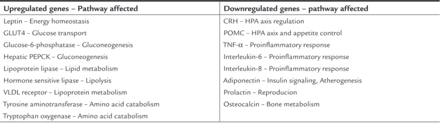
im-portant proteins that are directly or indirectly implicated in several metabolic pathways are rigorously regulated by GC (Table 1)14. 11β-Hydroxysteroid Dehydrogenase
TABLE 1 Selected glucocorticoid sensitive genes involved in important metabolic pathways
Upregulated genes – Pathway affected Downregulated genes – pathway affected
Leptin – Energy homeostasis GLUT4 – Glucose transport
Glucose-6-phosphatase – Gluconeogenesis Hepatic PEPCK – Gluconeogenesis Lipoprotein lipase – Lipid metabolism Hormone sensitive lipase – Lipolysis VLDL receptor – Lipoprotein metabolism
Tyrosine aminotransferase – Amino acid catabolism Tryptophan oxygenase – Amino acid catabolism
CRH – HPA axis regulation
POMC – HPA axix and appetite control TNF-α – Proinflammatory response Interleukin-6 – Proinflammatory response Interleukin-8 – Proinflammatory response Adiponectin – Insulin signaling, Atherogenesis Prolactin – Reproducion
Osteocalcin – Bone metabolism
GLUT4: Glucose transporter 4; PEPCK: Phoshpoonolpyruvate carboxylinase; VLDL: Very low density lipoprotein; CRH: Corticotropin hormone; HPA: Hupothalamic-pituitary-adrenal; POMC: Pro-oplomelanocortin; TNF-α: Tumor necrosis factor-α.
(11β-HSD) catalyzes the interconversion of inactive corti-sone to active cortisol, or vice-versa14. 11β-Hydroxysteroid
Dehydrogenase type 2 (11β-HSD2) predominates in renal tubules and protects the MR from excessive stimulation by cortisol. It has also been identified in colon, salivary glands and placenta18. The apparent mineralocorticoid
ex-cess syndrome results from defective 11β-HSD2. Indeed, its deficiency allows an increase of cortisol levels and MR activation inducing sodium retention, hypokalemia and hypertension19. 11β-Hydroxysteroid Dehydrogenase type
1 (11β-HSD1), which is mostly expressed in liver, fat, go-nadal tissue and central nervous system, is believed to func-tion as a reductase, generating active cortisol at a prerecep-tor level, thus enhancing the activation of GC20. The
co-localization of 11β-HSD1 with Hexose-6-phosphate de-hydrogenase (H6PDH) has an important role in providing nicotinamide adenine dinucleotide phosphate (NADPH) as cofactor to drive the direction to 11β-HSD1 reductase, rather than to dehydrogenase18.
P
HYSIOLOGICAL EFFECTS OFGLUCOCORTICOIDSGC are the final mediators of HPA axis activation, play-ing a key role in modulatplay-ing immunological and inflam-matory responses, energy metabolism and cardiovascu-lar homeostasis and general responses to stress18. In order
to face threats imposed by stressors, several responses are activated towards the restoration of homeostasis. Behav-ioral adaptations include increased arousal, euphoria and cognition, enhanced analgesia, and sleep inhibition. Phys-ical adaptations include increases in cardiovascular tone, respiratory rate, and metabolism. The activation of HPA axis promotes a redirection of energy, in order to deliver oxygen and nutrients to organs and tissues involved in the adaptation functional system. In the meantime, oth-er non-emoth-ergent functions, such as digestion, reproduc-tion, growth, and overall immunity, are temporarily sup-pressed3,12. All these acute effects increase the capacity for
generation of energy over a limited period of time im-proving the ability to ‘fight or flight’20. GC induce
lipol-ysis, even though they favor both abdominal and dorso-cervical fat accumulation17. Given that GC promote
delivery of fuel to skeletal muscles, the increase in triglyc-erides (TAG) stores in adipose tissue in clinical condi-tions with GC excess appears paradoxical. This paradox probably reflects the combination of elevated GC levels concomitantly with elevated insulin levels in individuals who are able to ingest unrestricted energy without con-suming it – for instance, through physical exercise. In these circumstances, fatty acid esterification predomi-nates over lipolysis and, combined with stimulation of
pre-adipocyte differentiation, promotes fat accumula-tion20,21, a mechanism that is most probably common to
what happens under chronic stress.
GC increase hepatic gluconeogenesis and decrease glucose uptake and insulin sensitivity, thus favoring hy-perglycemia. In order to provide amino acids as an addi-tional substrate for oxidative pathways, GC cause protein degradation at multiple tissues such as muscle, bone and skin. GC also antagonize the anabolic actions of growth and thyroid hormones, insulin, and sex steroids on their target tissues3,12. Globally, stress response is responsible
for a shift of normal metabolism, favoring a catabolic state which returns to normal after stress removal. Chron-ic exposure to stress, however, can be potentially damag-ing, as long exposure to GC can dysregulate multiple met-abolic pathways leading to a progressively increase in visceral adiposity, hyperglycemia, dyslipidemia, hyperten-sion and insulin resistance3,12. Elevated circulating GC
levels also result in myopathy, osteoporosis, osteonecro-sis, mental disturbances, increased susceptibility to in-fections and infertility12,20. GC actions in the central
ner-vous system are complex12. GC affect the capacity to
apprehend sensations and establish suitable reactions to stimuli22, modulate behavior and humor and interfere
with memory retention4. Short-term changes in
immu-nological function may be valuable, preventing the dam-age caused by sustained exposure to various cytokines3.
M
ETABOLICS
YNDROMEThe MS is a cluster of metabolic abnormalities that in-crease the risk for type 2 diabetes mellitus and cardiovas-cular disease. It can be defined as a state of disturbed met-abolic homeostasis characterized by the combination of central obesity, insulin resistance, dyslipidemia and hy-pertension17. The worldwide incidence of both obesity
and MS has been significantly rising in the last decades, threatening to become the new epidemic of this century. In fact, currently, MS affects ¼ of the adult Portuguese population23. It is noteworthy that these clinical
condi-tions often show a relacondi-tionship with indices of stress24.
Although obesity per se is not a required feature for the diagnosis of MS, several evidences suggest that both vis-ceral obesity and insulin resistance have a key role in the pathogenic mechanisms underlying this syndrome25. The
distribution of fat seems to be a powerful predictor of cardiometabolic disease26 and many prospective studies
have shown that central obesity is more often correlated to the features of MS25. This pathophysiologic
cortisol urinary metabolites excretion32. In accordance
with this, it has been shown that patients with obesity and MS show increased urinary excretion of free cortisol and its metabolites17. Interestingly, the excretion of
uri-nary free cortisol correlates with anthropometric param-eters of visceral fat distribution32. Overall, there appears
to be hyperactivity of the HPA axis in response to stress in patients with visceral obesity2,17, which is in accordance
with results that show a higher release of cortisol after stimulation with ACTH and greater ACTH release after CRH infusion30,33. Moreover, chronically active HPA axis
has an inadequate suppression by dexamethasone9.
Sev-as an indicator of obesity-related cardiovSev-ascular risk25. In
fact, even in normal-weight subjects, increased abdomi-nal circumference is associated with increased risk for car-diovascular disease27. The strong literature evidences
high-lighted in the next sections reinforce our opinion in that GC, released in response to chronic stress, induce an ac-cumulation of visceral fat that, when some conditions are met (such as a lifestyle characterized by excessive en-ergy intake, low physical activity and low quality food), will trigger the development of MS.
C
LINICAL ASSOCIATIONS BETWEEN HYPERCORTI-SOLISM AND METABOLIC SYNDROME
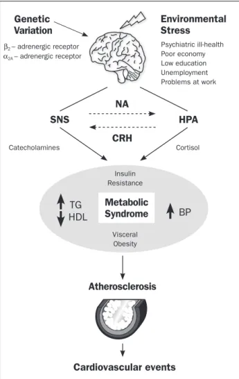
The similarities between the clinical features of Cushing syndrome and those of MS raised the hypothesis that MS is associated with GC excess9,14. Cortisol excess has been
implicated in the development of diabetes and obesity, highlighting the role of psychological stress on both con-ditions (Figure 1)17. Indeed, human studies have
docu-mented that abdominal obesity and its metabolic comor-bidities are significantly correlated with stress-related conditions such as adverse life events, psychological dis-turbances, and psychosocial problems2. In fact, the
indi-vidual inability to cope with long-term environmental adverse stressful events has been related to HPA axis hy-peractivation in obesity, particularly the visceral pheno-type9. Chronic work stress also seems to predict general
and central obesity during midlife1. Prolonged and
non-remitting stress may result in chronic hyperactivation of the HPA axis with resulting sustained GC release28, which
can progressively cause visceral fat accumulation and in-sulin resistance. In line with this, patients with MS seem to have HPA axis hyperactivity and a functional hyper-cortisolism14,29. On the contrary, in obese individuals, the
circulating GC levels have been reported as normal or even low30,31. Several hypotheses can explain an abnormal
secretion of cortisol in obesity. Firstly, the presence of a primary neuroendocrine abnormality can cause an irreg-ular central drive to CRH, ACTH and cortisol. Second, an altered peripheral metabolism of cortisol due to dysreg-ulation of 11β-HSD1 can also explain normal or low cor-tisol concentrations in obesity32. On the other hand, it
has been suggested that cortisol production rate may in-crease as the amount of visceral fat enlarges33, however,
as said before, in most cases cortisol plasma concentra-tions are normal or even low. This might be partly ex-plained by enhanced metabolic clearance of cortisol17, due
to a combination of enhanced 5α-redutase activity and impaired regeneration of cortisol from cortisone by 11β-HSD1 in the liver, which results in the increase of
Genetic Variation
NA
HPA SNS
TG
BP HDL
CRH
Metabolic Syndrome
Environmental Stress
β2 – adrenergic receptor
Cortisol
Insulin Resistance
Visceral Obesity Catecholamines
α2A – adrenergic receptor
Psychiatric ill-health Poor economy Low education Unemployment Problems at work
Atherosclerosis
Cardiovascular events
FIGURE 1 The stress response leads to activation of two major
neurohumoral systems, the hypothalamic-pituitary-adrenal (HPA) axis and the sympathetic-nervous-system (SNS) which, through the release of cortisol and catecholamines, respectively, exert crucial roles upon energy metabolism, ultimately leading to the development of features of the metabolic syndrome. TG – Triglycerides; HDL - High--density lipoprotein; BP – Blood pressure; NA-noradrenaline;
eral studies have shown that in obesity different stimu-lus such as some neuropeptides, psychological stress and mixed meal tests induce hyperactivation of HPA axis. In-dividuals that show elevated levels of cortisol in response to perceived stress show a higher association with central fat mass and signs of MS9. Animal studies demonstrated
that cynomolgus monkeys subjected to high stress levels comparably to controls secreted higher amounts of GC and were less sensitive to their negative feedback28.
An-other study reported that these animals undergoing phys-ical and psychologphys-ical stress presented higher basal cor-tisol levels and increased corcor-tisol release after ACTH stimulation, which was associated with greater visceral fat accumulation24. In adipose tissue, lipids are stored as
TAG. GC increase lipolysis in adipocytes, as a result of in-creased transcription and expression of the lypolytic pro-teins adipose triglyceride lipase and hormone-sensitive lipase, increasing the amount of fatty acids in circulation, which, in turn, contribute to triglyceride accumulation in other tissues34. In the liver, GC increase the expression
of fatty acid synthase increasing lipid production, thus favoring hepatic steatosis. GC also promote the secretion of lipoproteins14. TAG in circulation, as components of
both very low-density lipoproteins and chylomicrons, when hydrolyzed, release fatty acids that can be taken up by the surrounding tissues for use or storage, mainly in liver, muscle and central adipocytes34. GC promote the
differentiation of adipose stromal cells to mature adipo-cytes, increasing visceral fat accumulation34 and
redistrib-uting adipose tissue from peripheral to central depots, and increasing the size and number of fat cells17,35.
Adenosine monophosphate activated protein kinase (AMPK) is a sensor of cellular energy status and is activat-ed in response to a decrease of this state. When activatactivat-ed, AMPK stimulates appetite in the hypothalamus and
switch-es anabolic into catabolic pathways, such as glycolysis and fatty acid oxidation. GC inhibit the AMPK system thus contributing to central fat deposition36. GC increase
ca-loric and dietary fat intake34 and suppress thermogenesis9.
Growing evidence suggests that there is a relation-ship between type 2 diabetes and chronic stress disorders1.
In fact, circulating cortisol concentrations are higher in people with glucose intolerance and type 2 diabetes37. GC
raise blood glucose levels through several mechanisms38.
GC impair the insulin-dependent glucose uptake in pe-ripheral tissues, enhance glucose production in the liver and inhibit insulin secretion from pancreatic β-cells. Thus, cortisol excess can be correlated with diabetes mellitus in clinical settings38. Insulin stimulates translocation of the
GLUT4 glucose transporters from intracellular
compart-ments to plasma membrane, increasing the rate of glu-cose utilization, however this action is inhibited by high levels of GC14. In insulin sensitive tissues, such as liver
and skeletal muscle, GC also impair pathways involved in insulin receptor activation2,38. GC promote
gluconeo-genesis by stimulating the expression of phosphoenol-pyruvate carboxykinase (PEPCK) and glucose-6-phospha-tase (G6Pase), the rate-limiting enzymes of this pathway, resulting in increased hepatic glucose output and hyper-glycemia14,38. GC decrease GLUT2 expression in
pancre-atic β-cells and impair calcium disposal on insulin secre-tory process, thus affecting its secretion14.
Obesity seems to be implicated in the development of insulin resistance25,27. In fact, pathophysiological
ac-cumulation of lipids in the liver has been identified as an independent risk factor for insulin resistance and MS13.
Free fatty acids inhibit insulin secretion by pancreas and decrease glucose uptake39. GC release increases lipolysis
generating free fatty acids into the circulation, which in turn impair insulin signaling pathways downstream of its receptor, thus promoting insulin resistance27,39. In
hu-mans, the administration of dexamethasone decreases glucose oxidation, which can be due to an increase of free fatty acids plasmatic levels40. Thus, insulin resistance can
result from an excess of visceral fat27,41. This is in
accor-dance with our view, that visceral fat might be the main trigger for the development of MS. On the other hand, insulin resistance impairs lypogenesis, thus increasing the plasmatic levels of free fatty acids, creating a vicious circle39. Adipokines play an important role on fat
accu-mulation and insulin resistance27,39. Adiponectin
modu-lates a number of metabolic processes, including glucose homeostasis and fatty acid oxidation. It promotes insu-lin sensitivity and is negatively regulated by GC17.
Hypo-adiponectinemia is an independent risk factor for devel-oping MS and type 2 diabetes mellitus42. Leptin plays a
key role in regulating both energy intake and metabolism, and its circulating levels are directly proportional to body fat39. Activation of hypothalamic leptin receptors
sup-presses appetite, induces satiety and increases energy ex-penditure43. High sustained concentrations of leptin
re-leased from adipose tissue result in desensitization of leptin receptor leading to leptin resistance39,44.
Unusual-ly high circulating leptin levels and low adiponectin lev-els are generally exhibited by patients with obesity, insu-lin resistance and MS27. Visfatin is a recently discovered
adipokine with insulin-mimetic properties25. ACTH
ac-tivation seems to promote abdominal fat storage. GC stimulates NPY hypothalamic secretion and up-regulate the NPY Y2 receptor in visceral fat. NPY increases food intake and storage of energy as fat and NPY Y2 receptor activation stimulate fat angiogenesis, proliferation and differentiation of new adipocytes44.
GR signaling plays a significant role in metabolic regulation, and defects in this signaling pathway have been implicated in the development of several pheno-types of MS. In fact, insulin resistance is associated with increased GR expression in skeletal muscle38. Several
polymorphisms related to the HPA axis have been asso-ciated with HPA abnormal function and development of MS21,44,. Chronic stress disorders are commonly
associ-ated with behavioral changes which lead to weight gain and metabolic abnormalities. Chronic stress seems to promote unhealthy behaviors such as a sedentary life-style, alcohol consumption, smoking and overeating1.
Stress system affects the hypothalamic appetite-satiety centers therefore disturbing food intake3. The
relation-ship between stress and food ingestion has been exten-sively investigated but has led to conflicting results. In-creasing food intake during stress diminishes HPA axis response to stressors45, which might explain why various
individuals overeat when exposed to stress. Food inges-tion to relieve anxiety is a harmful coping strategy and can lead to undesirable weight gain and obesity46. On the
other hand, acute stress may be accompanied by a de-crease on food consumption. Indeed, acute elevations of CRH can cause anorexia and stimulate energy expendi-ture. As mentioned before, NPY increases food intake and storage of energy as fat, and acute stress inhibits NPY release3. On the contrary, chronic stressful
situa-tions have the opposite effect and people experiencing a high stress reactivity tend to have a greater caloric intake, preferably dense calories46. In fact, long-term exposure
to circulating GC seems to enhance consumption of high fat and highly palatable foods45. GC also decrease
ener-gy expenditure3 and diminish signs of satiety46. A high
caloric intake is a mechanism that seems natural in re-sponse to fasting. However, this is not the case of psy-chological stress conditions, in which food is used rath-er to relief anxiety than to ovrath-ercome fasting34,45.
11
β
-HSD1
ROLETissue-specific dysregulation of GC metabolism seems to be involved in the complex pathophysiology of obesity and the MS35. In fact, 11β-HSD1 expression positively correlates
with obesity and insulin resistance35. As mentioned before,
circulating GC concentrations are occasionally abnormal
in human obesity, however locally enhanced responsiveness to GC has also been implicated in MS development1,17, 19,38.
Obese individuals have a tissue-specific 11β-HSD1 dys-regulation32,37 in which 11β-HSD1 activity is found
selec-tively increased in visceral fat depots and decreased in liv-er20,29. The relationship between the 11β-HSD1 function
and metabolic disorders has been well established by stud-ies using genetically modified rodent models. In fact, up regulation of 11β-HSD1 expression selectively in adipose tissue leads to a model of MS in mice19, 38. Mice with a
sim-ilar degree of 11β-HSD1 over expression in the liver show an attenuated MS profile without visceral obesity29. On
the contrary, knockout mice lacking 11β-HSD1 exhibit protection for MS features27,29,37. Peroxisome
proliferator-activated receptors (PPAR) are a group of nuclear receptor proteins involved in adipocyte differentiation and fat re-distribution to the periphery47. 11β-HSD1 knockout mice
show higher expression of PPARγ receptor in all adipose tissue depots38.
O
BESITY AS A CHRONIC INFLAMMATORY STATEIn recent years, it has become clear that obesity is a state of chronic low-grade inflammation27. The association
be-tween insulin resistance and the other components of MS can be a consequence of their common outcomes as low-grade inflammation states27,48. Adipose tissue releases
cy-tokines that initiate a state of low-grade inflammation re-sulting in the metabolic, hemodynamic and vascular consequences of this state41,48. The uninterrupted release
of these pro-inflammatory adipokines is a chronic stim-ulus for HPA axis activation, creating a vicious cycle, in which hypercortisolemia promotes adipocyte growth and vice versa3. The pro-inflammatory cytokines, TNFα, IL-1
and IL-6 act synergistically activating HPA axis and in-creasing 11β-HSD1 expression in adipose tissue14,17. IL-6
concentrations have a strong correlation with visceral obe-sity and are associated with insulin resistant39,41. There is
a positive association between TNFα concentrations and BMI, and this cytokine seems to be implicated in insulin resistance development27,33,41. Thus, pro-inflammatory
cy-tokines are important players in this process probably starting with the dysfunction of visceral adipose tissue towards the development of other components of MS.
G
LUCOCORTICOIDS INHIBITORS AS POTENTIALTHERAPEUTIC TARGETS
To date, no single agent can ameliorate the underlying causes of MS27. Additional research is needed for novel
as an approach to treat some of the MS features14,
lead-ing to decreases in adiposity, glucose intolerance and insulin resistance, lowering fasting blood glucose and normalizing its postprandial levels20,29, 38. However,
long-term systemic treatment with a GR antagonist may not be a viable option, since it can excessively activate the HPA axis causing adrenal hyperplasia and undesirable increase in cortisol, androgens and mineralocorticoids19,38.
On the other hand, selective 11β-HSD1 inhibitors have shown considerable potential for MS treatment. Over the past years, clinical studies have been conducted for several 11β-HSD1 inhibitors29,38. The ability to decrease
intracellular cortisol levels in liver and adipose tissue, without altering circulating cortisol concentrations or responses to stress, might be an exciting therapeutic strategy for obesity, type 2 diabetes mellitus and MS treatment. In fact, these drugs have been shown to ame-liorate metabolic abnormalities, by improving lipid pro-file, insulin sensitivity, promoting glucose tolerance and blocking adipogenesis17,38. However, there are some
con-cerns regarding the use of these drugs. 11β-HSD1 inhi-bition on hippocampus might decrease central feedback inhibition, which may cause HPA axis activation with increased GC release and enhancement of its effects in peripheral tissues. Moreover, non-selective compounds can potently inhibit 11β-HSD1 causing apparent min-eralocorticoid excessive release with sodium retention, hypertension and hypokalemia38. Nevertheless, PPARα
and PPARγ agonists are able to down regulate 11β-HSD1 activity in liver and adipose tissue, respectively27,
con-stituting promising approaches. Emerging data suggest that dietary habits have a role on 11β-HSD1 modula-tion. For instance, naturally occurring 11β-HSD1 inhib-itors include polyphenols, such as flavones and querce-tin29. Coffee has been reported to have anti-diabetic
effects due to its ability to impair hepatic gluconeogen-esis and inhibit 11β-HSD1 function29. Dietary trans and
saturated fatty acids appear to be involved in the devel-opment of MS, through their ability to up regulate 11β-HSD1, thus, increasing local amplification of GC action in adipose tissue29. Sucrose promotes
simultane-ously a decrease in hepatic 11β-HSD1 and an increase in 11β-HSD1 in adipose tissue. Dietary sucrose increas-es H6PDH, which, in turn, may enhance 11β-HSD1 ac-tivity and intracellular GC. These observations support the fact that increased activity of 11β-HSD1 in response to sucrose ingestion is able to cause obesity29. The
anti-obesity effect of vitamin A supplementation might be, in part, due to its ability to decrease 11β-HSD1 activi-ty49. MS seems to be associated with vitamin D
deficien-cy and, accordingly, vitamin D status optimization seems to improve MS features50. A low-calcium diet alters GC
metabolism leading to hepatic up regulation of 11β-HSD129; instead a high intake of calcium is
associ-ated with a low prevalence of MS51.
O
UTLOOKThe pathogenesis of the MS is multiple and still poorly understood. No single factor has yet been identified as an underlying causal factor; however, we strongly believe that visceral obesity might be the key feature in the patho-genesis of this syndrome. As a matter of fact, visceral obe-sity seems to be the major predictor of MS incidence52. In
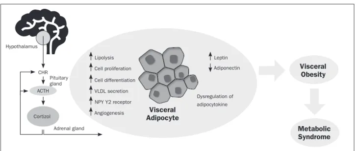
the context of stress response, GC direct effects are known for a long time12. Nevertheless the role of chronic stress
and the hyperactivity of the HPA axis in the development of the MS remain not fully understood. The main puta-tive mechanisms by which GC can promote visceral obe-sity are reviewed (Figure 2). Briefly, the release of GC in-duces lipolysis and the release of VLDL, increasing free fatty acids in circulation that are taken up for storage by other tissues, such as the visceral adipose tissue. GC pro-motes proliferation and differentiation of visceral adi-pose tissue via different mechanisms. For example, indi-rectly by enhancing the release of NYP, which is known by its action on adipocytes maturation, proliferation and angiogenic pathways. High levels of GC lead to an abnor-mal release of adipocytokines, such as leptin and adipo-nectin, which ultimately contributes to visceral fat accu-mulation. However, not all patients with MS present visceral obesity. As mentioned in a previous publication by our group, the size of abdominal adipocytes seems to be as important as the amount of accumulated fat in the abdominal cavity52. In fact, findings from our lab have
shown that large adipocytes are more prone to rupture, and cell rupture constitutes a focus of inflammation that incites metabolic and immune responses leading to a se-ries of pathogenic complications of obesity53. On the
oth-er hand, it is important to have in mind the ‘cross-talk’ between adipose tissue and other body systems, such as the immune system, since metabolic changes induced by GC release tend to be feed-forward and may, thus, lead to an endless spiral.
C
ONCLUSIONpathophys-iology of this syndrome remains partially understood, although available evidence hypothesized that hyperac-tivity of the HPA axis due to chronic stress and a state of functional hypercortisolism may have a central role in the pathogenesis of both abdominal obesity and in-sulin resistance. Data obtained from animal studies showed that exposure to both physical and psycholog-ical chronic stress was characterized by abnormal GC levels and several features of the MS. Although direct similar evidences are lacking in humans, epidemiologi-cal data have been providing evidence for a significant positive association between GC levels and the MS. In this review some of the underlying mechanisms sup-porting this hypothesis are highlighted. In our point of view, shared by others, one candidate that might be the link between chronic stress, GC and the MS, being prob-ably the main trigger for the development of this syn-drome, is visceral adiposity. Consequently, stress man-agement programs, capable of effectively decrease circulatory GC levels, may constitute an important tool in patients at risk or suffering from MS.
Acknowledgements
The authors are grateful to Eng. Joana Afonso, who contrib-uted with an original illustration for this paper (Figure 2).
R
ESUMOCortisol: o vilão na Síndrome Metabólica?
Objetivo: Este artigo revê o estado da arte
relativamen-te à associação entre as ações dos glicocorticoides e a obe-sidade e a resistência à insulina, dois dos principais com-ponentes da síndrome metabólica.
Métodos: Uma revisão da literatura nas bases de dados
PubMed e SciELO foi realizada usando os seguintes ter-mos: estresse, síndrome metabólica, glicocorticoides, obe-sidade, resistência à insulina, eixo hipotálamo-hipófise-suprarrenal e 11β-hidroxiesteróide desidrogenase.
Resultados: O estresse crônico, principalmente através
da desregulação do eixo hipotálamo-hipófise-suprarre-nal, promove a acumulação de gordura visceral. Recipro-camente, a obesidade promove um estado inflamatório sistêmico de baixo grau, mediado por alterações na secre-ção de adipocinas, que cronicamente podem estimular e perturbar o sistema de estresse. Esse círculo vicioso, pro-vavelmente iniciado pela disfunção do tecido adiposo vis-ceral, pode ser o mecanismo primário que conduz ao de-senvolvimento da síndrome metabólica.
Conclusões: Um conhecimento mais aprofundado
so-bre os mecanismos envolvidos na associação entre a libe-ração de glicocorticoides, a obesidade e o diabete tipo 2 pode ser útil na prevenção e tratamento da síndrome me-tabólica.
Unitermos: estresse; glicocorticoides; obesidade; resistên-cia à insulina; síndrome metabólica; 11β-hidroxiesteroide desidrogenase.
Hypothalamus
CHR
ACTH
Cortizol
Adrenal gland
Lipolysis Leptin
Adiponectin Cell proliferation
Cell differentiation
VLDL secretion
NPY Y2 receptor
Angiogenesis Visceral
Adipocyte
Dysregulation of
adipocytokine
Visceral Obesity
Metabolic Syndrome
FIGURE 2 Putative mechanisms involved in GC release, visceral adipose tissue accumulation and the pathogenesis of Metabolic Syndrome.
Cortisol, the major glucocorticoid in humans, is secreted in response to stressful events under the control of the hypothalamic– pituitary–adrenal axis. Cortisol exerts several cellular and metabolic effects in adipocytes, eventually promoting visceral obesity and the development of Metabolic Syndrome. CRH - corticotropin-releasing hormone; ACTH - adrenocorticotropic hormone.
R
EFERENCES1. Kyrou I, Tsigos C. Stress hormones: physiological stress and regulation of metabolism. Curr Opin Pharmacol. 2009;9:787-93.
2. Pasquali R, Vicennati V, Cacciari M, Pagotto U. The hypothalamic-pituitary-adrenal axis activity in obesity and the metabolic syndrome. Ann N Y Acad Sci. 2006;1083:111-28.
3. Kyrou I, Chrousos G, Tsigos C. Stress, visceral obesity, and metabolic complications. Ann N Y Acad Sci. 2006;1083:77-110.
4. McWwen BS. Stress, adaptation, and disease. Allostasis and allostatic load. Ann N Y Acad Sci. 1998;840:33-44.
5. Iwasaki Y, Takayasu S, Nishiyama M, Tsugita M, Taguchi T, Asai M, et al. Is the metabolic syndrome an intracellular Cushing state? Effects of multiple humoral factors on the transcriptional activity of the hepatic glucocorticoid-activating enzyme (11beta-hydroxysteroid dehydrogenase type 1) gene. Mol Cell Endocrinol. 2008;285:10-8.
6. Flegal KM, Graubard BI, Williamson DF, Gail MH. Excess deaths associated with underweight, overweight, and obesity. JAMA. 2005;293:1861-7. 7. Shomali M. Diabetes treatment in 2025: can scientific advances keep pace
with prevalence? Ther Adv Endocrinol Metab. 2012;3:163-73.
8. Brunner EJ, Hemingway H, Walker BR, Page M, Clarke P, Juneja M, et al. Adrenocortical, autonomic, and inflammatory causes of the metabolic syndrome: nested case-control study. Circulation. 2002;106:2659-65. 9. Vicennati V, Pasqui F, Cavazza C, Pagotto U, Pasquali R. Stress-related
development of obesity and cortisol in women. Obesity. 2009;17:1678-83. 10. Chandola T, Brunner E, Marmot M. Chronic stress at work and the metabolic
syndrome: prospective study. BMJ. 2006;332:521-5.
11. Rosmond R, Dallman MF, Björntorp P. Stress-related cortisol secretion in men: relationships with abdominal obesity and endocrine, metabolic and hemodynamic abnormalities. J Clin Endocrinol Metab. 1998;83:1853-9. 12. Charmandari E, Tsigos C, Chrousos G. Endocrinology of the stress response.
Annu Rev Physiol. 2005;67:259-84.
13. Vegiopoulos A, Herzig S. Glucocorticoids, metabolism and metabolic diseases. Mol Cell Endocrinol. 2007;275:43-61.
14. Wang M. The role of glucocorticoid action in the pathophysiology of the Metabolic Syndrome. Nutr Metab (Lond). 2005;2:3.
15. Tsigos C, Chrousos GP. Hypothalamic–pituitary–adrenal axis, neuroendocrine factors and stress. J Psychosom Res. 2002;53:865-71.
16. Torpy DJ, Ho JT. Corticosteroid-binding globulin gene polymorphisms: clinical implications and links to idiopathic chronic fatigue disorders. Clin Endocrinol (Oxf). 2007;67:161-7.
17. Anagnostis P, Athyros VG, Tziomalos K, Karagiannis A, Mikhailidis DP. Clinical review: the pathogenetic role of cortisol in the metabolic syndrome: a hypothesis. J Clin Endocrinol Metab. 2009;94:2692-701.
18. Wamil M, Seckl JR. Inhibition of 11beta-hydroxysteroid dehydrogenase type 1 as a promising therapeutic target. Drug Discov Today. 2007;12:504-20. 19. Walker BR, Andrew R. Tissue production of cortisol by 11beta-hydroxysteroid
dehydrogenase type 1 and metabolic disease. Ann N Y Acad Sci. 2006;1083:165–84.
20. Walker BR. Extra-adrenal regeneration of glucocorticoids by 11beta-hydroxysteroid dehydrogenase type 1: physiological regulator and pharmacological target for energy partitioning. Proc Nutr Soc. 2007;66:1-8. 21. Rosmond R. Role of stress in the pathogenesis of the metabolic syndrome.
Psychoneuroendocrinology. 2005;30:1-10.
22. Brown ES. Effects of glucocorticoids on mood, memory, and the hippocampus. Treatment and preventive therapy. Ann N Y Acad Sci. 2009;1179:41-55.
23. Fiuza M, Cortez-Dias N, Martins S, Belo A. Síndrome metabólica em Portugal: Prevalência e implicações no risco cardiovascular: resultados do Estudo Valsim. Rev Port Cardiol. 2008;27:1495-529.
24. Shively CA, Register TC, Clarkson TB. Social stress, visceral obesity, and coronary artery atherosclerosis: product of a primate adaptation. Am J Primatol. 2009;71:742–51.
25. Ribeiro Filho FF, Mariosa LS, Ferreira SR, Zanella MT. Gordura visceral e síndrome metabólica: mais que uma simples associação. Arq Bras Endocrinol Metab. 2006;50:230-8.
26. Haun D, Pitanga F, Lessa I. Razão cintura/estatura comparado a outros indicadores antropométricos de obesidade como preditor de risco coronariano elevado. Rev Assoc Med Bras. 2009;55:705-11.
27. Moller DE, Kaufman KD. Metabolic syndrome: a clinical and molecular perspective. Annu Rev Med. 2005;56:45-62.
28. Shively CA, Laber-Laird K, Anton RF. Behavior and physiology of social stress and depression in female cynomolgus monkeys. Biol Psychiatry. 1997;41:871-82.
29. Pereira CD, Azevedo I, Monteiro R, Martins MJ. 11β-Hydroxysteroid
dehydrogenase type 1: relevance of its modulation in the pathophysiology of obesity, the metabolic syndrome and type 2 diabetes mellitus. Diabetes Obes Metab. 2012;14:869-81.
30. Rask E, Walker B, Söderberg S, Livingstone DE, Eliasson M, Johnson O, et al. Tissue-specific changes in peripheral cortisol metabolism in obese women: increased adipose 11beta-hydroxysteroid dehydrogenase type 1 activity. J Clin Endocrinol Metab. 2002;87:3330-6.
31. Stewart PM, Boulton A, Kumar S, Clark PM, Shackleton CH. Cortisol metabolism in human obesity: impaired cortisone-->cortisol conversion in subjects with central adiposity. J Clin Endocrinol Metab. 1999;84:1022-7. 32. Rask E, Olsson T, Söderberg S, Andrew R, Livingstone DE, Johnson O, et
al. Tissue-specific dysregulation of cortisol metabolism in human obesity. J Clin Endocrinol Metab. 2001;86:1418-21.
33. Wajchenberg BL. Subcutaneous and visceral adipose tissue: their relation to the metabolic syndrome. Endocr Rev. 2000;21:697-738.
34. Peckett AJ, Wright DC, Riddell MC. The effects of glucocorticoids on adipose tissue lipid metabolism. Metabolism. 2011;60:1500-10.
35. Espíndola-Antunes D, Kater CE. Adipose tissue expression of 11beta-hydroxysteroid dehydrogenase type 1 in Cushing’s syndrome and in obesity. Arq Bras Endocrinol Metab. 2007;51:1397-403.
36. Christ-Crain M, Kola B, Lolli F, Fekete C, Seboek D, Wittmann G, et al. AMP-activated protein kinase mediates glucocorticoid induced metabolic changes: a novel mechanism in Cushing’s syndrome. FASEB. J 2008;22:1672-83. 37. Walker BR. Cortisol - cause or cure for metabolic syndrome? Diabet Med.
2006;23:1281-8.
38. Joharapurkar A, Dhanesha N, Shah G, Kharul R, Jain M. 11β-Hydroxysteroid dehydrogenase type 1: potential therapeutic target for metabolic syndrome. Pharmacol Rep. 2012;64:1055-65.
39. Costa J, Duarte J. Tecido adiposo e citocinas. Acta Med Port. 2006;19:251-6. 40. Dimitriadis G, Leighton B, Parry-Billings M, Sasson S, Young M, Krause U, et
al. Effects of glucocorticoid excess on the sensitivity of glucose transport and metabolism to insulin in rat skeletal muscle. Biochem J. 1997;321:707-12. 41. Ikeoka D, Madjer J, Pieber T. Adipose tissue, inflammation and cardiovascular
disease. Rev Assoc Med Bras. 2010;56:116-21.
42. Vasseur F, Meyre D, Froguel P. Adiponectin, type 2 diabetes and the metabolic syndrome: lessons from human genetic studies. Expert Rev Mol Med. 2006;20:1-12.
43. Hermsdorff HH, Monteiro JB. Gordura visceral, subcutânea ou intramuscular: onde está o problema? Arq Bras Endocrinol Metab. 2004;48:803-11. 44. Nieuwenhuizen AG, Rutters F. The hypothalamic-pituitary-adrenal-axis in
the regulation of energy balance. Physiol Behav. 2008;94:169-77. 45. Warne JP. Shaping the stress response: Interplay of palatable food choices,
glucocorticoids, insulin and abdominal obesity. Mol Cell Endocrinol. 2009;300:137-46.
46. Adam TC, Epel ES. Stress, eating and the reward system. Physiol Behav. 2007;91:449-58.
47. Seckl JR, Morton NM, Chapman KE, Walker BR. Glucocorticoids and 11beta-hydroxysteroid dehydrogenase in adipose tissue. Recent Prog Horm Res. 2004;59:359-93.
48. Wajchenberg BL, Nery M, Cunha MR, Silva ME. Adipose tissue at the crossroads in the development of the metabolic syndrome, inflammation and atherosclerosis. Arq Bras Endocrinol Metab. 2009;53:145-50. 49. Sakamuri VP, Ananthathmakula P, Veettil GN, Ayyalasomayajula V. Vitamin
A decreases pre-receptor amplification of glucocorticoids in obesity: study on the effect of vitamin A on 11beta-hydroxysteroid dehydrogenase type 1 activity in liver and visceral fat of WNIN/Ob obese rats. Nutr J. 2011;10:70. 50. Al-Daghri N, Alkharfy K, Al-Saleh Y, Al-Attas O, Alokail M, Al-Othman A, et al.
Modest reversal of metabolic syndrome manifestations with vitamin D status correction: a 12-month prospective study. Metabolism. 2012;61:661-6. 51. Liu S, Song Y, Ford ES, Manson JE, Buring JE, Ridker PM. Dietary calcium,
vitamin D, and the prevalence of metabolic syndrome in middle-aged and older U.S. women. Diabetes Care. 2005;28:2926-32.
52. Azevedo A, Santos A, Ribeiro L, Azevedo I. The metabolic syndrome. In: Soares R, Costa C, editors. Oxidative stress, inflammation and angiogenesis in the metabolic syndrome. Netherlands: Springer; 2009. p.1-19 53. Monteiro R, Castro P, Calhau C, Azevedo I. Adipocyte size and liability to