Prevalence of low bone mineral density in adolescents and adults
with cystic fibrosis
ROBERTA VANACOR1, FABIANA V. RAIMUNDO1, NATÁLIA A. MARCONDES1, BRUNO P. CORTE2, ALINE M. ASCOLI2, ALINE Z. DE AZAMBUJA2, LUCIANO SCOPEL2, PATRÍCIA V. DOS SANTOS2, PAULO T. R. DALCIN3, GUSTAVO A. M. FAULHABER2, TÂNIA W. FURLANETTO2
1-Postgraduate Program in Medicine, Medical Sciences - Federal University of Rio Grande do Sul, Porto Alegre, RS, Brazil 2-Division of Internal Medicine, Hospital de Clínicas de Porto Alegre - Federal University of Rio Grande do Sul, Porto Alegre, RS, Brazil 3-Division of Pneumology, Hospital de Clínicas de Porto Alegre - Federal University of Rio Grande do Sul, Porto Alegre, RS, Brazil
S
UMMARYArticle submitted: 01/25/13
Accepted for publication: 07/01/13
Correspondence:
Rua Guararapes, 82 Porto Alegre, RS – Brazil
ZIP Code: 90690-340 Phone/Fax: +55 51 3359-8152
[email protected] [email protected]
http://dx.doi.org/10.1590/1806-9282.60.01.012
Conflict of interest: none
Objective: The aim of this cross-sectional study was to evaluate the prevalence of low bone mass density in cystic fibrosis patients as well as to evaluate the fac-tors associated with bone mass in such patients.
Methods: Bone mass density was measured by dual-photon X-ray absorptio-metry of lumbar spine (L1-L4), in patients ≤19 years old, or lumbar spine and femur (total and neck) in patients ≥20 years old. Evaluations of nutritional sta-tus, biochemical parameters, and lung function were performed. Medication data were obtained from medical records.
Results: Fifty-eight patients were included in the study (25 males/ 33 females), mean age 23.9 years (16-53years). The prevalence of bone mass below the expec-ted range for age at any site was 20.7%. None of the subjects had history of frac-ture. Lumbar spine Z-score in cystic fibrosis patients correlated positively with body mass index (r= 0.3, p=0.001), and forced expiratory volume in the first se-cond (% predicted) (r=0.415, p=0.022). Mean lumbar spine Z-score was higher in women (p=0.001), in patients with no pancreatic insufficiency (p=0.032), and in patients with no hospitalization in the last 3 months (p=0.02). After multi-variate analysis, body mass index (p= 0.001) and sex (p=0.001) were independen-tly associated with Z-score in lumbar spine.
Conclusion: Low bone mass is a frequent problem in patients with CF, being independently associated with body mass index, and male sex.
Key words: cystic fibrosis, bone, bone density, bone loss, bone diseases, meta-bolic, risk factors.
I
NTRODUCTIONAdvances in the care of patients with cystic fibrosis (CF) increased survival. Bone health is important for the qua-lity of life in aging patients, and became more relevant in patients affected with CF1-4.
Several studies described lower bone mass density (BMD) in patients with CF, which increases fracture risk 5-8. Bone
fractures can cause pain, decrease respiratory status, and are a contraindication for lung transplantation in these patients9,10.
Cross-sectional studies in adults (age range from 16-60 years) with CF demonstrated that 6 to 68% had Z-scores ≤-2.08,11-16. Therefore, understanding the
me-chanisms of low BMD in CF patients is important to improve its prevention and treatment. Abnormal cal-cium homeostasis, poor nutritional status, chronic in-flammation, or inactivity associated with lung infec-tion exacerbainfec-tions could be responsible for bone abnormality5,8,12,17-19. Low BMD has been
independen-tly associated with malnutrition, male sex, ∆F508
mu-tation, and severe lung injury14. Other potential risk
In Brazil, just one study evaluated the prevalence of low BMD in patients with CF24. As bone mass is dependent on
ethnic factors25 and the prevalence of CFTR gene mutations
is not the same in different populations26, 27, it is important
to study how bone mass is affected in patients with cystic fi-brosis in different regions of the world. Therefore, the aim of this study was to evaluate the prevalence of low BMD in CF patients in a tertiary care facility in Porto Alegre, state of Rio Grande do Sul, Brazil, (30o S) as well as to evaluate the
factors associated with low bone mass in these patients.
M
ETHODSThe study was carried out in the outpatient program for ado-lescents and adults with CF at the Hospital de Clínicas de Por-to Alegre (HCPA), from February 25th to September 23th, 2011,
after approval by the institution’s Ethics Committee. Each patient gave written consent before inclusion in the study.
Inclusion criteria
Patients 16 years old or older with CF confirmed by sweat test (Cl-≥60 mEq/L) in two occasions or CFRT gene
mu-tation in both alleles were included. Exclusion criteria: Organ transplant.
Experimental design: Cross-sectional study. Data were obtained in the medical records and during interviews with the patients.
Evaluation of BMD
BMD was measured by dual-photon X-ray absorptiometry (DXA), with HOLOGIC QDR4500A, equipped with stan-dard density software (version 8.26) (4500 Acclaim den-sitometer, Hologic, Wattham, MA, USA). Regions evalua-ted were lumbar spine, L1- L4, in patients ≤19 years old, or lumbar spine and femur (total and neck) in patients ≥20 years old. Bone mass below the expected range for age was defined by Z-score as ≤ – 2.0 in premenopausal women and in men ≤ 50 years old. Osteoporosis was diag-nosed when the T-score was ≤-2.5 in perimenopausal or postmenopausal women or men > 50 years old28,29.
Evaluation of nutritional status and biochemical parameters
The nutritional status was evaluated by weight, height, and body mass index (BMI). In patients <20 years old, BMI ≥ percentile 50 for age and sex was considered appropria-te; in patients >20 years BMI was considered appropriate when it was ≥ 22kg/m2 for women and ≥23kg/m2 for
men30, 31. Calcium ingestion was estimated by food
fre-quency questionnaire. Serum samples were stored at -800C,
until measurements in the same assay run. Serum C-reac-tive protein (CRP), calcium, phosphate, magnesium, and
albumin levels were measured by routine methods. Serum 25(OH) vitamin D [25(OH)D] and parathyroid hormone (PTH) levels were measured, respectively, by chemilumi-niscence method (Liaison®, Diasorin, Stillwater, MN, USA;
intrassay coefficient of variation of 5.5%), and by sand-wich immunoassay to Intact PTH (iPTH, Siemens, Tarry-town, NY, USA; intrassay coefficient of variation 5.2%).
Evaluation of lung function
Lung function was evaluated through spirometry (Jaeger, Version 4.34, Würzburg, BY, Germany). Forced vital ca-pacity (FVC), forced expiratory volume in the first second (FEV1) and FEV1/FVC were measured three times, and the
best result was registered. All parameters were reported as absolute values and as percent of predicted for normal values 32. The number of exacerbations and hospital
ad-missions in the last year was determined. The assessment of the CF clinical severity and chest radiological severity were scored by a trained pulmonologist physician using the Shwachman-Kulczycki score (SK)33, and the score of
Brasfield34. Data from lung bacteria were collected.
Pharmacologic treatment
Medications in use were obtained from medical files and adherence to medications was confirmed during medical appointments.
Statistical analysis
The prevalence of bone mass below the expected range
for age, and osteoporosis were calculated. Factors asso-ciated with the Z-score in the lumbar spine were evalua-ted by the correlation tests of Pearson or Spearman, Stu-dent t or Mann-Whitney tests, when indicated. Backwards multiple linear regression, including factors associated with lumbar Z-score with p<0.2, was performed to iden-tify factors independently associated to it. All calculations were made in SPSS Software 16.0 (Chicago, IL, EUA).
R
ESULTSSixty-nine patients were eligible, and 58 agreed to parti-cipate and were included in the study. Their baseline cha-racteristics are shown in Table 1.
All patients had clinical aspects compatible with CF. The diagnosis was confirmed by sweat test in 53 (91%) pa-tients which had at least two positive test results; in five (9%) patients, the diagnosis was established by CFTR gene abnormalities. CFTR gene testing was performed in 33 (57%) patients: 8 (24%) were homozygous for ∆F508
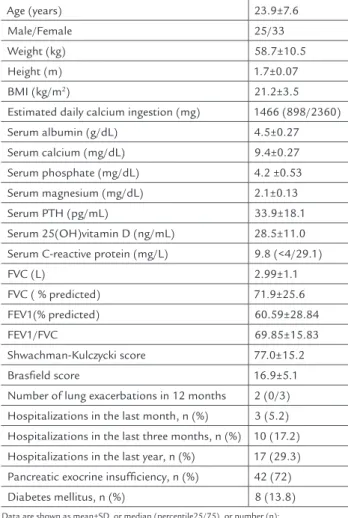
TABLE 1 Baseline characteristics of cystic fibrosis patients (n=58)
Age (years) 23.9±7.6
Male/Female 25/33
Weight (kg) 58.7±10.5
Height (m) 1.7±0.07
BMI (kg/m2) 21.2±3.5
Estimated daily calcium ingestion (mg) 1466 (898/2360) Serum albumin (g/dL) 4.5±0.27
Serum calcium (mg/dL) 9.4±0.27 Serum phosphate (mg/dL) 4.2 ±0.53 Serum magnesium (mg/dL) 2.1±0.13 Serum PTH (pg/mL) 33.9±18.1
Serum 25(OH)vitamin D (ng/mL) 28.5±11.0 Serum C-reactive protein (mg/L) 9.8 (<4/29.1)
FVC (L) 2.99±1.1
FVC ( % predicted) 71.9±25.6 FEV1(% predicted) 60.59±28.84
FEV1/FVC 69.85±15.83
Shwachman-Kulczycki score 77.0±15.2 Brasfield score 16.9±5.1 Number of lung exacerbations in 12 months 2 (0/3) Hospitalizations in the last month, n (%) 3 (5.2)
Hospitalizations in the last three months, n (%) 10 (17.2) Hospitalizations in the last year, n (%) 17 (29.3) Pancreatic exocrine insufficiency, n (%) 42 (72)
Diabetes mellitus, n (%) 8 (13.8) Data are shown as mean±SD, or median (percentile25/75), or number (n); BMI: Body mass index; PTH: parathyroid hormone; FVC: Forced vital capacity; FEV1: Forced expiratory volume in 1s.
All patients with pancreatic insufficiency were treated with pancreatic enzymes and vitamins A, D, E, and K, and seven patients received calcium supplementation. Three
(9.1%) women were using oral contraceptive. All women had normal menstrual periods, except one that had al-ready undergone menopause.
Inhaled corticosteroid therapy was used by 17 pa-tients (29.3%), and none used oral glucocorticosteroids. S. aureus, P. aeruginosa and B. cepacia were present in res-pectively, 45 (77.6%), 33 (56.9%), and 13 (22.4%) of the pa-tients. Sixteen patients (28%) had severe lung disease with FEV1 predicted below 40%. According to the Shwachman--Kulczycki score, 37 (64%) of the patients were in good or
excellent clinical condition.
BMD was assessed in the lumbar spine (L1-L4) in 58 patients; it was also measured in the proximal femur (to-tal and neck) in 38 patients aged 20 years or more. The pre-valence of bone mass below the expected range for age, Z--score <-2.0, at any site was 20.7% (9 males and 3 females); 9 patients were ≥ 20 years, so BMD was assessed in lumbar spine and proximal femur: 5 had low BMD only in L1-L4, one had low BMD only in proximal femur, and three had low BMD in both. One of the patients, a 53-year old post-menopausal woman, had osteoporosis. None of the sub-jects had history of fracture, and no vertebral fracture was described in the lateral chest X-ray. The mean Z-score for lumbar spine was -0.93±1.2, ranging from 1.6 to -3.2.
Serum total testosterone, bioavailable testosterone, and estradiol levels were measured in all male patients, and the means were, respectively, 4.7±2.1 ng/mL, 2.1±0.82 ng/ mL, and 29±14.2 pg/mL. Serum estradiol was positively correlated with serum testosterone levels (r = 0.550, p=0.004), and bioavailable testosterone levels (r = 0.477, p=0.016).
Lumbar spine Z-score in CF patients correlated posi-tively with BMI, and with FEV1 (% predicted), and mean lumbar spine Z-score was higher in women, in patients
Male
Female Sex A
p=0,001 0
-0,2 -0,4 -0,6 -0,8 -1 -1,2 -1,4 -1,6 -1,8 -2 -2,2
Z-score in lumbar spine
yes
no
Hospitalizarions in the last 3 months B
p= 0,02 0
-0,2 -0,4 -0,6 -0,8 -1 -1,2 -1,4 -1,6 -1,8 -2 -2,2
Z-score in lumbar spine
yes
no Pancreatic insufficiency C
p=0,032 0
-0,2 -0,4 -0,6 -0,8 -1 -1,2 -1,4 -1,6 -1,8 -2 -2,2
Z-score in lumbar spine
FIGURE 1 Mean lumbar spine Z-score in patients with cystic fibrosis and its association with sex (A), hospitalization in the last 3 months (B),
with no pancreatic insufficiency, and in patients with no hospitalization in the last 3 months, as shown in Figure 1. There was a positive correlation between lumbar Z--score and BMI (r=0.420, p=0.001) and lumbar ZZ--score and predicted FEV1(%) (r=0.300, p=0.02). There was no correlation of lumbar Z-score and age (r=-0.199, p=0.134), predicted FEV1(%)/FVC ratio (r=0.193, p=0.147), SK sco-re (r=0.202, p=0.129), Brasfield scosco-re (r=0.041, p=0.762), serum albumin (r=0.055, p=0.681), CRP (r=-0.165, p=0.732), calcium (r=0.046, p=0.216), phosphate (r=-0.063, p=0.638), magnesium (r=-0.036, p=0.638), PTH (r=0.005, p=0.971), and 25(OH)D (r=-0.181, p=0.173) levels, estimated cal-cium ingestion (r= -0.049, p=0.716), and lung exacerba-tions in 12 months (r=0.053, p=0.694).
Mean lumbar spine Z-score were similar in patients taking omeprazole (-1.29±1.25; n=10) or not (-0.82±1.16; n=48), p=0.255; using inhaled glucocorticosteroid (-0.74±1.14; n=17) or not (-1.01±1.21;n=41), p=0.421; with bacterial colonization for P. aeruginosa (-0.97±1.1, n=33) or not (-0.89±1.33; n=25), p=0.806; B. cepacea (-0.85±1.04, n=13)) or not (-0.96±1.24, n=45), 0.769; S. aureus (-0.86±1.08, n=45) or not (-1.20±1.52, n=13) p=0.344; with diabetes mellitus (-1.55±1.16, n=8) or not (-0.83±1.8, n=50), p=0.115; with hospitalization in the last year (-1.30± 1.2,n=17) or not (-0.78±1.17, n=41), p=0.126; and with hospitalization in the last month (-1.47±1.4, n= 3) or not (-0.90±1.19, n=55), p=0.430.
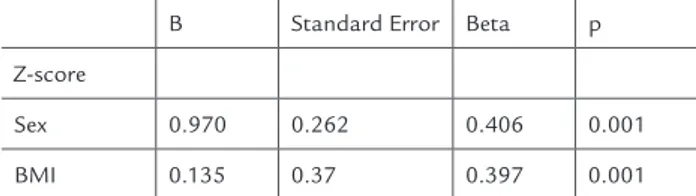
Multivariate stepwise backwards analysis revealed that BMI (p= 0.001), and sex (p=0.001) were independen-tly associated with lumbar spine Z-score (Table 2).
TABLE 2 Factors independently associated with lumbar spine Z-score by multivariate stepwise backwards analysis
B Standard Error Beta p
Z-score
Sex 0.970 0.262 0.406 0.001
BMI 0.135 0.37 0.397 0.001
Included in the model: Sex, BMI, FEV1 % predicted, number of hospitalizations in the last three months, pancreatic insufficiency and serum 25-hydroxyvitamin D levels.
D
ISCUSSIONIn the present study, 20.7% of the adolescent and adult CF outpatients had bone mass below the expected range
for age (Z-score ≤ 2.0 in lumbar spine and/or femur). In the univariate analysis, Z-score in lumbar spine was as-sociated with sex, BMI, FEV1 (% predicted), FVC%, num-ber of hospitalizations in the last three months, and pan-creatic insufficiency. After multivariate regression analysis including all the above, except CFV%, due to its
strong association with FEV1(%predicted), and serum 25(OH) levels, only sex and BMI were independently as-sociated with Z-score in lumbar spine.
The prevalence of bone mass Z-score<-2.0 in lumbar spine was 14.6%, in the patients studied by Lucidi et al 37,
and 25% in the patients studied by Dodd et al 12; these
small differences could be due to age, and clinical condi-tions of patients in different series. Lower prevalence of low bone mass in lumbar spine (1/17) was described by Street et al 13, but it cannot be excluded that this result
was due the small size of the sample. Despite the high prevalence of low bone mass, no fractures were observed in our study. As fractures were evaluated by history and routine lateral chest radiography in our patients, the pre-valence of fractures could have been underestimated.
Other studies described the prevalence of low bone mass in any site, lumbar spine, total body and femoral neck and/or total femur, despite of age below 20 years, and found higher prevalence from 23% to 68% 12, 15, 16, 18,
38-41. Proximal femur BMD measurement is not
conside-red appropriate for measurement of bone mass before skeletal maturity, because of higher variability and lack of reproducibility in this age group 35, 36.
Some factors have been associated with bone mass, like poor growth, delayed maturation, malnutrition, mus-cle deficits, decreased physical activity, chronic inflamma-tion, and use of medications such as glucocorticoids 2.
Several studies evaluated Z-score in lumbar spine and factors possibly associated with it 5, 6, 8, 12-14, 17, 21, 23, 24, 37, 39, 41, 42, nevertheless, some did not assess confounding.
Posi-tive factors independently associated with bone mass in lumbar spine were BMI, FEV1 (% predicted), fat mass, body weight, age of puberty, body cell mass in children and adolescents, SK score and serum leptin5, 6, 8, 14, 23, 39, 42.
Negative factors independently associated with bone mass were deltaF508, male sex, log of serum alkaline phospha-tase, enteral nutrition, number of days of hospitalization in the last year, number of hospital admissions in the last year, physical activity score, oral/inhaled corticoste-roids14, 17, 23, 39. Probably all those factors reflected disease
intensity, except for male sex. Why male subjects with CF had lower bone mass is unknown.
Haworth et al also found BMD significantly lower in men, despite comparable lung function, nutritional indi-ces, and no evidence of male hypogonadism 40. In our male
pa-tients, as described in another infectious disease 43, but all
patients had serum estradiol levels in the normal range. Of the nutritional factors associated with bone mass, the most important are vitamin D, calcium ingestion, and BMI 4.Although 25(OH)D was below 30ng/mL in 60.3%
of the patients (data not shown), there was no indepen-dent association between lumbar spine Z-score and se-rum 25(OH)D levels.
Glucocorticoids can decrease bone mass, when admi-nistered by the nasal route, if the amount absorbed is enough for systemic effects 44. In our study, there was no
association of inhaled steroids use with lumbar spine Z--score, as described previously 13, 37, probably due to low
dose and/or low absorption in these patients. Importan-tly, none of our patients used oral corticosteroids.
C
ONCLUSIONLow bone mass is a frequent problem in patients with CF, being associated with BMI, and male sex.
R
ESUMOPrevalência de densidade mineral óssea baixa em adoles-centes e adultos com fibrose cística.
Objetivo: Determinar a prevalência de massa óssea bai-xa em pacientes adolescentes e adultos com fibrose císti-ca e estudar os fatores potencialmente associados. Métodos: Densidade mineral óssea foi determinada por absorciometria por dupla emissão de raios X na coluna lombar em pacientes ≤ 19 anos e na coluna e no fêmur em pacientes ≥ 20 anos. Avaliações nutricionais, bioquí-micas e pulmonares foram realizadas. Dados referentes ao tratamento farmacológico foram coletados.
Resultados: 58 pacientes foram incluídos no estudo (25 homens/33 mulheres), média de idade de 23,9 anos (16-53). Massa óssea abaixo da esperada foi verificada em 20,7% dos pacientes. Não houve histórico de fratura. Z--score da coluna lombar associou-se positivamente com índice de massa corporal (r=0,3; p=0,022), volume expi-ratório forçado (% previsto) (r=0,415; p=0,001). A média do Z-score da coluna foi mais alta nas mulheres que nos homens (p=0,001), em pacientes que não possuíam insu-ficiência pancreática (p=0,02) e em pacientes que não ha-viam sido hospitalizados nos últimos três meses (p=0,032). Os fatores encontrados como preditores independentes de Z-score da coluna lombar foram sexo masculino (p=0,001) e índice de massa corporal (p=0,001).
Conclusão: Massa óssea baixa é frequente em pacientes com FC, estando associada independentemente com ín-dice de massa corporal e sexo masculino.
Unitermos: fibrose cística, densidade óssea, massa óssea, perdas ósseas, doenças ósseas.
Acknowledgments
This work was supported by the Incentive Fund for Re-search and Events of the Hospital de Clínicas de Porto Alegre, Porto Alegre, RS, Brazil, and the National
Coun-cil of Research, Brazil.
R
EFERENCES1. Buzzetti R, Salvatore D, Baldo E, Forneris MP, Lucidi V, Manunza D, et al. An overview of international literature from cystic fibrosis registries: 1. Mortality and survival studies in cystic fibrosis. J Cyst Fibros. 2009;8:229-37. 2. Aris RM, Merkel PA, Bachrach LK, Borowitz DS, Boyle MP, Elkin SL, et al.
Guide to bone health and disease in cystic fibrosis. J Clin Endocrinol Metab. 2005;90:1888-96.
3. UK Cystic Fibrosis Trust Bone Mineralisation Working Group; 2007. [cited 2012 Mar 27]. Available form: http://www.cftrust.org.uk/aboutcf/ publications/consensusdoc/Bone-Mineral-Booklet.pdf
4. Sermet-Gaudelus I, Bianchi ML, Garabedian M, Aris RM, Morton A, Hardin DS, et al. European cystic fibrosis bone mineralisation guidelines. J Cyst Fibros. 2011;10(Suppl 2):S16-23.
5. Legroux-Gerot I, Leroy S, Prudhomme C, Perez T, Flipo RM, Wallaert B, et al. Bone loss in adults with cystic fibrosis: prevalence, associated factors, and usefulness of biological markers. Joint Bone Spine. 2012;79:73-77. 6. Rossini M, Del Marco A, Dal Santo F, Gatti D, Braggion C, James G, et al.
Prevalence and correlates of vertebral fractures in adults with cystic fibrosis. Bone. 2004;35:771-6.
7. Wolfenden LL, Judd SE, Shah R, Sanyal R, Ziegler TR, Tangpricha V. Vitamin D and bone health in adults with cystic fibrosis. Clin Endocrinol (Oxf). 2008;69:374-81.
8. Elkin SL, Fairney A, Burnett S, Kemp M, Kyd P, Burgess J, et al. Vertebral deformities and low bone mineral density in adults with cystic fibrosis: a cross-sectional study. Osteoporos Int. 2001;12:366-72.
9. Paccou J, Zeboulon N, Combescure C, Gossec L, Cortet B. The prevalence of osteoporosis, osteopenia, and fractures among adults with cystic fibrosis: a systematic literature review with meta-analysis. Calcif Tissue Int. 2010;86:1-7. 10. Spira A, Gutierrez C, Chaparro C, Hutcheon MA, Chan CK. Osteoporosis
and lung transplantation: a prospective study. Chest. 2000;117:476-81. 11. Robertson J, Macdonald K. Prevalence of bone loss in a population with
cystic fibrosis. Br J Nurs. 2010;19:636-9.
12. Dodd JD, Barry SC, Barry RB, Cawood TJ, McKenna MJ, Gallagher CG. Bone mineral density in cystic fibrosis: benefit of exercise capacity. J Clin Densitom. 2008;11:537-42.
13. Street ME, Spaggiari C, Ziveri MA, Volta C, Federico G, Baroncelli GI, et al. Analysis of bone mineral density and turnover in patients with cystic fibrosis: associations between the IGF system and inflammatory cytokines. Horm Res. 2006;66:162-8.
14. King SJ, Topliss DJ, Kotsimbos T, Nyulasi IB, Bailey M, Ebeling PR, et al. Reduced bone density in cystic fibrosis: DeltaF508 mutation is an independent risk factor. Eur Respir J. 2005;25:54-61.
15. Frangolias DD, Pare PD, Kendler DL, Davidson AG, Wong L, Raboud J, et al. Role of exercise and nutrition status on bone mineral density in cystic fibrosis. J Cyst Fibros. 2003;2:163-70.
16. Bachrach LK, Loutit CW, Moss RB. Osteopenia in adults with cystic fibrosis. Am J Med. 1994;96:27-34.
17. Douros K, Loukou I, Nicolaidou P, Tzonou A, Doudounakis S. Bone mass density and associated factors in cystic fibrosis patients of young age. J Paediatr Child Health. 2008;44:681-5.
18. Cemlyn-Jones J, Gamboa F, Loureiro M, Fontes Baganha M. Evaluation of bone mineral density in cystic fibrosis patients. Rev Port Pneumol. 2008;14:625-34.
19. Haworth CS, Selby PL, Webb AK, Martin L, Elborn JS, Sharples LD, et al. Inflammatory related changes in bone mineral content in adults with cystic fibrosis. Thorax. 2004;59:613-7.
advanced cystic fibrosis lung disease. Am J Respir Crit Care Med. 1998;157:1892-9.
21. O’Reilly R, Fitzpatrick P, Leen G, Elnazir B, Greally P. Severe bone demineralisation is associated with higher mortality in children with cystic fibrosis. Ir Med J. 2009;102:47-9.
22. Flohr F, Lutz A, App EM, Matthys H, Reincke M. Bone mineral density and quantitative ultrasound in adults with cystic fibrosis. Eur J Endocrinol. 2002;146:531-6.
23. Conway SP, Morton AM, Oldroyd B, Truscott JG, White H, Smith AH, et al. Osteoporosis and osteopenia in adults and adolescents with cystic fibrosis: prevalence and associated factors. Thorax. 2000;55:798-804.
24. Caldeira RJ, Fonseca VM, Gomes SC Jr, Chaves CR. Prevalence of bone mineral disease among adolescents with cystic fibrosis. J Pediatr (Rio J). 2008;84:18-25.
25. Cauley JA, Lui LY, Ensrud KE, Zmuda JM, Stone KL, Hochberg MC, et al. Bone mineral density and the risk of incident nonspinal fractures in black and white women. JAMA. 2005;293:2102-8.
26. Bobadilla JL, Macek M Jr, Fine JP, Farrell PM. Cystic fibrosis: a worldwide analysis of CFTR mutations--correlation with incidence data and application to screening. Hum Mutat. 2002;19:575-606.
27. Streit C, Burlamaque-Neto AC, Abreu e Silva F, Giugliani R, Saraiva Pereira ML. CFTR gene: molecular analysis in patients from South Brazil. Mol Genet Metab. 2003;78:259-64.
28. ISCD: The ISCD’s official positions (updated 2007); Washington (DC): International Society for Clinical Densitometry; 2007.
29. WHO: Scientific group on the assessment of osteoporosis at primary health care level: Sumary Meeting Report Brussels, Belgium; 2004.
30. Borowitz D, Baker RD, Stallings V. Consensus report on nutrition for pediatric patients with cystic fibrosis. J Pediatr Gastroenterol Nutr. 2002;35:246-59.
31. Stallings VA, Stark LJ, Robinson KA, Feranchak AP, Quinton H. Evidence-based practice recommendations for nutrition-related management of children and adults with cystic fibrosis and pancreatic insufficiency: results of a systematic review. J Am Diet Assoc. 2008;108:832-9.
32. Pereira CA, Sato T, Rodrigues SC. New reference values for forced spirometry in white adults in Brazil. J Bras Pneumol. 2007;33:397-406.
33. Shwachman H, Kulczycki LL. Long-term study of one hundred five patients with cystic fibrosis; studies made over a five- to fourteen-year period. AMA J Dis Child. 1958;96:6-15.
34. Brasfield D, Hicks G, Soong S, Tiller RE. The chest roentgenogram in cystic fibrosis: a new scoring system. Pediatrics. 1979;63:24-9.
35. Baim S, Leonard MB, Bianchi M. American College of Radiology. Practice guideline for the performance of dual-energy x-ray absorptiometry (DXA). 2008 [cited 2012 Apr 15]. Available from: http://www.acr.org.
36. Baim S, Binkley N, Bilezikian JP, Kendler DL, Hans DB, Lewiecki EM, et al. Official Positions of the International Society for Clinical Densitometry and executive summary of the 2007 ISCD Position Development Conference. J Clin Densitom. 2008;11:75-91.
37. Lucidi V, Bizzarri C, Alghisi F, Bella S, Russo B, Ubertini G, Cappa M. Bone and body composition analyzed by Dual-energy X-ray Absorptiometry (DXA) in clinical and nutritional evaluation of young patients with Cystic Fibrosis: a cross-sectional study. BMC Pediatr. 2009;9:61.
38. Bianchi ML, Romano G, Saraifoger S, Costantini D, Limonta C, Colombo. BMD and body composition in children and young patients affected by cystic fibrosis. J Bone Miner Res. 2006;21:388-96.
39. Buntain HM, Greer RM, Schluter PJ, Wong JC, Batch JA, Potter JM, et al. Bone mineral density in Australian children, adolescents and adults with cystic fibrosis: a controlled cross sectional study. Thorax. 2004;59:149-55. 40. Haworth CS, Selby PL, Webb AK, Dodd ME, Musson H, Mc LNR, et al. Low
bone mineral density in adults with cystic fibrosis. Thorax. 1999;54:961-7. 41. Bhudhikanok GS, Lim J, Marcus R, Harkins A, Moss RB, Bachrach LK.
Correlates of osteopenia in patients with cystic fibrosis. Pediatrics. 1996;97:103-11.
42. Stephenson A, Jamal S, Dowdell T, Pearce D, Corey M, Tullis E. Prevalence of vertebral fractures in adults with cystic fibrosis and their relationship to bone mineral density. Chest. 2006;130:539-44.
43. Gennari L, Nuti R, Bilezikian JP. Aromatase activity and bone homeostasis in men. J Clin Endocrinol Metab. 2004;89:5898-907.