CLINICAL SCIENCE
Methicillin-resistant
staphylococcus aureus
(MRSA)
carriage in a dermatology unit
Renata L. Pacheco,IRenata D. Lobo,IIMaura S. Oliveira,IIElthon F. Farina,ICleide R. Santos,IISilvia F. Costa,I Maria Clara Padoveze,IIICilmara P. Garcia,IIPriscila A. Trindade,ILigia M. Quite´rio,IVEvandro A. Rivitti,IV Elsa M. Mamizuka,VAnna S. LevinI,II
IHospital das Clı´nicas da Faculdade de Medicina da Universidade de Sa˜o Paulo, Department of Infectious Diseases and LIM-54, Sa˜o Paulo/SP, Brazil. IIHospital das Clı´nicas da Faculdade de Medicina da Universidade de Sa˜o Paulo, Department of Infection Control of Sa˜o Paulo, Sa˜o Paulo/SP, Brazil. IIIUniversidade de Sa˜o Paulo, Nursing School, Department of Collective Health, Sa˜o Paulo/SP, Brazil.IVHospital das Clı´nicas da Faculdade de Medicina da Universidade de Sa˜o Paulo, Department and Division of Dermatology, Sa˜o Paulo/SP, Brazil.VUniversidade de Sa˜o Paulo, Department of Clinical Analyses, Faculdade de Cieˆncias Farmaceˆuticas, Sa˜o Paulo/SP Brazil.
OBJECTIVE:The aim of this study was to characterizeStaphylococcus aureus(MRSA) carriage in a dermatology unit.
METHODS:This was a prospective and descriptive study. Over the course of 26 weeks, surveillance cultures were collected weekly from the anterior nares and skin of all patients hospitalized in a 20-bed dermatology unit of a tertiary-care hospital. Samples from healthcare workers (HCWS) were cultured at the beginning and end of the study. Colonized patients were put under contact precautions, and basic infection control measures were enforced. Staphylococcus aureuscolonization pressure was determined monthly. Colonized and non-colonized patients were compared, and isolates were evaluated for antimicrobial susceptibility, SCCmec type, virulence factors, and type.
RESULTS: Of the 142 patients evaluated, 64 (45%) were colonized by MRSA (39% hospital acquired; 25% community acquired; 36% indeterminate). Despite isolation precautions, hospital-acquired Staphylococcus aureusoccurred in addition to the continuous entry ofStaphylococcus aureusfrom the community. Colonization pressure increased from 13% to 59%, and pemphigus and other bullous diseases were associated with MRSA colonization. Eleven out of 71 HCWs (15%) were Staphylococcus aureus carriers, although only one worker carried a persistent clone. Of the hospital-acquired MRSA cases, 14/28 (50%) were SCCmectype IV (3 PFGE types), 13 were SCCmectype III (46%), and one had an indeterminate type. These types were also present among the community-acquiredStaphylococcus aureusisolates. SSCmectype IV isolates were shown to be more susceptible than type III isolates. There were two cases of bloodstream infection, and the pvlandtstvirulence genes were absent from all isolates.
CONCLUSIONS:Dermatology patients were colonized by community- and hospital-acquiredStaphylococcus aureus. Half of the nosocomialStaphylococcus aureusisolates were SCCmectype IV. Despite the identification of colonized patients and the subsequent contact precautions and room placement, Staphylococcus aureus colonization continued to occur, and colonization pressure increased. Pemphigus and other bullous diseases were associated withStaphylococcus aureus.
KEYWORDS: Transmission; Surveillance culture; Molecular typing; MRSA; Hospital infection.
Pacheco RL, Lobo RD, Oliveira MS, Farina EF, Santos CR, Costa SF, et al. Methicillin-resistantstaphylococcus aureus(MRSA) carriage in a dermatology unit. Clinics. 2011;66(12):2071-2077.
Received for publication onJuly 3, 2011;First review completed onJuly 15, 2011;Accepted for publication onAugust 29, 2011 E-mail: gcih@hcnet.usp.br
Tel.: 55 11 2661-7066
INTRODUCTION
Staphylococcus aureus is a versatile pathogen capable of causing a wide variety of infections.1 The prevalence of nosocomial and community-acquired methicillin-resistant
Staphylococcus aureus (MRSA) infections has increased in recent years.2Methicillin resistance ofStaphylococcus aureus is mediated by a penicillin-binding protein (PBP2A) that has a low affinity forb-lactam antibiotics and is encoded by the mecA gene.3,4 This gene is carried on a mobile genetic element designated as Staphylococcal Cassette Chromo-some mec(SCCmec), which is integrated into the chromo-some. Currently, 11 types of SCCmechave been reported.5-7 In healthcare settings, patients who are colonized by or infected with MRSA serve as a reservoir and a source for the spread of this microorganism, which occurs mainly th-rough transiently colonized healthcare workers (HCWs).8,9
Copyrightß2011CLINICS– This is an Open Access article distributed under
the terms of the Creative Commons Attribution Non-Commercial License (http:// creativecommons.org/licenses/by-nc/3.0/) which permits unrestricted non-commercial use, distribution, and reproduction in any medium, provided the original work is properly cited.
Carriage of S. aureus is considered a risk factor for the development of infections, as infections are usually pre-ceded by a period of colonization.10
The objective of this study was to characterize MRSA carriage in a hospital dermatology unit.
METHODS
Hospital das Clı´nicas is a tertiary-care teaching hospital that is affiliated with the University of Sa˜o Paulo, Brazil. It has approximately 2,000 beds and is divided into 6 buildings. The dermatology unit has 20 beds, which are distributed across 10 rooms of one, two, or four beds each. This unit is used for the hospitalization of patients with severe dermatologic conditions. On average, there are 30 admissions per month and 4,500 patient-days each year.
Surveillance cultures of patients
Over a period of 26 weeks that began on May 31, 2005, weekly surveillance cultures for MRSA were obtained from all of the patients admitted to the unit. These samples were collected from the anterior nares and skin lesions. Patients who were positive for MRSA were not followed for further surveillance cultures. Patients who were negative for MRSA were cultured weekly until their discharge from the unit.
Due to the high proportion of patients who tested positive on the first surveillance culture, patients were cultured upon admission and then weekly from the 13th week of the study onward.
The swabs from the anterior nares were collected by introducing a swab into the nasal vestibulum and then rubbing with light pressure. The swabs were transported in sterile tubes containing culture medium and were sent immediately to the laboratory for further processing and analysis. The swabs from skin lesions were collected by gently scraping or rolling the swab across the lesion.
Definitions
Nosocomial acquisition of MRSA was defined as a positive culture in a patient who had been in the hospital for more than 48 hours and whose previous surveillance cultures at both sites had been negative (nares and skin).
For patients who were positive within the first 48 hours of hospitalization, MRSA was considered to have been present at the time of hospital admission. All other colonizations were considered indeterminate as to where MRSA acquisi-tion occurred.
The following patient data were registered upon admis-sion to the study: age, sex, date of admisadmis-sion to the hospital, underlying diseases, hospitalization within the previous 12 months, and use of antimicrobial drugs. The patients were followed until their discharge from the unit. We evaluated the colonization of the patients by dividing the study into six periods.
Control measures
All patients positive for MRSA were placed in separate rooms or were placed in a cohort with other MRSA carriers and put under contact precautions until hospital discharge or death. Hand hygiene was enforced by requiring the use of alcohol gel. There were two educational meetings with members of the staff in which the control measures and their objectives were discussed. Posters were spread
throughout the unit to report the weekly number of patients colonized by MRSA. Patients also received information regarding the contact precautions and their importance.
Surveillance cultures of healthcare workers (HCWs)
The anterior nares of the healthcare workers were cultured on two occasions: at the beginning and at the end of the study. HCWs who tested positive were not treated.
Ethics
The study protocol followed the ethical guidelines of the 1975 Declaration of Helsinki and was approved by the Ethics Commission of the Hospital das Clı´nicas (approval number: 1072/04).
Analysis of data
Continuous variables are herein presented as the means
¡ standard deviation or median and range. Frequencies
were calculated for the categorical variables. MRSA-colo-nized patients were compared to non-coloMRSA-colo-nized patients using the x2 test for categorical variables and the Mann Whitney test for continuous variables. The data were analyzed using Epi Info 6.04 software (CDC, Atlanta, USA). A p-value of 0.05 was considered statistically significant.
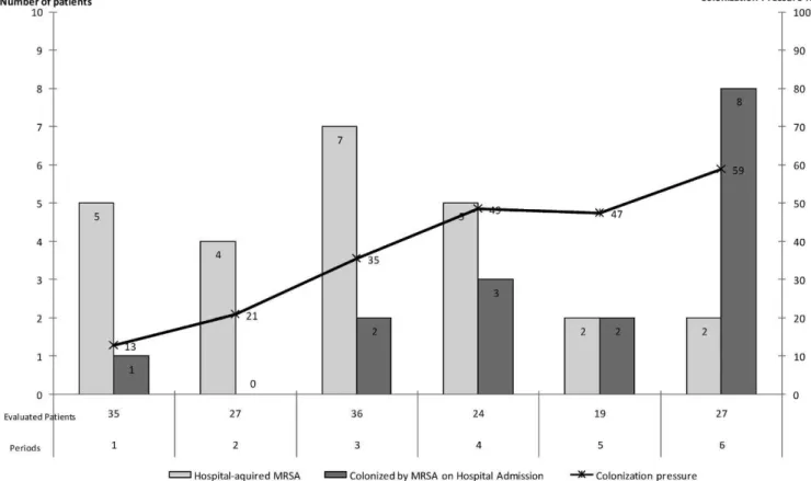
For each period, we determined the total number of patient-days and the number of MRSA patient-days (by summing the days that each MRSA-colonized patient remained in the unit after the day that MRSA colonization was first identified). Colonization pressure was also determined for each period by dividing the number of MRSA patient-days by the total number of patient-days (expressed as a percentage).
Microbiological methods
Detection of MRSA. Samples were plated onto mannitol salt agar (MSA) and inoculated into brain heart infusion (BHI) broth. Mannitol-fermenting colonies were subcul-tured onto 5% sheep blood agar plates. If the initial MSA culture was negative, a subculture from the BHI broth was carried out on MSA plates to increase the sensitivity of detection. S. aureus was identified by Gram staining and DNAse and catalase tests. Methicillin resistance was determined using Mueller Hinton agar supplemented with 4% NaCl and 6mg/mL oxacillin according to guidelines of the Clinical Laboratory Standards Institute (CLSI).11If there was growth of at least one colony-forming unit, the culture was considered positive.S. aureusATCC 29213 and NCTC 10442 were used as controls.
The phenotypic identification was confirmed by poly-merase chain reaction (PCR), amplifying 117 bp and 214 bp fragments from the coagulase andmecA genes, respectively, using the NCL-SA-PS kit (Novo Castra, United Kingdom), as previously described by Kearns et al.12
Susceptibility testing. Minimal inhibitory concentrations (MICs) were determined using the broth microdilution me-thod and were interpreted according to CLSI guidelines11,13 for oxacillin, penicillin, chloramphenicol, ciprofloxacin, clinda-mycin, erythroclinda-mycin, gentamicin, rifampicin, sulfametho-xazole/trimethoprim, tetracycline and vancomycin.
enzyme (Fermentas Life Sciences, Canada) and then determining the fragment-size patterns obtained with pulsed-field gel electrophoresis (PFGE) using a CHEF DR-II apparatus (Bio-Rad Laboratories, USA).14 The patterns were analyzed as recommended by Tenover et al.15Types were defined as isolates that differed by at least seven fragments and were identified using letters. Subtypes of a given clonal type were defined as those isolates that differed by fewer than seven fragments and were identified using numbers.
Multilocus sequence typing was performed for 10 isolates according to methods described elsewhere.16This sequence typing was performed for one isolate of each PFGE type (seven isolates), one isolate for which the SCCmectype could not be determined and two isolates in whichmecA could not be detected.
SCCmectyping. The determination of the SCCmec type was performed using the multiplex PCR method, as described by Oliveira & Lencastre.17 DNA was extracted using a commercial kit (Genomic Prep Cells and Tissue DNA Isolation, Amersham Pharmacia, Biotech, Germany). PCR amplifications were performed using a GeneAmp PCR System 2400 thermocycler (Perkin-Elmer, Waltham, MA, USA). The MRSA strains NTCT 10442, N315, 85/2082 and JSCS 1968, which belong to SCCmectypes I, II, III, and IV, respectively, were used as positive controls.
Detection of genes for virulence factors. One isolate from each PFGE subtype was evaluated for the Panton Valentine leukocidin (pvl), LukE-LukD leukocidin ( lukE-lukD) and toxic shock syndrome toxin-1 (tst) virulence genes by PCR using primers described elsewhere.18 Briefly,
samples were denatured for 5 minutes at 94
˚
C; subjected to 30 cycles of denaturation at 95˚
C for 30 seconds, annealing at 55˚
C for 1 minute, and extension at 72˚
C for 2 min; and then subjected to a final extension at 72˚
C for 5 minutes. The MR108 (positive forpvl) and N315 (positive for lukE-lukDandtst) isolates were used as controls.RESULTS
During the study period, 153 patients were admitted to the dermatology unit. Of these, 11 were lost to follow-up, and 142 were evaluated until discharge and included in the analysis.
Of the 142 patients, 64 (45%) were colonized by MRSA. There were 26 patients who were positive only from the culture of the anterior nares (one patient harbored two isolates), 11 who were positive only from skin culture and 27 who had tested positive from both sites (two patients each harbored three isolates). Thus, 94 MRSA isolates from 64 patients were microbiologically evaluated.
Among the 64 patients (45%) who were colonized by MRSA, 25 (39%) had hospital-acquired infections, 16 (25%) were colonized at the time of admission and 23 patients (36%) had an indeterminate site of acquisition. The distributions over time of patients who were colonized at the time of admission and of those who acquired MRSA in the hospital are presented in Figure 1. Despite the control measures, the nosocomial acquisition of MRSA occurred during the entire study period. In addition, during the entire study period, there was a continuous entry of patients who were MRSA positive at the time of admission, and the
colonization pressure also increased over the six-month study period (Figure 1).
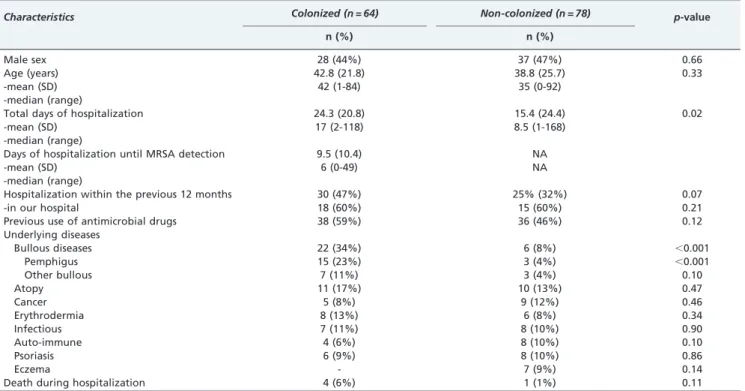
The characteristics of the MRSA-colonized and non-colonized patient populations are presented in Table 1. Bullous diseases, such as pemphigus and others, were significantly more frequent among colonized patients.
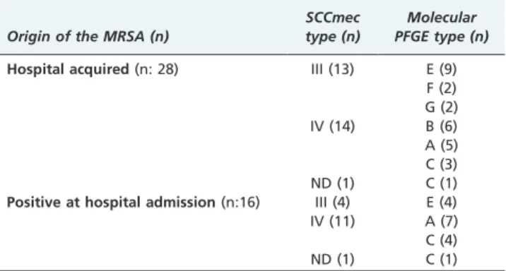
The SCCmectypes and molecular types of the 94 isolates were determined. When a patient harbored more than one isolate of the same SCCmectype and PFGE type, only one isolate for that patient was included in the final analysis. Therefore, 67 isolates from 64 patients were analyzed. There were seven molecular PFGE types (A to G) that were divided into 30 subtypes. The distribution of the patient isolates according to the origin of acquisition, SCCmectype and molecular type is presented in Table 2. The results of the MLST for isolates of each PFGE type (A to G) are presented in Table 3.
The SCCmec type IV isolates belonged to four different PFGE molecular types (A-D) and four different sequence types (ST5, ST8, ST97 and ST1176). However, the PFGE type and ST did not correlate completely, as ST8 belonged to two different PFGE types (B and D), and there were 2 ST (ST97 and ST5) among the isolates of PFGE type C.
Of the SCCmec type III isolates, there were 3 PFGE molecular types (E-G), all of which belonged to ST239, as does the multiresistant Brazilian Endemic Clone (BEC).
Of the isolates obtained at the time of patient admission, most were SCCmectype IV, but there were also four isolates that were similar to the BEC.
The hospital-acquired isolates were evenly distributed among SCCmectypes IV and III.
Among the 30 isolates tested (all carried SCCmectype IV), the pvl and tst virulence factors were absent from all of them, and thelukD-lukEgene was present in all of them.
During the study period, there were two cases of bloodstream infections. These were caused by MRSA of SCCmectype IV and PFGE type A. These patients had been colonized by the same subtypes at earlier time points (25 and 40 days prior).
Thirty-seven healthcare workers (HCWs) were cultured at the beginning of the study, and 34 were cultured at the end of the study. Of these, five (14%) were positive on the first occasion, and six (18%) were positive on the second. The distribution of the isolates from healthcare workers accord-ing to SCCmec type and molecular type is presented in Table 4. Fifteen HCWs were cultured on both occasions, and three were positive on both occasions, but only one healthcare worker (belonging to the housekeeping staff) presented the same subtype twice (C3). However, none of the patients presented the C3 subtype.
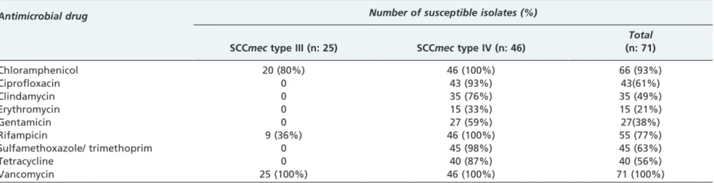
The antimicrobial susceptibilities of the isolates that colonized 60 patients and 11 healthcare workers are shown in Table 5.
DISCUSSION
Colonization with MRSA was prevalent among patients hospitalized for dermatological diseases (45%), and this colonization was due to both hospital acquisition and colonization prior to admission.
This study began by examining routine surveillance cultures to determine the extent of the problem and to identify patients who should be placed under contact precautions, such as placement in a private room or in a patient cohort. Through the enforcement of these measures, we expected to halt the nosocomial transmission of MRSA. Community-acquired MRSA (CA-MRSA) has been rarely described in Brazil and has been shown to occur mainly in
Table 1- Characteristics of MRSA-colonized patients and non-colonized patients in the dermatology unit over a 6-month period.
Characteristics Colonized (n = 64) Non-colonized (n = 78) p-value
n (%) n (%)
Male sex 28 (44%) 37 (47%) 0.66
Age (years) -mean (SD) -median (range)
42.8 (21.8) 42 (1-84)
38.8 (25.7) 35 (0-92)
0.33
Total days of hospitalization -mean (SD)
-median (range)
24.3 (20.8) 17 (2-118)
15.4 (24.4) 8.5 (1-168)
0.02
Days of hospitalization until MRSA detection -mean (SD)
-median (range)
9.5 (10.4) 6 (0-49)
NA NA
Hospitalization within the previous 12 months -in our hospital
30 (47%) 18 (60%)
25% (32%) 15 (60%)
0.07 0.21
Previous use of antimicrobial drugs 38 (59%) 36 (46%) 0.12
Underlying diseases
Bullous diseases 22 (34%) 6 (8%) ,0.001
Pemphigus 15 (23%) 3 (4%) ,0.001
Other bullous 7 (11%) 3 (4%) 0.10
Atopy 11 (17%) 10 (13%) 0.47
Cancer 5 (8%) 9 (12%) 0.46
Erythrodermia 8 (13%) 6 (8%) 0.34
Infectious 7 (11%) 8 (10%) 0.90
Auto-immune 4 (6%) 8 (10%) 0.10
Psoriasis 6 (9%) 8 (10%) 0.86
Eczema - 7 (9%) 0.14
Death during hospitalization 4 (6%) 1 (1%) 0.11
the southern region of the country19, 20near the border with Uruguay, which is a country in which CA-MRSA is a significant problem. In addition, in a previous study of MRSA isolates obtained from bloodstream infections,21 there were six cases of hospital-acquired MRSA bacteremia the dermatology unit. Thus, we expected most of the cases of MRSA colonization to be hospital acquired. However, in our dermatology unit, we observed that approximately half of the colonization cases were hospital-acquired and that a significant proportion of patients were positive for MRSA at the time of admission. Our results suggest that many patients are colonized by community-acquired MRSA or are colonized at other healthcare facilities and may be a source of MRSA transmission to other patients in the hospital. There was a significantly higher proportion of patients with pemphigus and other bullous diseases among MRSA carriers, which suggested that disseminated bullous disease is a risk factor for MRSA colonization.
During the entire study period, nosocomial transmission of MRSA occurred as well as admission of colonized patients. This likely generated or increased colonization pressure and made control more difficult despite the efforts directed to prevent nosocomial MRSA transmission. Over the six-month study period, the frequency of nosocomial-acquired cases tended to decrease, but the colonization pressure increased from 13% to 59%. We attributed this
decrease in the nosocomial acquisition of MRSA to the extensive efforts of the staff, which included improving the isolation conditions and the infection control measures. Our original goal was the complete eradication of nosocomial transmission, but this goal was not achieved. The coloniza-tion pressure was first evaluated for vancomycin-resistant enterococci.22 This type of resistance has been studied in MRSA and has been shown to play a role as a risk factor for nosocomial transmission.23 In one study, a colonization pressure above 30% led to a 5-fold increase in the risk of MRSA colonization.24
Due to the large number of patients who are colonized at the time of admission, the use of routine surveillance cultures and the placement of colonized patients into cohorts may not affect the rates of colonization unless all patients are immediately placed under contact precautions even before the surveillance cultures are analyzed. Furthermore, the success of MRSA control that is based mainly on the identification of carriers through the analysis of surveillance cultures depends on the sensitivity of these cultures, which may not be sufficiently high to detect all colonized patients. Some authors have reported that the lack of throat culturing may lead to an underestimation of the percentage of MRSA carriers by as much as 8 to 18%.25, 26
The clinical impact and the relevance of routine patient culturing may be contested because, despite a high proportion of colonized patients, there were only two cases of bloodstream infection in our six-month study period. Routine culturing and the use of patient cohorts and private rooms for positive patients are expensive and labor-intensive measures. In our hospital, most rooms are meant to hold two or four patients, and there are very few private rooms. During patient isolation, the rooms are not used at full capacity, which is a problem because there is a nationwide shortage of hospital beds. In addition, although the carriage of S. aureus is considered a risk factor for the development of infection,10 one prospective study found that nasal colonization with MRSA was a poor predictor of the subsequent occurrence of MRSA respiratory tract or bloodstream infection.27
This study also addressed the role of HCWs in the transmission of MRSA. Among HCWs, MRSA was observed in 15% of the cultures. Only three HCWs were positive on both occasions, but two HCWs harbored different molecular types of MRSA at each culture, suggesting transient carriage. Interestingly, half of the HCWs who were positive for MRSA harbored isolates of subtypes that were not present among the patients. This result suggests that a large Table 2- SCCmectype and molecular type of 67 MRSA
isolates according to the origin of acquisition.
Origin of the MRSA (n)
SCCmec type (n)
Molecular PFGE type (n)
Hospital acquired(n: 28) III (13) E (9)
F (2) G (2)
IV (14) B (6)
A (5) C (3)
ND (1) C (1)
Positive at hospital admission(n:16) III (4) E (4)
IV (11) A (7)
C (4)
ND (1) C (1)
MRSA: methicillin-resistantS. aureus; PFGE: pulsed-field gel electrophoresis; ND: not determined.
Table 3- Multilocus sequence typing results for 10 methicillin-resistantS. aureussubtypes that colonized patients hospitalized in the dermatology unit over a period of 6 months.
Subtype determined by PFGE Sequence type SCCmec type
A1 1176 IV
B1 8 IV
C1 97 IV
C6 5 ND
D 8 IV
E1 239 III
E8 239
-E9 239
-F 239 III
G 239 III
PFGE: pulsed-field gel electrophoresis; SCCmec:staphylococcal chromosomal cassettemec; ND: not determined; -: absence ofmecA.
Table 4- The SCCmecand molecular types of 11 MRSA isolates obtained from healthcare workers at the beginning and the end of the study.
Healthcare workers
SCCmec type (n)
Molecular PFGE type (n)
Beginning of study(n: 37) 5 (14%) positive for MRSA
IV (5) C (3)
B (1) A (1) End of study(n: 34) 6 (18%) positive for
MRSA
IV (5)
III (1)
A (1) B (3) C (1) E (1)
proportion of the HCW colonizations were acquired outside of the hospital, either in the community or at other institutions.
MRSA with SCCmectype III was multidrug resistant and belonged to the Brazilian Endemic Clone (BEC). Molecular typing by pulsed-field gel electrophoresis (PFGE) revealed three different types and one predominant type (79%). Within this predominant type, there were nine different subtypes. The BEC has been endemic in Brazil for more than a decade.28 During this time, it is likely that changes occurred that made Tenover’s criteria17 inadequate to interpret its clonality. In this situation, multilocus sequence typing (MLST) is more useful because it evaluates seven housekeeping genes that are more stable over time.29 BEC presents as sequence type ST-239,28and all of the SCCmec type III isolates tested in our study also belonged to this ST. The presence of MRSA with SCCmec type IV at our hospital is a more recent phenomenon than the existence of the BEC. Therefore, the PFGE typing reflected clonality that was similar although not identical to the MLST.21 Furthermore, we expected that the SCCmec type IV would be mainly community acquired. However, similar to other geographic areas,30it seems as though this type has crossed hospital borders and circulates not only in the community but also in the hospital environment.
As observed in other studies conducted at our hospi-tal,21,31SCCmec type IV MRSA isolates from dermatologic patients did not express the Panton-Valentine leukocidin (PVL) virulence factor. Thetstgene was also absent from all of the isolates. This finding is interesting because PVL-producing clones have been associated with skin and soft tissue infections32,33and because TSST-1 is the cause of skin lesions. On the other hand, in a hospital environment, where patients are severely ill and have many diseases, a virulent strain ofS. aureuswould probably not be successful because it would cause severe infection and high mortality, as it does in the community.
Our study has limitations. First, it was our original intention to use the CDC definitions, but the necessary information was not available for a large proportion of the included patients. Because of this lack of information, we had to introduce the ‘‘indeterminate’’ category. There was a large proportion of patients for which the origin of colonization was deemed indeterminate, which was in part due to the fact that surveillance culturing immediately upon admission was only initiated halfway into the study. In addition, we did not observe SCCmectypes other than types
I to IV, and there were a few isolates for which we could not determine the type. Finally, it was not possible to perform MLST analyses on each of the PFGE subtypes.
In summary, almost half of the dermatology patients in this study were colonized by MRSA at the time of hospital admission or acquired MRSA while in the hospital. Half of the nosocomial MRSA cases were SCCmec type IV. Nosocomial MRSA colonization continued throughout the study period despite the identification of colonized patients, the use of contact precautions and patient cohorting or the placement of patients in private rooms based on their surveillance cultures. Colonization pressure continuously increased during the study period, which may partly explain the difficulty in controlling the spread of MRSA. Pemphigus and other bullous diseases were significantly associated with MRSA colonization. The healthcare workers were predominantly colonized by SCCmec type IV MRSA, and this colonization was found to be transient and predominantly community acquired.
ACKNOWLEDGMENTS
This study was funded by grants from the Fundac¸a˜o de Amparo a` Pesquisa do Estado de Sa˜o Paulo (FAPESP)- 06/03003-7; 05/5754-0 and 06/ 00569-0.
We thank Robson E. Soares for his suggestions in the laboratory and with the manuscript.
AUTHOR CONTRIBUTIONS
Pacheco RL was responsible for the collection of patient data and clinical specimens, laboratory procedures and analysis of data. Lobo RD was responsible for the collection of data and specimens, implementation of control measures and contributed to the writing of the manuscript. Oliveira MS was responsible for the collection of data and specimens, implementa-tion of control measures and contributed to the writing of the manuscript. Farina EF was responsible for the laboratory procedures and analysis of data. Santos CR was responsible for the collection of data and specimens and implementation of control measures. Costa SF supervised the laboratory work. Padoveze MC and Garcia CP contributed to the writing of the manuscript. Trindade PA was responsible for the laboratory procedures. Quite´rio LM and Rivitti EA were responsible for the organization of the unit and implementation of control measures. Mamizuka EM was responsible for the laboratory procedures. Levin AS was responsible for the general supervision and the writing of the manuscript.
REFERENCES
1. Gordon RJ, Lowy FD. Pathogenesis of methicillin-resistant Staphylococcus aureus infection. Clin Infect Dis. 2008;46:S350-9, doi: 10.1086/533591. Table 5- Antimicrobial susceptibility according to SCCmectype of 71 MRSA isolates that colonized 60 patients and 11 healthcare workers in the dermatology unit over a period of 6 months.
Antimicrobial drug Number of susceptible isolates (%)
SCCmectype III (n: 25) SCCmectype IV (n: 46)
Total (n: 71)
Chloramphenicol 20 (80%) 46 (100%) 66 (93%)
Ciprofloxacin 0 43 (93%) 43(61%)
Clindamycin 0 35 (76%) 35 (49%)
Erythromycin 0 15 (33%) 15 (21%)
Gentamicin 0 27 (59%) 27(38%)
Rifampicin 9 (36%) 46 (100%) 55 (77%)
Sulfamethoxazole/ trimethoprim 0 45 (98%) 45 (63%)
Tetracycline 0 40 (87%) 40 (56%)
2. Boucher HW, Corey GR. Epidemiology of methicillin-resistant Staphylococcus aureus. Clin Infect Dis. 2008;46:S344-9, doi: 10.1086/ 533590.
3. Ito T, Hiramatsu K. Acquisition of methicilin resistance and progression of multiantibiotic resistance in methicilin-resistant Staphylococcus aureus. Yonsei Med J. 1998;39:526-33.
4. Hiramatsu K, Ito T, Hanaki H. Mechanisms of methicillin and vancomycin in Staphylococcus aureus. Clin Infect Dis. 1999;5:221-42. 5. Shore AC, Deasy EC, Slickers P, Brennan G, O’Connell B, Monecke S,
et al. Detection of Staphylococcal Cassette Chromosome mec Type XI Carrying Highly Divergent mecA, mecI, mecR1, blaZ, and ccr Genes in Human Clinical Isolates of Clonal Complex 130 Methicillin-Resistant Staphylococcus aureus. Antimicrob Agents Chemother. 2011;55:3765-73, doi: 10.1128/AAC.00187-11.
6. Ito T, Ma XX, Takeuchi F, Okuma K, Yuzawa H, Hiramatsu K. Novel type V staphylococcal cassette chromosome mec driven by a novel cassette chromosome recombinase, ccrC. Antimicrob Agents Chemother. 2004;48:2637-51, doi: 10.1128/AAC.48.7.2637-2651.2004.
7. Oliveira DC, Milheirico C, Lencastre H. Redefining a structural variant of staphylococcal cassette chromosome mec, SCCmec type VI. Antimicrob Agents Chemother. 2006;50:3457-9, doi: 10.1128/AAC.00629-06. 8. Harbarth S, Masuet-Aumatell C, Schrenzel J, Francois P, Akakpo C, Renzi
G, et al. Evaluation of rapid screening and pre-emptive contact isolation for detecting and controlling methicillin-resistant Staphylococcus aureus in critical care: an interventional cohort study. Crit Care. 2006;10:R25, doi: 10.1186/cc3982.
9. Albrich WC, Harbarth S. Health-care workers: source, vector, or victim of MRSA? Lancet Infect Dis. 2008;8:289-301, doi: 10.1016/S1473-3099(08)70097-5.
10. Kluitmans J, Wertheim HF. Nasal carriage of Staphylococcus aureus: and prevention of nosocomial infections. Infection. 2005; 33:3-8, doi: 10.1007/ s15010-005-4012-9.
11. Clinical and Laboratory Standards Institute, Performance standard for antimicrobial susceptibility testing. Document M100–S17. Wayne, Pennsylvania: CLSI, 2007.
12. Kearns AM, Seiders PR, Wheeler J, Freeman R, Steward M. Rapid detection of methicillin-resistant staphylococci by multiplex PCR. J Hosp Infect. 1999;43:33-7, doi: 10.1053/jhin.1999.0631.
13. National Committee for Clinical Laboratory Standards: Methods for dilution antimicrobial susceptibility test for bacteria that grow aero-bically. In Approved standard M7-A5 5th edition. NCCLS. Wayne, Pa, 2002.
14. Bannerman TL, Hancock GA, Tenover FC, Miller JM. Pulsed-field gel electrophoresis as a replacement for bacteriophage typing of Staphylococcus aureus. J Clin Microbiol. 1995;33:551-5.
15. Tenover FC, Arbeit RD, Goering RV, Mickelsen PA, Murray BE, Persing DH, et al. Interpreting chromosomal DNA restriction patterns produced by pulsed-field gel electrophoresis: criteria for bacterial strain typing. J Clin Microbiol. 1995;33:2233-9.
16. Enright MC, Day NP, Davies CE Peacock SJ, Spratt BG. Multilocus sequence typing for characterization of methicillin-resistant and methi-cillin-susceptile clones of Stapylococcus aureus. J Clin Microbiol. 2000;38:1008-15.
17. Oliveira DC, de Lencastre H. Multiplex PCR strategy for rapid identification of structural types and variants of the mec element in methicillin-resistant Staphylococcus aureus. Antimicrob. Agents Chemother. 2002;46:2155-61, doi: 10.1128/AAC.46.7.2155-2161.2002. 18. Jarraud S, Mougel C, Thioulouse J, Lina G, Meugnier H, Forey F, et al.
Relationships between Staphylococcus aureus genetic background, virulence factors, agr groups (alleles), and human disease. Infect Immun. 2002;70:631-41, doi: 10.1128/IAI.70.2.631-641.2002.
19. Ribeiro A, Coronado AZ, Silva-Carvalho MC, Ferreira-Carvalho BT, Dias C, Rozenbaum R, et al. Detection and characterization of international community-acquired infections by methicillin-resistant Staphylococcus
aureus clones in Rio de Janeiro and Porto Alegre cities causing both community- and hospital- associated diseases. Diagn Microbiol Infect Dis. 2007; 59:339-45, doi: 10.1016/j.diagmicrobio.2007.05.007.
20. Ribeiro A Dias C, Silva-Carvalho MC, Berquo´ L, Ferreira FA, Santos RN, Ferreira-Carvalho BT, et al. First report of infection with community-acquired methicillin-resistant Staphylococcus aureus in South America. J Clin Microbiol. 2005;43:1985-8, doi: 10.1128/JCM.43.4.1985-1988.2005. 21. Trindade PDA, Pacheco RL, Costa SF, Rossi F, Barone AA, Mamizuka
EM, et al. Prevalence of SCCmec Type IV in Nosocomial Bloodstream Isolate of Methicillin-Resistant Staphylococcus aureus Clone. J Clin Microbiol. 2005;43:3435-7, doi: 10.1128/JCM.43.7.3435-3437.2005. 22. Bonten MJ, Slaughter S, Ambergen AW, Hayden MK, van Voorhis J,
Nathan C, et al. The role of ‘‘colonization pressure’’ in the spread of vancomycin-Resistant enterococci-an important infection control vari-able. Arch Intern Med. 1998;158:1127-32, doi: 10.1001/archinte.158.10. 1127.
23. Williams VR, Callery S, Vearncombe M, Simor AE. The role of colonization pressure in nosocomial transmission of methicillinresistant Staphylococcus aureus. Am J Infect Control. 2009;37:106-110, doi: 10. 1016/j.ajic.2008.05.007.
24. Merrer J, Santoli F, Appe´re´ de Vecchi C, Tran B, De Jonghe B, Outin H. ‘‘Colonization pressure’’ and risk of acquisition of methicillin-resistant Staphylococcus aureus in a medical intensive care unit. Infect Control Hosp Epidemiol. 2000;21:718-23, doi: 10.1086/501721.
25. Mertz D, Frei R, Jaussi B, Tietz A, Stebler C, Fluckiger U, et al. Throat Swabs Are Necessary to Reliably Detect Carriers of Staphylococcus aureus. Clin Infect Dis. 2007;45:475–7, doi: 10.1086/520016.
26. Nilsson P, Ripa T. Staphylococcus aureus throat colonization is more frequent than colonization in the anterior nares. J Clin Microbiol. 2006;44:3334–9, doi: 10.1128/JCM.00880-06.
27. Sarikonda KV, Micek ST, Doherty JA, Reichley RM, Warren D, Kollef MH. Methicillin-resistant Staphylococcus aureus nasal colonization is a poor predictor of intensive care unit-acquired methicillin-resistant Staphylococcus aureus infections requiring antibiotic treatment. Crit Care Med. 2010;38:1991-5.
28. Vivoni AM, Diep BA, de Gouveia Magalha˜es AC, Santos KR, Riley LW, Sensabaugh GF, et al. Clonal composition of Staphylococcus aureus isolates at a Brazilian university hospital: identification of international circulating lineages. J Clin Microbiol. 2006;44:1686-91, doi: 10.1128/JCM. 44.5.1686-1691.2006.
29. Faria NA, Carrico JA, Oliveira DC, Ramirez M, de Lencastre H. Analysis of typing methods for epidemiological surveillance of both methicillin-resistant and methicillin-susceptible Staphylococcus aureus strains. J Clin Microbiol. 2008;46:136-44, doi: 10.1128/JCM.01684-07.
30. Schuenck RP, Noue´r SA, Winter Cde O, Cavalcante FS, Scotti TD, Ferreira AL, et al. Polyclonal presence of non-multiresistant methicillin-resistant Staphylococcus aureus isolates carrying SCCmec IV in health care-associated infections in a hospital in Rio de Janeiro, Brazil. Diagn Microbiol Infect Dis. 2009;64:434-41, doi: 10.1016/j.diagmicrobio.2009.04. 007.
31. Vidal PM, Trindade PA, Garcia TO, Pacheco RL, Costa SF, Reinert C, et al. Differences between ‘‘classical’’ risk factors for infections caused by methicillin-resistant Staphylococcus aureus (MRSA) and risk factors for nosocomial bloodstream infections caused by multiple clones of the staphylococcal cassette chromosome mec type IV MRSA strain. Infect Control Hosp Epidemiol. 2009;30:139-45, doi: 10.1086/593954. 32. Boyle-Vavra S, Daum RS. Community-acquired methicillin-resistant
Staphylococcus aureus: the role of Panton-Valentine leukocidin. Lab Invest. 2007;87:3-9, doi: 10.1038/labinvest.3700501.