CASE REPORT
Changing pattern of gonadotropins in a patient with
an adrenal-androgen secreting tumor
Jose´ Antonio Miguel Marcondes,ICristiano RG Barcellos,I Michelle P Rocha,IHe´lio BisiII
IEndocrinology - Hospital das Clı´nicas de Sa˜o Paulo, Sa˜o Paulo, Sa˜o Paulo, Brazil.IIPatologia - Faculdade de Medicina da Universidade de Sa˜o Paulo,
Sa˜o Paulo, SP, Brazil.
Email: [email protected] Tel.: 55 11 3159-0582
INTRODUCTION
There are a few studies about the secretion of gonado-tropins in women. Acute testosterone infusion to normal women with and without testolactone inhibits LH sssecre-tion in a dose-dependent manner, without no effect on FSH secretion,1 while short-term administration of testosterone to normal women and eugonadal female-to-male transsex-uals did not alter either gonadotropin levels, or affect the pulsatile secretion of LH.2
Regarding the effect of chronic hyperandrogenemia, in polycystic ovary syndrome a specific pattern of gonado-tropins secretion has been described, with an increased concentration of basal LH and diminished FSH and with an increase in the frequency of LH pulses,3while in adrenal or ovarian androgen-secreting tumors, gonadotropin levels vary from high,4-6 normal for age7-9 or suppressed.7,10-11 The reason for the different gonadotropins pattern in androgen-secreting tumors is not clear. It is possible that the level of testosterone and the time exposure could influence the secretion of gonadotropins, as postulated by Spinder et al in transsexual2and Bachelotet alin virilizing
ovarian tumors.10
We report one patient with an adrenal androgen-secreting tumor (adrenal adenoma), where we document for the first time the transition of gonadotropin levels from normal to a suppressed state.
CASE REPORT
A 49-year-old female with a 4-year history of amenorrhea, excessive hair growth, hair loss, and deepening of the voice was admitted to the hospital. She had had normal menstrual cycling since menarche and denied hot flushes. At pre-sentation she denied using any medication in the previous 6 months.
Physical examination revealed a normal weight (body mass index of 22.6 kg/m2), parietal temporal baldness, and marked hirsutism. Her muscle mass was slightly enlarged. Blood pressure was 120670 mmHg and heart rate was 76 beats/ min. Breasts were hypoatrophic, without masses or galactor-rhea. The abdomen was normal, and a pelvic examination revealed clitoromegaly. No anexial mass was palpable.
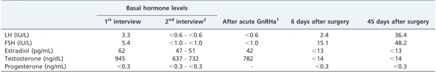
The initial laboratory investigation revealed a normal blood count. Liver and renal functions were within normal range. Basal hormone evaluation revealed an increased level of total testosterone with a level of DHEAS, estradiol and gonadotropins concentration normal for age (Table 1). Dexamethasone suppression test (0.5 mg, q.i.d. for 5 days) failed to suppress testosterone levels, whereas cortisol and DHEAS were suppressed normally. Cushing syndrome was ruled out by an overnight dexamethasone suppression test (1.0 mg - cortisol,1.0mg/dL).
Pelvic ultrasonography showed a right ovary of 6.8 cc and a left ovary of 5.8 cc, without nodules or cysts. Computed tomography of the abdomen disclosed a normal right adrenal gland. A solid nodule of 1.58 cm was seen on the left adrenal gland.
The patient refused further evaluation at that time, but appeared for a new evaluation 2 years later. Her physical examination was largely unchanged, except for progression of temporal balding. Basal hormone levels of total testoster-one and estradiol did not change significantly (Table 1), while basal gonadotropin levels were suppressed in two separated samples. After acute GnRH administration, gonadotropin levels remained suppressed.
Pelvic ultrasonography showed a right ovary of 2.7 cc and a left ovary of 1.8 cc, without nodules or cysts. On Computed Tomography of the abdomen, the solid nodule on the left adrenal gland was unchanged.
The patient underwent a left adrenalectomy by laparo-scopy. The tumor measured 4.564.062.5 cm and weighed 45 g. Macroscopically, the tumor was encapsulated with a thin, smooth, fibrous capsule. The tumor had a firm consistency with a variable brown to yellow colour on cross-section. There were no haemorrhages, necrosis or cystic degenerations. On microscopical examination, the tumor consisted of large polygonal cell with abundant eosinophilic cytoplasm, which showed focal peripherical basophilic granules and sometimes fine clear, vacuolation. Occasionally, there was some lipofuchsin pigment.
The patient was submitted to blood collection 6 and 45
days and re-examined 60 days after the surgical procedure. Total testosterone and estradiol decreased while gonado-tropins increase to postmenopausal range. She reported hot flushes and reduced libido, however male pattern baldness and hirsutism were unchanged.
DISCUSSION
The patient in this case study is a 49-year-old woman with a history of virilization for four years and an adrenal
Copyrightß2011CLINICS– This is an Open Access article distributed under the terms of the Creative Commons Attribution Non-Commercial License (http:// creativecommons.org/licenses/by-nc/3.0/) which permits unrestricted non-commercial use, distribution, and reproduction in any medium, provided the original work is properly cited.
CLINICS 2011;66(2):355-357 DOI:10.1590/S1807-59322011000200029
adenoma. Upon first examination, her basal gonadotropin levels were normal. The patient refused further examination or treatment, and was not seen in our out-patient clinic for two years. Upon her return, basal and GnRH-stimulated gonadotropin levels were suppressed, without significant change in total testosterone and estradiol levels. After surgery, gonadotropins increased to postmenopausal range. As gonadotropin levels in adrenal and ovarian androgen-secreting tumors have been described as variable,4-11 this case led us to question whether the time of exposure of the gonadotrophic axis could be one factor that regulates gonadotropin levels in virilizing syndrome.
In patients with polycystic ovary syndrome, a state of chronic mild hyperandrogenemia, dynamic abnormalities of gonadotropins secretion are some of the most prominent findings.3 High basal LH levels and increased LH pulse
amplitude have been described and may be related to elevated oestrogen levels3. This same pattern of abnormality was also described in a patient with a luteinized thecoma of the ovary.12
In the other hand, there is not a specific pattern of gonadotropin levels in patients with virilizing tumors, except for inhibin-secreting ovarian tumors, where FSH is suppressed and LH can be normal or low.13 As a rule, gonadotropin levels in androgen-secreting ovarian tumors have been described as normal or suppressed.7-11 Additionally, mean basal LH levels and LH pulse number and amplitude were lower in patients with ovarian tumors than compared to those with PCOS or normal women,10 suggesting different mechanisms of hypothalamic or pitui-tary feedback. In the same way, gonadotropin levels in patients with adrenal androgen-secreting tumors have also been reported as normal or suppressed, and significantly lower than in patients with non-tumors forms of hyperan-drogenism.5
As long-term administration of androgens to women leads to virilizing effects, some of them irreversible, the effects of testosterone on gonadotropins secretion in normal women have been studied in female-to-male transsexual subjects.2 Administration of testosterone undecanoate for six-weeks to eugonadal transsexuals, or for three-months to agonadal transsexuals, did not affect the pulsatile secretion of LH or the basal levels of LH and FSH, despite the fact that this regimen resulted in a biologically effective androgen milieu. On the other hand, testosterone-esters administered to agonadal female-to-male transsexuals for at least six months, resulting in higher testosterone levels, were asso-ciated with a significant decrease in mean basal LH and FSH levels.2It is interesting to note that while acute testosterone
infusion inhibited LH secretion in a dose-dependent manner,
without any effect on FSH secretion1, long term administra-tion was associated with a decrease in both LH and FSH levels2. According to the authors, this could be a consequence
of the duration of testosterone exposure.
To our knowledge, this is the first documented case of changing patterns of gonadotropins secretion observed in a patient with Virilizing Syndrome. This cannot be attributed to spontaneous fluctuation of gonadotropins secretion because responses to acute GnRH stimuli were blunted. In the same way, there was no change in the androgen milieu, as total testosterone and estradiol levels were unchanged. It could be that the gonadotropins pattern in virilizing states changes with time, as in female-to-male transsexuals.2 In fact, an inhibitory effect of testosterone and dihydrotestos-terone on the LH response of GnRH in rat pituitary cells has been reported14. According to this, when the tumor was removed and the testosterone level felt, there was a gradual release of the gonadotrophic axis, with increase of the gonadotropins levels to the status of menopause.
CONCLUSION
We report a patient who showed a changing pattern of gonadotropin levels that appeared to be time-dependent. As there were no changes in testosterone and estradiol levels, we postulate that exposure of the hypothalamus-pituitary to an androgen milieu over time can regulate gonadotropin levels in patients with androgen-secreting tumors.
REFERENCES
1. Serafini P, Silva PD, Paulson RJ, Elkind-Hirsch K, Hernandez M, Lobo RA, et al. Acute modulation of the hypothalamic-pituitary axis by intravenous testosterone in normal women. Am J Obst Gynecol. 1986; 155:1288-92.
2. Spinder T, Spijkstra JJ, Gooren LJ, Hompes PG, van Kessel H.Spijkstra JJ, et al. Effects of long–term testosterone administration on gonadotropin secretion in agonadal female to male transsexual compared with hypogonadal and normal women. J Clin Endocrinol Metab. 1989;68:200-7, doi: 10.1210/jcem-68-1-200.
3. Minanni SL, Marcondes JA, Wajchenberg BL, Cavaleiro AM, Fortes MA, Rego MA, et al. Analysis of gonadotropin pulsatility in hirsute women with normal menstrual cycles and in women with polycystic ovary syndrome. Fertil Steril. 1999;71:675-83, doi: 10.1016/S0015-0282(98)00514-7. 4. Kaltsas GA, Isidori AM, Kola BP, Skelly RH, Chew SL, Jenkins PJ, et al. The value of the low-dose dexamethasone suppression test in the differential diagnosis of hyperandrogenism in women. J Clin Endocrinol Metab. 2003; 88:2634-43, doi: 10.1210/jc.2002-020922.
5. Taylor HC, Velasco ME, Flores SG, Berg G, Brown TR. Amenorrhea and failure to virilize ina patient with a testosterone secreting granulosa cell tumor. Clin Endocrinol. 1982;17:557-67, doi: 10.1111/j.1365-2265.1982. tb01628.x.
6. Munemura M, Inoue S, Himeno R, Koyama N, Mizumoto J, Maeyama M, et al. Endocrine studies on ovarian androblastomas (Sertoli-Leydig tumors). J Endocrinol Investigation. 1984;7:615-22.
7. d’Alva CB, Abiven-Lepage G, Viallon V, Groussin L, Dugue MA, Bertagna X, et al. Sex steroids in androgen-secreting adrenocortical
Table 1 -Hormone levels under basal and dynamic condition and after surgery.
Basal hormone levels
After acute GnRHa1 6 days after surgery 45 days after surgery 1stinterview 2ndinterview2
LH (IU/L) 3.3 ,0.6 -,0.6 ,0.6 2.4 36.4
FSH (IU/L) 5.4 ,1.0 -,1.0 ,1.0 15.1 48.2
Estradiol (pg/mL) 62 47 - 51 42 ,13 ,13
Testosterone (ng/dL) 945 637 - 732 782 ,14 ,14
Progesterone (ng/mL) ,0.3 ,0.3 -,0.3 - ,0.3 ,0.3
LH: luteinizing hormone, FSH: follicle stimulating hormone. 1After the administration of 200
mg by IV route.
2Two years later
Gonadotropins in virilizing tumor
Marcondes JAM et al. CLINICS 2011;66(2):355-357
tumors: clinical and hormonal features in comparison with non-tumoral causes of androgen excess. European J Endocrinol. 2008;159: 641-7. 8. Fragoso MC, Latronico AC, Carvalho FM, Zerbini MC, Marcondes JA,
Araujo LM, et al. Activating mutation of the stimulatory G protein (gsp) as a putative cause of ovarian and testicular human stromal Leydig cell tumors. J Clin Endocrinol Metab. 1998;83:2074-8, doi: 10.1210/jc.83.6.2074. 9. Marcondes JA, Nery M, Mendonc¸a BB, Hayashida SA, Halbe HW, Carvalho FM, et al. A virilizing Leydig cell tumor of the ovary associated with stromal hyperplasia under gonadotropin control. J Endocrinol Investigation. 1997; 20:685-9.
10. Bachelot A, Laborde K, Bresson JL, Plu-Bureau G, Raynaud A, Bertagna X, et al. Luteinizing hormone pulsatility in patients with major ovarian hyperandrogenism. J Endocrinol Investigation. 2007;30:636-46.
11. Tita P, Spina A, Briguglia G, Magro A, Gallo D, Finocchiaro C, et al. Clinical features and hormonal characteristics in a case of ovarian arrhenoblastoma. J Endocrinol Investigation. 1996;19:484-7.
12. Dunaif A, Scully RE, Andersen RN, Chapin DS, Crowley WF Jr. The effects of continuous androgen secretion on the hypothalamic-pituitary axis in woman: evidence from a luteinized thecoma of the ovary. J Clin Endocrinol Metab. 1984;59:389-93, doi: 10.1210/jcem-59-3-389. 13. Lappo¨hn RE, Burger HG, Bouma J, Bangah M, Krans M, de Bruijn HW.
Inhibin as a marker for granulosa-cell tumors. N Engl J Med. 1989;321: 790-3.
14. Turgeon JL, Waring DW. Androgen modulation of luteinizing hormone secretion by female rat gonadotropes. Endocrinology. 1999;140:1767-74, doi: 10.1210/en.140.4.1767.
CLINICS 2011;66(2):355-357 Gonadotropins in virilizing tumor
Marcondes JAM et al.