Early assessment of percutaneous coronary
interven-tions for chronic total occlusions analyzed by novel
echocardiographic techniques
Ercan Erdogan,IMehmet Akkaya,IAhmet Bacaksiz,IAbdurrrahman Tasal,IOsman So¨nmez,IMehmet Ali Elbey,IISeref Kul,IMehmet Akif Vatankulu,IMurat Turfan,IO¨ mer Go¨ktekinI
IBezmialem Foundation University, Faculty of Medicine, Department of Cardiology, Istanbul/Turkey.IIDicle University, School of Medicine, Department of Cardiology, Diyarbakır/Turkey.
OBJECTIVE: Successful revascularization of chronic total occlusions has been associated with improved left ventricular systolic function, reduced anginal symptoms, increased exercise capacity, and increased survival. This study was conducted to determine the impact of revascularization in chronic total occlusion on left ventricular function using novel echocardiographic techniques.
METHODS:A total of 129 patients with chronic total occlusion who underwent revascularization between April 2011 and November 2012 were included in this study. Echocardiographic assessments with two-dimensional speckle tracking echocardiography and real-time three-dimensional echocardiography were performed before the procedure and one month after the procedure. The left ventricular ejection fraction, left ventricular volumes, and three-dimensional systolic dyssynchrony index were quantified.
RESULTS: An immediate procedural success was obtained in 118 patients (91.5%). There were no acute or subacute stent thromboses during follow-up. The mean left ventricular ejection fraction significantly increased (p,0.001), while the left ventricular end-diastolic and end-systolic volumes significantly decreased (p= 0.001
and p,0.001, respectively). The three-dimensional systolic dyssynchrony index also decreased significantly (p,0.001). The global longitudinal strain showed a significant increase after successful revascularization (p,0.001). An increase in the global longitudinal strain was correlated with an increase in the left ventricular ejection fraction (r = 0.27, p= 0.02). The patients with a left ventricular ejection fraction $50% displayed a greater improvement in the global longitudinal strain, and the patients with diabetes showed less improvement.
CONCLUSIONS: Using novel echocardiographic techniques, our results showed that restoring the coronary blood flow in chronic total occlusion patients reduces the left ventricular volumes and improves the left ventricular ejection fraction and the global longitudinal strain of hibernating myocardium.
KEYWORDS: Chronic Total Occlusions; Left Ventricular Function; Percutaneous Coronary Intervention.
Erdogan E, Akkaya M, Bacaksiz A, Tasal A, So¨nmez O, Elbey MA, et al. Early assessment of percutaneous coronary interventions for chronic total occlusions analyzed by novel echocardiographic techniques. Clinics. 2013;68(10):1333-1337.
Received for publication onJune 14, 2013;First review completed onJuly 22, 2013;Accepted for publication onJuly 27, 2013
E-mail: [email protected]
Tel.:+90 212 453 17 00
& INTRODUCTION
Coronary artery chronic total occlusion (CTO) is one of the most challenging obstacles faced by interventional cardiologists. Approximately one-third to one-half of the patients with significant coronary artery disease (CAD) on angiography have at least one CTO (1,2). Although
collaterals are capable of maintaining myocardial viability at rest, they may fail to provide adequate blood flow during exercise, which can result in angina. Successful percuta-neous coronary interventions (PCIs) of CTOs have been shown to improve the left ventricular (LV) systolic function, reduce angina, increase exercise capacity, and reduce the need for late bypass surgery (3-5).
An accurate assessment of LV function by determining the LV volumes and the ejection fraction (EF) is important in evaluating the prognoses of patients with CAD. Two-dimensional speckle tracking echocardiography (2D-STE) can assess the global LV function, and it is superior for EF measurements because it is angle-independent, less subject to artifacts, and easier to conduct than Doppler-derived tissue velocity imaging (6,7). Based on 2D-STE, automated Copyrightß2013CLINICS– This is an Open Access article distributed under
the terms of the Creative Commons Attribution Non-Commercial License (http:// creativecommons.org/licenses/by-nc/3.0/) which permits unrestricted non-commercial use, distribution, and reproduction in any medium, provided the original work is properly cited.
No potential conflict of interest was reported.
function imaging is used to reflect the systolic LV function by assessment of the LV global longitudinal strain (GLS). Longitudinal tissue deformation is evaluated by frame-by-frame tracking of the individual speckles throughout the cardiac cycle. This imaging technique discriminates between active and passive myocardial motion and enables angle-independent quantification of myocardial deforma-tion in two dimensions (8,9). 2D-STE is based on tracking the characteristic speckle patterns created by interference of ultrasound beams in the myocardium, and its accuracy has been confirmed using sonomicrometry and magnetic resonance imaging (MRI) as reference methods (10).
The aim of this study was to investigate the changes in the LV volumes, LVEF, and GLS of patients with CTO, before and one month after PCI, using novel echocardiographic methods.
& MATERIALS AND METHODS
Patient selection
A total of 129 patients with CTO who had attempted PCI at Bezmialem Foundation University Hospital between April 2011 and November 2012 were screened for inclusion in this study. Eleven patients who had failed PCI were excluded, and 118 patients who had been successfully revascularized were included in the study. All of the patients underwent physical examination, chest X-ray, 12-lead electrocardiography (ECG), and transthoracic echocar-diographic (2DE) evaluations. Echocardiography was also repeated one month after the revascularization procedure. CTO was defined as lumen compromise resulting in either Thrombolysis In Myocardial Infarction (TIMI) flow grade 0
or 1, with a likely duration of .3 months (11). All of the
patients included had a native vessel occlusion estimated to be of at least three months in duration based on a history of sudden chest pain, a previous myocardial infarction (MI) in the same target vessel territory, or the time between diagnosis made on coronary angiography and PCI. All of the patients had symptomatic angina and/or a positive functional ischemia study. PCI and stent implantation were performed in a standard manner. Drug-eluting stents (DESs) were used in all of the angioplasty procedures. Heparin was administered to maintain an activated clotting
time.250 seconds. A PCI of the CTO was performed with
modern techniques such as bilateral injections; specialized hydrophilic, tapered tip, and stiff wires; parallel wires; microcatheters; and a retrograde approach when possible. After the PCI, all of the patients were prescribed lifelong aspirin. Clopidogrel was prescribed for at least 12 months in all of the patients. The patients were followed prospectively by a telephone interview or an outpatient visit after 30 days. Procedural success was defined as the successful recanali-zation and dilation of at least one CTO per patient with or
without stent implantation, a residual stenosis of,50%, and
a TIMI flow.2. The procedure time was defined as the time
difference between the patient’s entry and exit from the catheterization room.
The study protocol was approved by the institutional clinical research and ethics committee of Bezmialem Foundation University Hospital, and all of the patients provided written informed consent.
2DE and 2D-STE were performed on the subjects at rest in the left lateral decubitus position with synchronized electrocardiography by two professional cardiologists,
who were blinded to the clinical data, with a commercially available system (Philips iE33, Bothell, WA, USA) equipped with a broadband S5-1 transducer (frequency transmitted: 1.7 MHz; received: 3.4 MHz). The parasternal long-axis views were used to derive the M-Mode measurements of the left atrial (LA) size and the LV end-diastolic (LVEDD) and end-systolic (LVESD) dimensions. The endocardial borders were traced in the end-systolic frame of the 2D images from the three apical views. Four consecutive end-expiratory cardiac cycles using high-frame-rate (50 Hz or more) harmonic imaging in each echocardiographic view were acquired. The speckles were tracked frame by-frame throughout the LV wall during the cardiac cycle, and basal, mid, and apical regions of interest were created. The operator manually adjusted the segments that failed to be tracked. All of the measurements were made blinded to the other results and the clinical details.
Real-time three-dimensional echocardiography (RT3DE) images were obtained from an apical window with the patient in the same position as for 2D-STE. Full-volume images were gathered over four cardiac cycles using a
matrix array transducer (64 transducer; Philips iE33,
Andover, MA). Measurements of the 3D-LV volumes and the 3D-EF were performed off-line (QLAB workstation using 3D-Advanced Quantification, Philips). The systolic dyssynchrony index (SDI) was defined as the standard deviation of the time to minimum systolic volume of the 16 LV segments, expressed as a percent of the RR duration. A higher SDI indicated greater LV dyssynchrony.
Intra-observer variability was determined by the observer repeating the measurement of the GLS in 20 randomly selected patients ten days after the first measurement. Inter-observer variability was determined by another Inter-observer measuring these variables in the same database. The intra-and inter- observer reproducibility of GLS parameter was shown acceptable. The intra- and inter- observer variations were 5.4% and 6.7% for GLS, respectively.
Statistical analysis
The continuous variables are reported as the mean ¡
standard deviations (SD), and the categorical variables are expressed as percentages. Comparisons of the categorical and continuous variables between the two groups were
performed using the x2 test and an unpaired t-test,
respectively. The GLS and velocities were compared using
an independent two samplet-test. The correlation between
the GLS and LVEF variables was tested using a correlation analysis. The delta values are defined as the difference between the first month and the preprocedure value. A
value ofp,0.05 was considered statistically significant. The
SPSS 15.0 for Windows statistical software package program (SPSS Inc., Chicago, IL, USA) was used for the statistical analysis.
& RESULTS
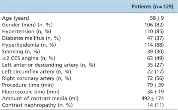
Table 1 shows the baseline demographic, angiographic, and procedural characteristics of the study patients. The
mean age of the patients was 58¡9 years. There were no
mean procedure time was 79¡39 min, the fluoroscopy time
was 34¡19 min, and the amount of contrast was 492¡174 ml.
Contrast-induced nephropathy was developed in 14 (11%) patients, and one patient underwent hemodialysis.
Half of the patients (63 patients, 48.8%) suffered from
angina pectoris ($2 CCS) before the procedure, and the
occurrence of angina pectoris decreased significantly after PCI. Compared with the measurements before the proce-dure, the LVEDV and LVESV decreased significantly
(p= 0.001 and p,0.001, respectively) and the mean LVEF
increased significantly (p,0.001) after the procedure. The
SDI derived from RT3DE decreased significantly after
successful revascularization (p,0.001). The GLS increased
significantly one month after PCI (p,0.001) (Table 2). We
found that the increase in the GLS was correlated with an
improvement in the LVEF (r= 0.27,p= 0.02; Figure 1). The
patients with an LVEF $50% displayed greater
improve-ment in the GLS compared with the patients with an LVEF
,50% (1.1¡0.9 vs. 0.12¡0.3, p,0.001). The patients with
diabetes mellitus (DM) showed a lower degree of GLS improvement compared with the patients without DM
(1.1¡1.0vs.0.62¡0.6,p= 0.07).
& DISCUSSION
In this study, we used 2DSTE and RT3DE to determine that successful revascularization of CTO of the coronary arteries may improve the LV contractile function. We found
that the improvement in the myocardial contractile function based on strain analysis seems to be more significant in the
patients with a LVEF $50% and less significant in the
patients with DM. To the best of our knowledge, this is the first study showing the benefit of revascularization of CTOs on LV function using 2DSTE and RT3DE.
LV systolic function is most commonly assessed by the LVEF using 2DE. The assessment of patients with CTO may be challenging with 2DE because many of these patients suffer from long-term myocardial ischemia or MI and develop aneurysmal formation or advanced ventricular remodeling. Strain and strain rate imaging using the 2D-STE method is a novel echocardiographic technique for the evaluation of regional and global myocardial function, is relatively free from angle dependency and the frame-rate limitations of tissue Doppler imaging, and enables calcula-tions to be made more easily. The global LV contractile function, as assessed by 2D-STE, was shown to be superior to EF measurements by 2DE (6,7). RT3DE is the other new echocardiographic technique for calculating the actual LV volume based on the actual LV shape, rather than on geometrical assumptions. It has good reproducibility even in cases of heart cavity deformation or segmental wall motion abnormalities. It is more accurate than 2DE, and its accuracy approaches that of the gold standard, namely MRI (12,13).
Despite the presence of coronary collaterals, the majority of patients with a CTO show various degrees of LV dysfunction. The possibility of a functional recovery and its beneficial effect on survival are the rationale for the often technically demanding attempt to recanalize a CTO (3-5,14-18). Recovery of LV function in chronically ischemic myocardium depends on the presence of hibernating or stunned but viable myocardium (19). When reperfusion is acquired, the hibernating myocardium at least partially restores the contractile function, resulting in regional and global LV function recovery (20). Baks et al. showed the beneficial effect of successful CTO revascularization on the end-systolic and end-diastolic volumes. They showed that the extent of dysfunctional but viable myocardium before revascularization was related to an improvement in the end-systolic volume and LVEF (21). In the present study, we found a reduction in LV volumes; however, we used RT3DE, which is more robust than 2DE. Cheng et al. used contrast-enhanced MRI to demonstrate that successful CTO-PCI results in improved LV function and attenuated LV remodeling if the vessel patency is maintained (22). The improvements in LVEF and myocardial function in our study, based on 2DSTE and RT3DE, are consistent with the results of Cheng et al. The improvements in the LVEF and GLS observed in the current study may be a result of the recovered hibernating myocardium.
Multiple retrospective studies have shown the potential benefit of PCI in patients with CTO. Successful treatment improves anginal symptoms, exercise tolerance, and LV function. The prospective Total Occlusion Angioplasty Study (TOAST-GISE) showed that revascularization of a CTO is associated with relieved angina and reduces the 12-month incidence of cardiac death or MI and the need for CABG (5). Joyal et al. recently identified 13 observational studies comparing the outcomes after successful versus failed CTO recanalizations, showing that successful CTO recanalization was associated with a 44% reduction in mortality, a 78% reduction in the subsequent need for CABG, and a 55% reduction in residual or recurrent angina
Table 1 -The demographic, angiographic, and procedural characteristics of the patients.
Patients (n = 129)
Age (years) 58¡9
Gender (men) (n, %) 106 (82)
Hypertension (n, %) 110 (85)
Diabetes mellitus (n, %) 47 (37)
Hyperlipidemia (n, %) 114 (88)
Smoking (n, %) 39 (30)
$2 CCS angina (n, %) 63 (49) Left anterior descending artery (n, %) 35 (27) Left circumflex artery (n, %) 22 (17)
Right coronary artery (n, %) 72 (56)
Procedure time (min) 79¡39
Fluoroscopic time (min) 34¡19
Amount of contrast media (ml) 492¡174
Contrast nephropathy (n, %) 14 (11)
CCS: Canadian Cardiovascular Society.
Table 2 -Echocardiographic measures of the successfully revascularized patients before and one month after the procedure (n = 118).
Preprocedure One month p-value
LVEDD (mm) 52.6¡4.9 52.3¡5.2 0.03
LVESD (mm) 33.5¡5.8 33.4¡5.9 0.3
3D end-diastolic volume (ml) 76.22¡22.55 71.98¡22.64 0.001 3D end-systolic volume (ml) 34.54¡16.49 31.63¡16.22 ,0.001 Mean 3D-LVEF (%) 56.31¡9.81 58.32¡10.2 ,0.001
3D-SDI (%) 4.2¡3.1 3.4¡2.3 ,0.001
Global longitudinal strain (%)
-12.51¡1.81 -13.23¡2.29 ,0.001
(23). In accordance with the above mentioned studies, successful revascularization of CTOs in our study resulted in a significant improvement in anginal symptoms. Approximately half of the patients did suffer from angina CCS 2 or greater before the procedure, and none of them had CCS 2 or greater after the procedure.
Numerous experimental studies have demonstrated that ischemia depresses the regional systolic function, resulting in severe systolic dyssynchrony, prolonged tension devel-opment in the ischemic regions, and impaired global relaxation (24,25). Bonow et al. reported that CAD was associated with dyssynchrony, which improved after mechanical revascularization (26). In the present study, the SDI derived by RT3DE, which is a marker of dyssynchrony, was improved one month after CTO revascularization. The improvement in dyssynchrony may be related to an improvement in the hibernating myocardium and in decreased LV volumes.
Endothelial dysfunction, structural changes of the micro-circulation, and a negative influence on the development and prognosis of CAD are well-established features of patients with DM (27-29). Patients with DM have a greater extent of CAD and are prone to impaired clinical outcomes compared with nondiabetic patients (30-32). A previous observational study about CTO PCI in diabetics was
performed by Safley et al. They reported that DM patients do not seem to have the same survival benefits from successful PCI of a CTO as patients without DM (33). In our study, we found relatively less improvement in the GLS in diabetic patients. It is still not clear why diabetics benefit less from PCI of a CTO, but we speculate that the extensive atherosclerosis and impaired microvascular circulation may explain the worse outcomes in this population.
Limitations
There are several limitations to this study. The main limitation is its observational nature and the fact that it was nonrandomized. Additionally, the duration of occlusion was not known with certainty in some of the cases. A lack of follow-up data beyond the hospital stay is another limita-tion.
Our results showed that restoring the blood flow to dysfunctional but viable myocardium led to a moderate improvement in LV function and a reduction in the adverse remodeling in the majority of patients with a CTO. Angiographical success was accompanied by anginal relief.
& AUTHOR CONTRIBUTIONS
Erdogan E contributed to the study design, data collection and interpretation, statistical analysis, manuscript preparation, and literature
search. Akkaya M contributed to the study design, data collection and interpretation, and literature search. Bacaksiz A contributed to the data interpretation, manuscript preparation, and literature search. Tasal A, So¨nmez O, Elbey MA, Kul S, Turfan M, Vatankulu MA, and Go¨ktekin O contributed to the data collection and interpretation and statistical analysis.
& REFERENCES
1. Christofferson RD, Lehmann KG, Martin GV, Every N, Caldwell JH, Kapadia SR. Effect of chronic total occlusion on treatment strategy. Am J Cardiol. 2005;95(9):1088-91, http://dx.doi.org/10.1016/j.amjcard. 2004.12.065.
2. Srinivas VS, Brooks MM, Detre KM, King SB, Jacobs AK, Johnston J, et al. Contemporary percutaneous coronary intervention versus balloon angioplasty for multivessel coronary artery disease: a comparison of the National Heart, Lung and Blood Institute Dynamic Registry and the Bypass Angioplasty Revascularisation Investigation (BARI) study. Circulation. 2002;106(13):1627-33, http://dx.doi.org/10.1161/01.CIR. 0000031570.27023.79.
3. Melchior JP, Doriot PA, Chatelain P, Meier B, Urban P, Finci L, et al. Improvement of left ventricular contraction and relaxation synchronism after recanalization of chronic total coronary occlusion by angioplasty. J Am Coll Cardiol. 1987;9(4):763-8, http://dx.doi.org/10.1016/S0735-1097(87)80230-9.
4. Suero J, Marso SP, Jones PG, Laster SB, Huber KC, Giorgi LV, et al. Procedural outcomes and long term survival among patients undergoing percutaneous coronary intervention of a chronic total occlusion in native coronary arteries: a 20 year experience. J Am Coll Cardiol. 2001;38(2):409-14, http://dx.doi.org/10.1016/S0735-1097(01)01349-3.
5. Olivari Z, Rubartelli P, Pisicone F, Ettori F, Fontanelli A, Salemme L, et al. On behalf of TOAST-GISE Investigators. Immediate results and one-year clinical outcome after percutaneous coronary interventions in chronic total occlusions, (TOAST-GISSE). J Am Coll Cardiol. 2003;41(10):1672-8, http://dx.doi.org/10.1016/S0735-1097(03)00312-7.
6. Marwick TH. Should we be evaluating the ventricle or the myocardium? Advances in tissue characterization. J Am Soc Echocardiogr. 2004;17(2):168-72, http://dx.doi.org/10.1016/j.echo.2003.10.021 7. Leitman M, Lysyansky P, Sidenko S, Shir V, Peleg E, Binenbaum M, et al.
Two-dimensional strain-a novel software for real-time quantitative echocardiographic assessment of myocardial function. J Am Soc Echocardiogr. 2004;17(10):1021-9, http://dx.doi.org/10.1016/j.echo.2004. 06.019
8. Reisner SA, Lysyansky P, Agmon Y, Mutlak D, Lessick J, Friedman Z. Global longitudinal strain: a novel index of left ventricular systolic function. J Am Soc Echocardiogr. 2004;17(6):630-3, http://dx.doi.org/10. 1016/j.echo.2004.02.011.
9. Delgado V, Mollema SA, Ypenburg C, Tops LF, van der Wall EE, Schalij MJ, et al. Relation between global left ventricular longitudinal strain assessed with novel automated function imaging and biplane left ventricular ejection fraction in patients with coronary artery disease. J Am Soc Echocardiogr. 2008;21(11):1244-50, http://dx.doi.org/10.1016/ j.echo.2008.08.010.
10. Amundsen BH, Helle-Valle T, Edvardsen T, Torp H, Crosby J, Lyseggen E, et al. Noninvasive myocardial strain measurement by speckle tracking echocardiography: Validation against sonomicrometry and tagged magnetic resonance imaging. J Am Coll Cardiol. 2006;47(4):789-93, http://dx.doi.org/10.1016/j.jacc.2005.10.040.
11. Stone GW, Kandzari DE, Mehran R, Colombo A, Schwartz RS, Bailey S, et al. Percutaneous recanalization of chronically occluded coronary arteries: a consensus document: part I. Circulation. 2005;112(15):2364-72, http://dx.doi.org/10.1161/CIRCULATIONAHA.104.481283.
12. Bauer F, Shiota T, Qin JX, White RD, Thomas JD. Measurement of left atrial and ventricular volume in real-time 3D echocardiography: Validation by nuclear magnetic resonance. Arch Mal Coeur Vaiss. 2001;94(1):31-8.
13. Jenkins C, Bricknell K, Hanekom L, Marwick TH. Reproducibilityand accuracy of echocardiographic measurements of left ventricular para-meters using real-time three-dimensional echocardiography. J Am Coll Cardiol. 2004;44(4):878-86, http://dx.doi.org/10.1016/j.jacc.2004.05.050. 14. Engelstein E, Terres W, Hofmann D, Hansen L, Hamm CW. Improved
global and regional left ventricular function after angioplasty for chronic coronary occlusion. Clin Investig. 1994;72(6):442-7.
15. Danchin N, Angioi M, Cador R, Tricoche O, Dibon O, Juillie`re Y, et al. Effect of late percutaneous angioplastic recanalization of total coronary
artery occlusion on left ventricular remodeling, ejection fraction, and regional wall motion. J Am Coll Cardiol. 1996;78(7):729-35.
16. Van Belle E, Blouard P, McFadden EP, Lablanche JM, Bauters C, Bertrand ME. Effects of stenting of recent or chronic coronary occlusions on late vessel patency and left ventricular function. J Am Coll Cardiol. 1997;80(9):1150-4.
17. Sirnes PA, Myreng Y, Molstad P, Bonarjee V, Golf S. Improvement in left ventricular ejection fraction and wall motion after successful recanaliza-tion of chronic coronary occlusions. Eur Heart J. 1998;19(2):273-81, http://dx.doi.org/10.1053/euhj.1997.0617.
18. Dzavik V, Carere RG, Mancini GBJ, Cohen EA, Catellier D, Anderson TE, et al. Predictors of improvement in left ventricular function after percutaneous revascularization of occluded coronary arteries: a report from the Total Occlusion Study of Canada (TOSCA ). Am Heart J. 2001;142(2):301-8, http://dx.doi.org/10.1067/mhj.2001.116960. 19. Wijns W, Vatner SF, Camici PG. Hibernating myocardium. N Engl J Med.
1998;339(3):173-81.
20. Bax JJ, Visser FC, Poldermans D, Elhendy A, Cornel JH, Boersma E, et al. Relationship between preoperative viability and postoperative improve-ment in LVEF and heart failure symptoms. J Nucl Med. 2001;42(1):79-86. 21. Baks T, van Geuns RJ, Duncker DJ, Cademartiri F, Mollet NR, Krestin GP, et al. Prediction of left ventricular function after drug-eluting stent implantation for chronic total coronary occlusions. J Am Coll Cardiol. 2006;47(4):721-5, http://dx.doi.org/10.1016/j.jacc.2005.10.042. 22. Cheng ASH, Selvanayagam JB, Jerosch-Herold M, van Gaal WJ,
Karamitsos TD, Neubauer S, et al. Percutaneous treatment of chronic total coronary occlusions improves regional hyperemic myocardial blood flow and contractility: insights from quantitative cardiovascular magnetic resonance imaging. JACC Cardiovasc Interv. 2008;1(1):44-53, http://dx.doi.org/10.1016/j.jcin.2007.11.003.
23. Joyal D, Afilalo J, Rinfret S. Effectiveness of recanalization of chronic total occlusions: a systematic review and meta-analysis. Am Heart J. 2010;160(1):179-87, http://dx.doi.org/10.1016/j.ahj.2010.04.015. 24. Kumada T, Karliner JS, Pouleur H, Gallagher KP, Shirato K, Ross J Jr.
Effects of coronary occlusion on early ventricular diastolic events in conscious dogs. Am J Physiol. 1979;237(5):H542-9.
25. Green MV, Jones-Collins BA, Bacharach SL, Findley SL, Patterson RE, Larson SM. Scintigraphic quantitation of asynchronous myocardial motion during the left ventricular isovolumic relaxation period: a study in the dog during acute ischemia. J Am Coll Cardiol. 1984;4(1):72-9, http://dx.doi.org/10.1016/S0735-1097(84)80321-6.
26. Bonow RO, Vitale DF, Bacharach SL, Frederick TM, Kent KM, Green MV. Asynchronous left ventricular regional function and impaired global diastolic filling in patients with coronary artery disease: reversal after coronary angioplasty. Circulation. 1985;71(2):297-307, http://dx.doi.org/ 10.1161/01.CIR.71.2.297.
27. Schofield I, Mlik R, Izzard A, Austin C, Heagerty A. Vascular structural and functional changes in type 2 diabetes mellitus: evidence for the roles of abnormal myogenic responsiveness and dyslipidemia. Circulation. 2002;106(24):3037-43, http://dx.doi.org/10.1161/01.CIR.0000041432.80615. A5.
28. Watts GF, O’Brien SF, Silvester W, Millar JA. Impaired endothelium-dependent and inendothelium-dependent dilatation of forearm resistance arteries in men with diettreated non-insulin-dependent diabetes: role of dyslipi-daemia. Clin Sci (Lond). 1996;91(5):567-73.
29. Haffner SM, Lehto S, Ro¨nnemaa T, Pyo¨ra¨la¨ K, Laakso M. Mortality from coronary heart disease in subjects with type 2 diabetes and in non-diabetic subjects with and without prior myocardial infarction. N Engl J Med. 1998;339(4):229-34.
30. Malmberg K, Yusuf S, Gerstein HC, Brown J, Zhao F, Hunt D, et al. Impact of diabetes on long-term prognosis in patients with unstable angina and non-Q-wave myocardial infarction: results of the OASIS (Organization to Assess Strategies for Ischemic Syndromes) Registry. Circulation. 2000;102(9):1014-9, http://dx.doi.org/10.1161/01.CIR.102.9. 1014.
31. Comparison of coronary bypass surgery with angioplasty in patients with multivessel disease. The Bypass Angioplasty Revascularization Investigation (BARI) Investigators. N Engl J Med. 1996;335(4):217-25. 32. Gu K, Cowie CC, Harris MI. Diabetes and decline in heart disease