(1) Doutora em Ciências Pneumológicas, Unidade de Microbiologia, Universitat Rovira i Virgili, Reus, Espanha.
(2) PPG, Ciências Pneumológicas, Mestrado e Doutorado, Universidade Federal do Rio Grande do Sul (UFRGS), Porto Alegre, RS, Brasil. (3) Laboratório de Micologia/Hospital Santa Rita/Santa Casa-Complexo Hospitalar, Porto Alegre, RS, Brasil.
(4) Departamento de Patologia/Santa Casa-Complexo Hospitalar, Porto Alegre, RS, Brazil.
(5) Departamento de Medicina Interna, Universidade Federal do Rio Grande do Sul (UFRGS), Porto Alegre, RS, Brazil. (6) Pesquisador nível 1B CNPq (Conselho Nacional de Desenvolvimento Científico e Tecnológico).
Instituição financiadora: Coordenação de Aperfeiçoameneto de Pessoal de Nível Superior (CAPES). BEX: 0060/09-7
Correspondence to: Dr. Luiz Carlos Severo. Laboratório de Micologia/Hospital Santa Rita, Santa Casa-Complexo Hospitalar, Anne Dias 295, 90020-090 Porto Alegre, RS, Brasil, Phone:
HISTOPATHOLOGY, SEROLOGY AND CULTURES IN THE DIAGNOSIS OF CRYPTOCOCCOSIS
Alexandra Flávia GAZZONI(1), Cecília Bittencourt SEVERO(2,3), Emily Ferreira SALLES(2,4) & Luiz Carlos SEVERO(3,5,6)
SUMMARY
Cryptococcosis is one of the most common opportunistic fungal infections in patients with acquired immunodeficiency syndrome (AIDS). We report 13 cases of cryptococcal infection based on histopathology, serology and cultures. Epidemiological analysis, histochemical techniques of hematoxilin and eosin (HE) and Grocot’s silver (GMS), as well special histochemical techniques such as Mayer’s mucicarmine (MM) and Fontana-Masson (FM), cryptococcal antigen test (CrAg) and isolation on fungal media: Sabouraud’s (SAB), brain-heart infusion agar (BHI) and canavanine-glycine-bromothymol blue (CGB) agar were analyzed. Unsatisfactory staining results by MM stain associated to negative titers by CrAg test, which FM stain confirmed that capsule-deficient Cryptococcus infections
were observed in four cases. Eight isolated cases were identified as follows: six cases were infection with Cryptococcus neoformans
and two cases were Cryptococcus gattii.
KEYWORDS: Budding index; Carminophilic index; Mayer’s mucicarmine stain; Fontana-Masson stain; Cryptococcal antigen test; Cultures.
INTRODUCTION
Cryptococcosis is a systemic infection caused by naturally
encapsulated basidiomycetous of genus Cryptococcus7,13. This yeast
causes human infection ranging from asymptomatic pulmonary colonization to meningitis and disseminated disease6. Cryptococcosis is
caused by two species: Cryptococcus gattii is typically found in tropical
and subtropical climates zones, and usually causes disease in apparently normal hosts5,8,23,25, whereas the C. neoformans present in urban pigeon
droppings, has a worlwide distribution and is a common opportunistic infection5,6,23,25. Specifically, conditions predisposing to a change in
cellular immunity have been associated with a significant increase in risk to obtain the cryptococcosis, and these include lymphoproliferative disorders, organ transplantation, and receiving immunosuppressive therapy23. Cryptococcosis is also one of the most common opportunistic
fungal infections in acquired immunodeficiency syndrome (AIDS) patients, and, in that group, it is associated with a high mortality rate23.
Microscopically, Cryptococcus has spherical to oval yeast cells, 5-10
µm in diameter that are surrounded by a polysaccharide capsule2,7, which
is a major virulence factor and the substrate detected by cryptococcal
antigen tests (CrAg)4,7. However, capsule-deficient Cryptococcus
infections may result in false-negative results using the CrAg test13,15,29.
Cryptococcosis laboratory diagnosis includes conventional methods (direct microscopic examination and histopathology)5,11,12,13,
usually associated to serology3,4,20 and to isolation of the organism in
culture3,5,23. Histopathologic identification in biopsy specimens is based
on the micromorphological characteristics of Cryptococcus, and include
histochemical techniques of hematoxilin and eosin (HE) and Grocott’s silver (GMS), as well special histochemical techniques such as Mayer’s mucicarmine (MM), which stains the capsule magenta, and Fontana-Masson (FM), which stains fungal melanin reddish-brown1,12,13 .
In the Schwartz classification, infection is divided into two major histological categories, based upon reactions of the tissues24. The reactive
pattern presents intense granulomatous inflammatory response, composed of macrophages, multinucleated giant cells, and lymphocytes5,13,24; the
yeasts are found in an intracelullar location (phagocytozed)15,23. In the
paucireactive pattern, yeasts are found extracellularly, associated to a minimal or absent inflammatory response, and tissue destruction results from compression necrosis by masses of cryptococcal tecidual6,23,24,26,27.
To evaluate the inflammatory response of Cryptococcus infections,
two morphologic parameters are analyzed6,24. The viability of the
organisms is estimated using the Carminophilic Index (CI), which indicates capsular synthesis. The divisional or mitotic activity of yeast is estimated by the Budding Index (BI), and indicates replication in vivo24.
This histopathological study has two main purposes: (a) to characterize the inflammatory response of Cryptococcus; (b) to showthe
MATERIAL AND METHODS
A retrospective study was conduced by reviewing the medical records of patients who were diagnosed with cryptococcal infection based on histopathological identification, serological and cultures at the Micology Laboratory of Santa Casa Complexo Hospitalar (Porto Alegre, RS), in Southern Brazil. The data reviewed included age, gender, predisposing factors, host immune status, sites of infections, CrAg titer, and clinical presentation (localized or disseminated).
Culture: The identification of Cryptococcus was confirmed by the
isolation of yeasts with white mucoid colonies (depending on the capsule thickness) after cultivation on fungal media (within 48-72 h), namely Sabouraud’s (SAB) at 25 °C, and brain-heart infusion (BHI) agar at 35 °C; and by microscope by demonstrating the presence of spherical to oval yeast cells, budding on a narrow base, with a surrounding capsular structure. The species were discriminated by a color reaction when grown on canavanine-glycine-bromothymol blue (CGB) agar.
Analysis of microscopic pathology: Biopsy specimens were submitted to standard histological processing1. Tissue sections were
stained by histochemical techniques of HE18 and GMS14,18, sections
stained by special histochemical techniques of MM13,19 were also included.
The histological reactions to HE2,24, the measurement of BI to GMS24,
and the presence of magenta capsular structure12,13,19 and measurement of
CI to MM stain24 were determined. Special histochemical techniques of
FM stain13,17,18,19 were applied in those cases that showed negative results
by the MM stain, and negative titers by the CrAg test.
CrAg detection: The IMMY test - the commercial kit used in this
study - has a vital component in Detacher Enzyme (DE), Pronase. DE
digests antibodies in immune complexes (the antigen is not affected), which can mask the detection of antigens. DE also eliminates the rheumatoid factor, that can cause false positives3,4.
RESULTS
Patients characteristics: During the 28-year period between 1980 and 2008, there were 13 patients diagnosed with cryptococcosis based on histopathology, serology and cultures. Out of those, nine (70%) patients were men and four (30%) women. Their age ranged from 10 - 56 years old (average of 37.5 years). The median age was 36 years for men and 41 for women.
The majority of patients (77%) were considered to be immunocompromised with at least one identifiable risk factor, including infection with human immunodeficiency virus (HIV) in five (39%) patients, solid organ transplantation in four (31%) patients - renal (three
Table 1
Histologic pattern, measurements of BI and CI, CrAg titers and identification of Cryptococcus species complex in 13 patients
Cases Sex, Age Predisposing
factors
Histologic pattern BI CI CrAg titers Specimen, CGBa
Immunocompetent
01 M, 10 None Reactive
(Fibrotic nodules) (Cryptococcomas)
0 +++ 1:128 Cerebrospinal fluid, C. gattii
02* F, 42 None Reactive 15 0 Negative Axillary tumor biopsy, C. gattii
03 M, 56 None Reactive 12 ++++ 1:5112 Not done
Immunocompromised
04 M, 13 Tx pulmonary Paucireactive
(Pulmonary nodules)
2 ++++ Negative Lung tissue biopsy, C. neoformans
05 M, 29 AIDS Paucireactive 4 +++ 1: 1049.536 Lymph node biopsy, C. neoformans
06 F, 30 AIDS Paucireactive 2 + 1:2 Not done
07 M, 31 AIDS Paucireactive 10 ++++ 1:5112 Cerebrospinal fluid, C. neoformans
08 M, 40 Tx renal Reactive 3 + 1:8 Cutaneous tissue biopsy, C.
neoformans
09 M, 42 AIDS Reactive 4 + 1:128 Not done
10* F, 42 Tx renal Reactive 5 0 Negative Not done
11* M, 50 AIDS Reactive 8 0 Negative Lung tissue biopsy, C. neoformans
12 F, 50 Tx renal Paucireactive 3 +++ Negative Not done
13* M, 53
Immunosupres-sive drugs
Reactive 2 0 Negative Lung tissue biopsy, C. neoformans
cases) and lung (one case) - and user of immunosuppressive drugs in one (8%) patient. Three patients were considered immunocompetent: one presented infection by a capsule-deficient strain, one exhibited an axillary tumoral form, and the other one was a 10 year-old child.
A total of 30% (n = 4) presented localized pulmonary infection; all were HIV-uninfected. Disseminated infection was found in 70% (n = 9); of these, 78% (n = 7) presented of the involvement central nervous system (CNS), these five were HIV-positive patients. Extraneural, extrapulmonary manifestations were observed in seven (54%) cases, including mediastinal lymphadenopathy (four cases), concomitant to oral cavity involvement (one case), hepatic (one case) and cutaneous (one case).
It was possible to identify the Cryptococcus species complex in eight cases: six (46%) cases of Cryptococcus neoformans and two
(15%) cases of Cryptococcus gattii. All C. neoformans isolated were
immunocompromised hosts; two C. gattii isolated were in apparently
imunocompetent patients. As shown in Table 1, the Cryptococcus species were isolated on fungal media, namely SAB, BHI and CGB.
Histological pattern
Reactive: Circumscribed granulomas were seen in seven (54%) patients. The lesions were composed of compactly aggregated histiocytes and multinucleated giant cells, including both Langerhans and foreign body type, with numerous intracytoplasmatic yeasts (phagocytozed) (Fig. 1A). Central area of necrosis associated to marginal fibrosis was observed (Fig. 1B). In one case, fibrotic nodules were identified.
Paucireactive: Six (46%) cases presented numerous extracellular organisms, associated with a minimal to absent inflammatory response. Typical cells were spherical, oval, or ellipsoid, and surrounded by optically clear, smoothly spherical zones, or halos that are unstained capsules. In three (23%) cases chronic lymphadenitis were identified showing destruction of the architecture by compact masses of
encapsulated yeasts (Fig. 1C). One case revealed organizing pneumonia. Table 1 summarizes the histologic patterns, measurements of CI and BI, and CrAg titers in 13 patients.
CI: Ranged from one-plus to four-plus (+ to ++++). Paucireactive
pattern had the highest measurements of CI; the reactive pattern has shown marked variation among cases, ranging from one-plus to three-plus (+ to +++). A total of nine (70%) patients exhibited a magenta capsule stained by MM stain (cases 1, 3, 4, 5, 6, 7, 8, 9, 12) (Fig. 2A); four (30%) patients did not show a reactive carminophilic staining (cases 2, 10, 11, 13); all presented the reactive pattern of the infection.
BI: Both patterns showed great variation in this index (measurements
0 to 15) (Fig. 2B).
CrAg test: Seven (54%) out of 13 patients had a positive serology, including samples from serum, cerebrospinal fluid and urine. Among those patients, the cases 1, 3, 5, 7 presented major measurements of CI. Of the six (46%) patients that presented negative results (cases 2, 4, 10, 11, 12, 13), four (30%) did not show the presence of a carminophilic capsule after staining with MM thus preventing the measurements of CI (cases 2, 10, 11, 13). Two (15%) transplant recipients presented localized pulmonary infection and showed negative results to the CRAg test (cases 4, 12).
Fontana-Masson stain: Cases 2, 10, 11, 13 did not show the presence of a mucicarminophilic capsule after staining with MM stain and presented negative results by CrAg test; in all cases the FM stain detected fungal melanin in the cell wall (Fig. 2C).
DISCUSSION
Before the AIDS epidemic, cryptococcosis was considered an unusual infection with low incidence rate6,23. After the recognition of AIDS,
the diseases emerged as an important opportunistic infection16,20. The
association with organ transplantation (four cases in our group) and use of immunosuppressive drugs has been established6,9,23.
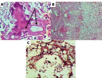
Fig. 1 - Section of the lung stained with HE. Histologic patterns of cryptococcosis. A, multinucleated giant cell of foreign body type (case 2) (x100). B, granulomatous inflammation consisting of necrosis, macrophages, lymphocytes and fibroblastic activity (case 2) (x10). C, section of the lymph node with complete effacement of lymphoid tissue by compact extracellular yeasts (case 5) (x100).
Epidemiological analysis revealed a male predominance, with an
average age similar to those in Colombia21 and France10. Among men
and women, the average age showed differences in agreement with previous reports, suggesting that the average age is higher in women10,20.
Although rarely, cryptococcosis can affect children8. In our sample, it was
present in two children (15%). One was immunocompetent and lived in a biogeoclimatic endemic zone for Cryptococcus gattii in Brazil (Pará)8,
and the other was a lung transplant recipient.
Among organ systems, the respiratory tract is the most frequent site of entry involved6,16,22. The organism is trophic to the CNS, and
the majority of cases were related to meningitis9,10, most commonly in
HIV-positive patients. In contrast, pulmonary involvement was found
to occur more frequently in HIV-negative patients. Cryptococcus may
undergo hematogenous spread to other organ systems6,12. Extrapulmonary
and extraneural manifestations were observed in 54% of patients. The following body sites are considered to be manifestations of disseminated disease: lymph node (lymphadenopathy), head and neck (involvement of oral cavity), skin and gastrointestines (hepatic)12,24.
C. gattii is likely to cause disease in healthy hosts and behaves as
a primary pathogen, whereas C. neoformans affects predominantly
immunocompromised individuals, especially those infected by HIV5,8,25.
It is emphasized that the biopsy specimens should be in saline (0.9% NaCl) solution for fungal isolation, as well identification of species, since formalin fixation causes death of fungal agent preventing its growth on culture media.
Paucireactive cryptococcosis results from the impaired capability to successfully mount an effective inflammatory response. Cell-mediated immunity has a role in the defense mechanism24; in its absence, organisms
proliferate extracellularly associated with destruction of tissue13,16,26.
Histological analysis of our cases is consistent with these features. In the paucireactive infection, the BI varied greatly, and most cases showed less than 5% budding forms. When compared with the reactive infection, organisms showed highest measurements of CI. There is apparently an inverse correlation between capsular production and intense inflammatory responses6,24.
A reactive pattern results in hosts having an active cell-mediated
immune response24. The histology is characterized by a granulomatous
response composed of histiocytes, giant cells, and lymphocytes associated with fibroblastic activity16,24,26,27,28. Our cases demonstrated these
histologic features in different degrees. When compared to paucireactive infection, the yeasts cells were less abundant, and both CI and BI showed a greater spectrum of variation.
The fibrotic nodules, better known as the cryptococcomas, represent an infection in immunocompetent persons24. Similar lesions are presented
in histoplasmosis24 and tuberculosis24,28. Case 1 contained scarce
organisms having a CI of +++ and BI of zero.
The CrAg were first described by BLOOMFIELD et al.4 in the sera
and cerebrospinal fluid (CSF) of seven from a total of nine patients. The CrAg test is approximately 95% sensitive and specific3.
False-negative results reported were due to a poorly encapsulated strain6,23.
Although commercial kits differ, the CrAg test detects at least 10 ng of polysaccharide per mL of biological fluid6. These false-negatives
are due to capsular deficiency as this strain does not produce enough polysaccharides antigens to be detected by the CrAg test. In fact, the size of the polysaccharides of cells of the deficient-capsule strain is smaller than those of the typical strain. Patients with primary pulmonary cryptococcosis without dissemination may have false-negative serum tests, since yeasts have not yet disseminated from the lung. Once antigen is detected in the serum of a patient with cryptococcosis, this is a sign that infection has disseminated from its pulmonary location6. On the basis
of these observations, in false-negative titers and unsatisfactory staining results by MM stain, the special techniques of the FM stain for fungal melanin confirm infections by capsule-deficient Cryptococcus strains.
RESUMO
Histopatologia, sorologia e cultivo no diagnóstico da criptococose
A criptococose é a mais comum infecção fúngica oportunística observada em pacientes com síndrome da imunodeficiência adquirida (AIDS). Relatamos 13 casos da infecção baseados no diagnóstico histopatológico, sorológico e cultivo. Foram analisadas: a epidemiologia, as técnicas histoquímicas básicas de hematoxilina-eosina (HE) e coloração pela prata (GMS), bem como as técnicas histoquímicas especiais de mucicarmim de Mayer (MM) e Fontana-Masson (FM), o teste do antígeno criptocóccico (CrAg) e o isolamento em cultivos em ágar-Sabouraud (SAB), ágar infusão de cérebro-coração (BHI) e meio com canavanina azul de bromotimol (CGB). Em quatro casos, resultados tintoriais insatisfatórios pela coloração de MM associados a títulos negativos pelo teste do CrAg, a coloração de FM confirmou a
infecção pelo Cryptococcus deficiente de cápsula. Oito isolados foram
identificados: seis casos apresentaram a infecção por Cryptococcus neoformans e dois casos apresentaram a infecção por Cryptococcus gattii.
REFERENCES
1. ARTAL, E.M. - Diagnóstico histopatológico de las micosis. Rev. iberoamer. Micol., 212: 1-9, 2004.
2. BAKER, R.D. & HAUGEN, R.K. - Tissue changes and tissue diagnosis in cryptococcosis: a study of 26 cases. Amer. J. clin. Path., 25: 14-24, 1955.
3. BERLIN, L. & PINCUS, J.H. - Cryptococcal meningitis. False-negative antigen test results and cultures in nonimmunosuppressed patients. Arch. Neurol., 46: 1312-1316, 1989. 4. BLOOMFIELD, N.; GORDON, M.A. & ELMENDORF Jr., D.F. - Detection of
Cryptococcus neoformans antigen in body fluids by latex particle agglutination. Proc. Soc. exp. biol. Med., 114: 64-67, 1963.
5. BOVERS, M.; HAGEN, F. & BOEKHOUT, T. - Diversity of the Cryptococcus neoformans-Cryptococcus gattii species complex. Rev. iberoamer. Micol., 25(suppl.): S4-S12, 2008.
6. CASADEVALL, A. & PERFECT, J.R. - Cryptococcus neoformans. Washington, ASM Press, 1998.
7. CHANDLER, F.W. & WATTS, J.C. - Cryptococcosis. In: CONNOR, D.H.; CHANDLER, F.W.; SCHWARTZ, D.A. et al., ed. Pathology of infectious diseases. Stamford, Appleton & Lange, 1997. p. 989-997.
8. CÔRREA, M.P.S.C.; OLIVEIRA, E.C.; DUARTE, R.R.B.S. et al. - Criptococcose em crianças no Estado do Pará, Brasil. Rev. Soc. bras. Med. trop., 32: 505-508, 1999. 9. DIAMOND, R.D. & BENNETT, J.E. - Prognostic factors in cryptococcal meningitis. A
10. DROMER, F.; MATHOULIN-PELISSIER, S.; FONTANET, A. et al. - Epidemiology of HIV-associated cryptococcosis in France (1985-2001): comparison of the pre- and post-HAART eras. AIDS, 18: 555-562, 2004.
11. GAL, A.A.; KOSS, M.N.; HAWKINS, J.; EVANS, S. & EINSTEIN, H. - The pathology of pulmonary cryptococcal infections in the acquired immunodeficiency syndrome. Arch. Path. Lab. Med., 110: 502-507, 1986.
12. GAZZONI, A.F.; PEGAS, K.L. & SEVERO, L.C. - Histopathological techniques in the diagnosis of cryptococcosis due to capsule-deficient Cryptococcus: report of a case. Rev. Soc. bras. Med. trop., 41: 76-78, 2008.
13. GAZZONI, A.F.; SEVERO, C.B.; BARRA, M.B. & SEVERO, L.C. - Atypical micromorphology and uncommon location of cryptococcosis: a histopathologic study using special histochemical techniques (one case report). Mycopathologia, 167: 197-202, 2009.
14. GROCOTT, R.G. - A stain for fungi in tissue sections and smears using Gomori’s methenamine-silver nitrate technic. Amer. J. clin. Path., 25: 975-979, 1955. 15. HARDING, S.A.; SCHELD, W.M.; FELDMAN, P.S. & SANDE, M.A. - Pulmonary
infection with capsule-deficient Cryptococcus neoformans. Virchows Arch. A. Path.. Anat. Histol., 382: 113-118, 1979.
16. JARVIS, J.N. & HARRISON, T.S. - Pulmonary cryptococcosis. Semin. Respir. crit. Care Med., 29: 141-150, 2008.
17. KWON-CHUNG, K.J.; HILL, W.B. & BENNETT, J.E. - New, special for histopathological diagnosis of cryptococcosis. J. clin. Microbiol., 13: 383-387, 1981.
18. LACAZ, C.S.; PORTO, E.; MARTINS, J.E.C.; HEINS-VACCARI, E.M. & MELO, N.T. - Tratado de Micologia médica. São Paulo, Sarvier, 1998.
19. LAZCANO, O.; SPEIGHTS Jr., V.O.; BILBAO, J.; BECKER, J. & DIAZ, J. - Combined Fontana-Masson-Mucin staining of Cryptococcus neoformans. Arch. Path. Lab. Med., 115: 1145-1149, 1991.
20. LEAL, A.L.; FAGANELLO, J.; FUENTEFRIA, A.M. et al. - Epidemiological profile of cryptococcal meningitis patients in Rio Grande do Sul, Brazil. Mycopathologia, 166: 71-75, 2008.
21. LIZARAZO, J.; LINARES, M.; BEDOUT, C. et al. - Estudio clínico y epidemiológico de la criptococosis en Colombia: resultados de nueve años de la encuesta nacional, 1997-2005. Biomédica, 27: 94-109, 2007.
22. PAPPAS, P.G.; PERFECT, J.R.; CLOUD, G.A. et al. - Cryptococcosis in HIV-negative patients in the era of effective azole therapy. Clin. infect. Dis., 33: 690-699, 2001. 23. PERFECT, J.R. & CASADEVALL, A. - Cryptococcosis. Infect. Dis. Clin. N. Amer.,
16: 837-874, 2002.
24. SCHWARTZ, D.A. - Characterization of the biological activity of Cryptococcus infections in surgical pathology. The budding index and carminophilic index. Ann. clin. Lab. Sci., 18: 388-397, 1988.
25. SEVERO, L.C.; OLIVEIRA, F.M. & LONDERO, A.T. - Cryptococcosis due to
Cryptococcus neoformans var. gattii in Brazilian patients with AIDS. Report of three cases. Rev. iberoamer. Micol., 16: 152-154, 1999.
26. SHIBUYA, K.; COULSON, W.F.; WOLLMAN, J.S. et al. - Histopathology of cryptococcosis and other fungal infections in patients with acquired immunodeficiency syndrome. Int. J. infect. Dis., 5: 78-85, 2001.
27. SHIBUYA, K.; COULSON, W.F. & NAOE, S. - Histopathology of deep-seated fungal infections and detailed examination of granulomatous response against cryptococci in patients with acquired immunodeficiency syndrome. Jap. J. med. Mycol., 43: 143-151, 2002.
28. SHIBUYA, K.; HIRATA, A.; OMUTA, J. et al. - Granuloma and cryptococcosis. J. infect. Chemother., 11: 115-122, 2005.
29. SUGIURA, Y.; HOMMA, M. & YAMAMOTO, T. - Difficulty in diagnosis chronic meningitis caused by capsule-deficient Cryptococcus neoformans. J. Neurol. Neurosurg. Psychiat., 76: 1460-1461, 2005.