Cardiovascular responses produced by central injection
of hydrogen peroxide in conscious rats
Leonardo M´aximo Cardoso
a
, D´ebora Sim˜oes de Almeida Colombari
b
, Jos´e Vanderlei Menani
b
,
Deocl´ecio Alves Chianca Jr.
c
, Eduardo Colombari
a
,
b
,
∗
aDepartment of Physiology, Federal University of S˜ao Paulo (UNIFESP), Rua Botucatu, 862, 04023-060 S˜ao Paulo, SP, Brazil bDepartment of Physiology and Pathology, State University of S˜ao Paulo (UNESP), Araraquara, SP, Brazil
cDepartment of Biological Sciences, Federal University of Ouro Preto (UFOP), Ouro Preto, MG, Brazil
Received 22 December 2005; received in revised form 22 July 2006; accepted 24 July 2006 Available online 14 August 2006
Abstract
Reactive oxygen species (ROS) have been shown to modulate neuronal synaptic transmission and may play a role on the autonomic control of
the cardiovascular system. In this study we investigated the effects produced by hydrogen peroxide (H
2O
2) injected alone or combined with the
anti-oxidant agent
N
-acetil-
l
-cysteine (NAC) or catalase into the fourth brain ventricle (4th V) on mean arterial pressure and heart rate of conscious
rats. Moreover the involvement of the autonomic nervous system on the cardiovascular responses to H
2O
2into the 4th V was also investigated.
Male Holtzman rats (280–320 g) with a stainless steel cannula implanted into the 4th V and polyethylene cannulas inserted into the femoral
artery and vein were used. Injections of H
2O
2(0.5, 1.0 and 1.5
mol/0.2
L,
n
= 6) into the 4th V produced transient (for 10 min) dose-dependent
pressor responses. The 1.0 and 1.5
mol doses of H
2O
2also produced a long lasting bradycardia (at least 24 h with the high dose of H
2O
2). Prior
injection of
N
-acetyl-
l
-cysteine (250 nmol/1
L/rat) into the 4th V blockade the pressor response and attenuated the bradycardic response to H
2O
2(1
mol/0.5
L/rat,
n
= 7) into the 4th V. Intravenous (
i.v.
) atropine methyl bromide (1.0 mg/kg,
n
= 11) abolished the bradycardia but did not affect
the pressor response to H
2O
2. Prazosin hydrochloride (1.0 mg/kg,
n
= 6)
i.v.
abolished the pressor response but did not affect the bradycardia. The
increase in the catalase activity (500 UEA/1
L/rat injected into the 4th V) also abolished both, pressor and bradycardic responses to H
2O
2. The
results suggest that increased ROS availability into 4th V simultaneously activate sympathetic and parasympathetic outflow inducing pressor and
bradycardic responses.
© 2006 Elsevier Inc. All rights reserved.
Keywords: Hydrogen peroxide; Blood pressure; Heart rate; Prazosin; Methyl atropine;N-Acetyl-l-cysteine; Anti-oxidant
1. Introduction
Considerable evidence suggests that reactive oxygen species
(ROS) such as superoxide anion (O
2•−), hydrogen peroxide
(H
2O
2) and hydroxyl radical (HO
•) may act as cellular
sig-naling molecules to regulate biological function (reviewed in
[1,34]
). ROS are the result of incomplete reduction of oxygen
to O
2•−which is spontaneously or enzymatically dismutated to
H
2O
2[19]
. Different types of cells can produce O
2•−and H
2O
2Abbreviations: CVLM, caudal ventrolateral medulla; HR, heart rate; MAP, mean arterial pressure; NTS, nucleus of the tract solitary; NAC,N-acetil-l -cysteine; RVLM, rostral ventrolateral medulla; SOD, superoxide dismutase; 4th V, fourth brain ventricle; UEA, units of enzymatic activity
∗Corresponding author. Tel.: +55 11 5084 9554.
E-mail address:[email protected](E. Colombari).
in response to a variety of extracellular stimuli, like cytokines,
peptide growth factors, agonists of heterotrimeric G
protein-coupled receptors (angiotensin II, thrombin, lysophosphatidic
acid, sphingosine 1-phosphate, histamine and bradykinin) and
sheer stress (reviewed in
[34]
). It was previously demonstrated
that H
2O
2in the central nervous system (CNS) modulates
synap-tic transmission
[16,31]
. The reversibility of H
2O
2effects on
synaptic transmission and the demonstration that similar effects
are seen with endogenously generated, as well as exogenously
added H
2O
2[3,9]
have implicated the H
2O
2as an endogenous
neuromodulator
[4]
.
A select group of brainstem nuclei play critical roles in the
maintenance of cardiovascular homeostasis and in the
patho-physiology of the hypertension
[13,33]
. Recent finds suggest
that endogenously generated ROS in medullary neurons could
play a role in the autonomic control of the blood pressure as
indicated by the co-localization of angiotensinergic receptors
(AT
1) and the gp91
phoxsubunit of the O
2•−generating enzyme
NADPH oxidase in somatodendrids and axons of neurons in the
nucleus of the solitary tract (NTS)
[40]
. Furthermore, the activity
of neurons in the rostroventrolateral medulla (RVLM), an
impor-tant source of sympathetic output to cardiovascular system, is
suggested to be modulated by ROS
[21,22,43]
. Results with
injections of superoxide dismutase (SOD)
[43]
, SOD mimetics
like tempol
[22]
or genetic manipulations that induce
overex-pression of SOD in the RVLM
[22]
have suggested that O
2•−is a pivotal ROS in the generation/maintenance of sympathetic
output. However, the role of the H
2O
2or the effects of a possible
interaction between H
2O
2and O
2•−into the medulla remain to
be investigated.
The increase in sympathetic activity is believed to play an
important role in the development and maintenance of the
hyper-tension
[11]
. Experimental
[5,20,22,24,32,39,41]
and clinical
tests
[8,17,23]
have suggested that disruptions of the
reduc-tion/oxidation (redox) state may be associated with
hyperten-sion. For instance, clinical studies reported that hypertensive
patients exhibited significantly higher production of blood H
2O
2than normotensive subjects and among normotensive, those
sub-jects with a family history of hypertension had increased
pro-duction of blood H
2O
2[23]
. However, the relationship between
ROS and hypertension is still not well established as well as
the mechanisms by which alterations in the redox state could be
linked to hypertension or other cardiovascular diseases.
The understanding on how central H
2O
2and redox state
can modulate cardiovascular function is an important step for a
best interpretation on how anti-oxidant species might be applied
in therapeutic profiles. Since endogenous systems generating
O
2•−, and consequently H
2O
2, were identified into the medulla
[40]
, we hypothesized that H
2O
2acting in medullary networks
could affect sympathetic and/or parasympathetic output
control-ling cardiovascular system. Therefore, in this study, we
investi-gated the possible mechanisms activated by an oxidative burst
produced by injections of H
2O
2into the fourth ventricle to
induce cardiovascular responses in unanaesthetized rats.
2. Materials and methods
2.1. Animals
Studies were performed in male Holtzman rats (51 animals), weighing 280–320 g, from the main breeding stock of animal facility from Dentistry School, State University of S˜ao Paulo (UNESP). Animals were housed in indi-vidual cages in a room with controlled temperature (22±3◦C) and humidity (40–60%) and received rat chow (Guabi Rat Chow, Paulinia, SP, Brazil) and waterad libitum. Lights were on from 7 a.m. to 7 p.m. All experiments were done in accordance with the Brazilian Society for Neuroscience and Behavior Guidelines for Animal Experimentation and had the approval of the institutional animal care and use committee of the Federal University of S˜ao Paulo/Escola Paulista de Medicina (process no. 0670/04). All efforts were made to minimize animal suffering and limit the number of animals used for these experiments.
2.2. Drugs
Hydrogen peroxide, catalase (from bovine liver, 2860 UEA/mg of powder) atropine methyl bromide, prazosin HCl andN-acetil-l-cysteine (NAC) were purchased from Sigma–Aldrich Co. Hydrogen peroxide, atropine methyl
bro-mide and prazosin HCl were diluted in phosphate buffer saline (PBS, pH 7.2). N-Acetil-l-cysteine was neutralized with bicarbonate (1 mol/L) and the final volume completed with PBS right before the experiments. Catalase was diluted in PBS right before the injections.
2.3. Cerebral surgery
Rats were anesthetized with i.p. injections of ketamine (80 mg/kg, body weight) combined with xilazine (7 mg/kg, body weight) (Cristalia Produtos Qu´ımicos e Farmacˆeuticos, Itapira, SP) and placed in a Stoelting stereotaxic instrument. An incision was made through the skin on the skull to expose bregma and lambda that were positioned at the same horizontal plane. A stainless steel cannula (12.0 mm×0.6 mm o.d.) was implanted in the midline, 13.0 mm cau-dal to bregma and 6.0 mm below the skull surface, directed to the fourth 4th V. Two jeweler screws were implanted in the skull, and the cannula was fixed to the screws with acrylic cement. At the end of the surgery, rats received an intramuscular injection with 30,000 IU of penicillin (Fort Dodge Sa´ude Animal Ltda, Campinas, SP), and they were placed in individual cages with chow and waterad libitum.
2.4. Arterial pressure and heart rate recording
Three days after brain surgery, under ketamine plus xilazine anesthesia, a polyethylene catheter (PE-10 connected to PE-50, Clay Adams, Parsippany, NJ, USA), filled with heparinized saline (125 IU/mL), was inserted into the aorta through the right femoral artery for measurement of pulsatile arterial pressure (PAP). A second catheter was inserted into the inferior vena cava through the right femoral vein for administration of drugs. The free ends of the catheters were tunneled subcutaneously and exteriorized at the back of the neck. During the experiments, cannulas were connected to a swivel and this to a Stathan Gould pressure transducer connected to an analog-to-digital data acquisition system (PowerLab 16Sp; ADInstruments, Australia). Data were collected at a 400 Hz sampling rate. Heart rate (HR) and mean arterial pressure (MAP) were derived on-line from the pulsatile arterial pressure signal with Chart 4.12 for windows software (ADInstruments, Australia). All experiments were performed in unanesthetized freely moving rats, approximately 24 h after the cannulation surgery.
2.5. Injections into the fourth ventricle
Injections into the 4th V were made with 10l Hamilton syringes connected
by polyethylene tubing (PE-10) to an injector needle. The injector, when com-pletely inserted, protruded 2 mm beyond the tip of the guide cannula. Injections in the 4th V were 0.2–1.0l for about 5–10 s.
2.6. Dose–response curve for H
2O
2After 20 min of arterial pressure and HR recording, six animals received injections of PBS and H2O2at the doses of 0.1, 0.5 and 1.0mol/0.2L/rat.
PBS or the different doses of H2O2were randomly injected into 4th V with
an interval of 15 min between injections (a period enough for the returning of arterial pressure to the baseline pre-injection value after one injection). To compare with the effects of H2O2injections, a separated group of five animals
received just five injections of PBS (vehicle group). In these two groups of animals, baseline arterial pressure and HR were also measured 24 h after the injections. An additional group of eight animals received PBS and H2O2at the
dose of 1.5mol/rat into the 4th V. In this group, baseline arterial pressure and
HR were also measured 24 and 48 h after the injections. For all three groups we evaluated the maximum changes in MAP and HR induced by the injections of H2O2.
2.7. Treatment with atropine methyl bromide and prazosin
hydrochloride
Injections of H2O2(1mol/rat) into the 4th V were carried out 1 h before
(1.0 mg/kg of body weight) or prazosin hydrochloride (1.0 mg/kg of body weight). To quantify the effect of the muscarinic and␣1adrenergic blockades,
we compared the maximum changes in MAP and HR induced by injections of H2O2into the 4th V before and afteri.v.injections of the atropine and prazosin,
as well as the changes induced by H2O2after injection of vehicle.
2.8. Treatment with catalase
Catalase enzyme (500 UEA), an enzymatic scavenger of H2O2, or
vehi-cle were injected into the 4th V 2–4 min before an injection of H2O2
(1.0mol/0.5L/rat). Forty eight hours later the animals were tested again in
a counterbalanced design i.e., those that had received catalase in the first test received vehicle in the second test and vice-versa.
2.9. Treatment with N-acetil-
l
-cysteine (NAC)
In a group of seven animals, NAC (250 nmol/1L/rat), a thiol
anti-oxidant, was injected into the 4th V 2–5 min before an injection of H2O2
(1.0mol/0.5L/rat). In an another group of seven animals, vehicle (PBS) was
injected into the 4th V 2–5 min before the injection of H2O2.
2.10. Histology
At the end of the experiments, 0.2L of 2% Evans blue solution was injected
into the 4th V. Immediately after the injection, the animals were deeply anes-thetized with sodium thiopental (70 mg/kg of body weight,i.v.), and perfused through the heart with 0.9% NaCl followed by 10% buffered formalin. The brains were removed, frozen, cut coronally in 70m sections, stained with Giemsa stain
and analyzed by light microscopy to confirm the presence of dye in the 4th V. Fig. 1is a photomicrograph of a hindbrain slice showing the site of injection into the 4th V in one rat of the rats tested.
2.11. Statistical analysis
The results were reported as means±standard error of means (S.E.M.). Analysis of variance (ANOVA) for repeated measures followed by post hoc pairwise multiple comparisons Student–Newman–Keuls test or paired and non-pairedt-tests were used. For dose–response evaluation, the Bonferroni correction was used to adjust Type I error values for the post hoc tests. Differences were considered significant when the probability of a Type I error was less than 5% (p< 0.05).
Fig. 1. (A) Schematic draw of a rat brain coronal slice showing the medulla and cerebellum (13.3 mm caudal to bregma). ST, solitary tract; NTS, nucleus tractus solitari; DMNV, dorsal motor nucleus of vagus; HN, hypoglossal nucleus; 4th V, fourth ventricle. The rectangle in (A) corresponds to the area presented in the photomicrograph (B) at the same coronal section level showing the tract of injector needle directed to the 4th V (arrow). Schematic draw adapted from Paxinos and Watson[29].
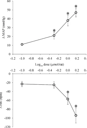
Fig. 2. Changes in MAP (MAP) and HR (HR) produced by H2O2 (0.1,
0.5, 1.0 and 1.5mol) injected into the 4th V. Abscissa represents the log dose
of H2O2. ‘*’ Different from vehicle (ANOVA followed by Bonferronit-test,
p< 0.05);n= 6.
3. Results
3.1. Effects of H
2O
2injected into the 4th V on MAP and HR
Injections of H
2O
2(0.5, 1.0 and 1.5
mol/rat) into the 4th
V produced a dose-dependent increase in MAP (
Fig. 2
). Mean
arterial pressure peaked from 20 to 100 s after the injections of
H
2O
2into the 4th V and returned to baseline pre-injection level
from 5 to 10 min after the injections.
Injections of H
2O
2(1.0 and 1.5
mol/rat) into the 4th V also
produced bradycardia that peaked between 2 and 5 min after the
injections (
Fig. 2
). The bradycardia was maintained for at least
24 h (but not for 48 h) with the 1.5
mol dose of H
2O
2into the
4th V, while with the 1
mol dose, HR had already returned to
baseline pre-injection level 24 h after the injection (
Table 1
).
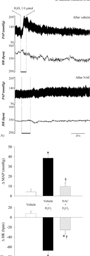
3.2. Effect of the pre-treatment with NAC into 4th V on
central H
2O
2-induced pressor and bradycardic responses
The treatment with NAC (250 nmol/1
L/rat) into the 4th
Table 1
Baseline level of mean arterial pressure (MAP) and heart rate (HR) before, 24 and 48 h after the injections of H2O2(0.1, 0.5, 1.0 and 1.5mol) or PBS into 4th V
Treatment Before injections 24 h after injections 48 h after injections
MAP (mmHg) HR (bpm) MAP (mmHg) HR (bpm) MAP (mmHg) HR (bpm)
Vehicle (PBS) 115±4 381±13 112±4 372±13 – –
0.1–1.0mol 111±3 349±12 117±4 363±13 – –
1.5mol 113±3 366±11 118±4 292±8* 128±9 370±13
Rats treated with vehicle received four injections of PBS. Rats treated with H2O20.1–1.0mol received one injection of PBS and injections of H2O2at the doses
0.1, 0.5 and 1.0mol and rats treated with H2O21.5mol received one injection of PBS and one injection of H2O2at the dose of 1.5mol. ‘*’ Different from
before the injection of H2O2or from vehicle treated rats (ANOVA followed by Student–Newman–Keuls test for comparisons in different treatments or pairedt-test
for comparisons before and after the treatment,p< 0.05). vehicle,n= 5, H2O2, 0.1–1.0mol,n= 6, H2O21.5mol,n= 8 (48 h after injections,n= 4).
(354
±
14 bpm before versus 353
±
13 after;
p
= 0.854;
non-paired
t-test).
The treatment with NAC abolished the pressor response
(vehicle: 38
±
4 mmHg versus NAC: 10
±
4 mmHg;
p
< 0.001;
n
= 7; non-paired
t-test), and attenuated the bradycardic
response by 61% (vehicle:
−67
±
7 bpm versus NAC:
−26
±
8 bpm;
p
= 0.002;
n
= 7; non-paired
t-test) induced by
H
2O
2(1
mol/0.5
L/rat) into the 4th V (
Fig. 3
).
3.3. Effect of the pre-treatment with catalase into the 4th V
on central H
2O
2-induced pressor and bradycardic
responses
Catalase (500 UEA/1
L/rat) into the 4th V did not affect
baseline MAP (118
±
3 mmHg before versus 116
±
5 mmHg
after, paired
t-test;
n
= 6;
p
= 0.685) but slightly reduced
base-line HR (377
±
19 bpm before versus 345
±
14 bpm after;
n
= 6;
paired
t-test,
p
= 0.005). The pre-treatment with catalase
abol-ished the pressor response (vehicle: 38
±
5 mmHg versus
cata-lase: 9
±
2 mmHg;
p
< 0.001;
n
= 6; ANOVA one way
fol-lowed by Student–Newman–Keuls) and bradycardic response
(vehicle:
−
51
±
12 bpm versus catalase:
−
6
±
4 bpm;
p
= 0.004;
n
= 6; ANOVA one way followed by Student–Newman–Keuls)
induced by H
2O
2(1.0
mol/0.5
L/rat) into the 4th V (
Fig. 4
).
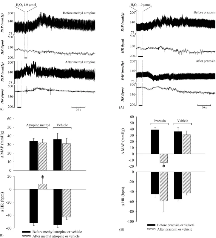
3.4. Effect of i.v. atropine on central H
2O
2-induced pressor
and bradycardic responses
Intravenous injection of the muscarinic receptor blocker
atropine methyl bromide (1.0 mg/kg of body weight) increased
HR (331
±
9 bpm before versus 428
±
9 bpm after atropine,
n
= 11; paired
t-test,
p
< 0.001) and MAP (117
±
13 mmHg
before versus 125
±
3 mmHg after atropine,
n
= 11; paired
t-test,
p
= 0.024). Pre-treatment with atropine completely blocked the
bradycardic response to injection of 1
mol of H
2O
2into the
4th V, but did not affect the pressor response (
Fig. 5
).
3.5. Effect of i.v. prazosin on central H
2O
2-induced pressor
and bradycardic responses
Intravenous administration of the
␣1
adrenergic receptor
blocker prazosin (1.0 mg/kg of body weight) increased HR (from
312
±
15 to 430
±
22 bpm,
n
= 6; paired
t-test,
p
= 0.002)
with-out changing baseline MAP (from 108
±
1 to 102
±
2 mmHg;
paired
t-test,
p
= 0.142). Instead of the pressor responses
pro-duced by H
2O
2into the 4th V in control tests, after
i.v.
prazosin,
H
2O
2into the 4th V reduced MAP (
Fig. 6
). The bradycardia to
H
2O
2in the 4th V was not affected by
i.v.
prazosin (
Fig. 6
).
4. Discussion
The present results show that H
2O
2injected into the 4th
V simultaneous and through independent mechanisms
acti-vates sympathetic and parasympathetic systems inducing
pres-sor and bradycardic responses. The prespres-sor responses are
tran-sitory (less than 1 h), while bradycardia with the high dose
(1.5
mol/rat) lasted for 24 h. These responses were abolished
(pressor response) or reduced (bradycardia) by pre-treatment
with the anti-oxidant NAC suggesting that they depend on
cen-tral increases in ROS.
The peripheral blockade of muscarinic receptors with
atropine abolished H
2O
2-induced bradycardia, without
chang-ing the pressor response, which suggests that H
2O
2-induced
bradycardia is totally dependent on increases in vagal
dis-charges. On the other hand, peripheral blockade of
␣1
adreno-ceptors with prazosin abolished the pressor response elicited by
H
2O
2, suggesting that increased ROS in the brainstem stimulate
sympathetic discharges causing vasoconstriction and increase in
arterial pressure. In spite of the blockade of the pressor response,
the bradycardia produced by H
2O
2was not modified by
i.v.
pra-zosin, which suggests that the bradycardia is not the result of
baroreflex activation. Moreover, after prazosin, H
2O
2into the
4th V induced a small hypotension, probably a consequence of
the bradycardia still present.
Differently from the transitory (less than 1 h) pressor
responses, the bradycardia with the high dose of H
2O
2into the
4th was still present 24 h later, which suggests that
parasympa-thetic tone remains modified for at least 24 h. The bradycardia
is completely reversed and HR return to control levels 48 h after
H
2O
2injections, which suggests that bradycardia could result
from an irreversible oxidation of pivotal structures involved in
the neurotransmission that are replaced by turnover from 24 to
48 h after H
2O
2injections. The different time-course of pressor
and bradycardic responses and the effects of
i.v.
prazosin and
atropine suggest that independent mechanisms are activated by
H
2O
2into the 4th V to produce these two responses.
Fig. 3. (A) Recordings of pulsatile arterial pressure (PAP) and HR in a rat of the group that received NAC (250 nmol/1L/rat) into the 4th V prior to H2O2
(1.0mol/0.5L/rat) in the same place; (B) changes in MAP (MAP) and HR
(HR) produced by H2O2(1.0mol/0.5L/rat) injected into the 4th V after
vehicle or NAC injection in the same place. NAC:n= 6; vehicle:n= 6. * Different from the vehicle;†different from H2O2after vehicle (p< 0.05, ANOVA one way
followed by Student–Newman–Keuls test).
Fig. 4. (A) Recordings of pulsatile arterial pressure (PAP) and HR in a rat of the group that received catalase (500 UAE/1L/rat) into the 4th V prior to
H2O2(1.0mol/0.5L/rat) in the same place; (B) changes in MAP (MAP)
and HR (HR) produced by H2O2(1.0mol/0.5L/rat) injected into the 4th
V before and after injection of catalase or vehicle in the same place. Cata-lase:n= 6; vehicle:n= 6.‘*’ Different from the vehicle;†different from H
2O2
Fig. 5. (A) Recordings of pulsatile arterial pressure (PAP) and HR in a rat of the group that received atropinei.v.prior to H2O2 (1.0mol/0.5L/rat)
into the 4th V; (B) changes in MAP (MAP) and HR (HR) produced by H2O2(1.0mol/0.2L/rat) injected into the 4th V before and after injections
of atropine methyl (1 mg/kg b.w.) or vehiclei.v. Atropine: n= 11; vehicle:
n= 6. ‘*’ Different from the responses before atropine methyl and after
pre-treatment with vehicle (p< 0.05, pairedt-test and ANOVA one way followed by Student–Newman–Keuls test, respectively).
RVLM, caudal ventrolateral medulla (CVLM) and the nucleus
ambiguous. The RVLM controls the sympathetic discharges
activating pre-ganglionic sympathetic neurons in the spinal cord,
while the nucleus ambiguous controls parasympathetic activity
Fig. 6. (A) Recordings of pulsatile arterial pressure (PAP) and HR in a rat of the group that received prazosini.v.prior to H2O2 (1.0mol/0.5L/rat)
into the 4th V; (B) changes in MAP (MAP) and HR (HR) produced by H2O2(1.0mol/0.2L/rat) injected into the 4th V before and after injections of
prazosin (1 mg/kg b.w.) or vehiclei.v. Prazosin:n= 6; vehicle:n= 6. ‘*’ Different
from the responses before prazosin and after pre-treatment with vehicle (p< 0.05, pairedt-test and ANOVA one way followed by Student–Newman–Keuls test, respectively).
[36]
. The RVLM and nucleus ambiguous are influenced by
sig-nals arising from the NTS, the site of the first synapse of baro
and chemoreceptor afferents
[26,27,37]
. The H
2O
2injected into
nucleus of the vagus (see
Fig. 1
). The stimulation of these areas
by H
2O
2or any other ROS that result from H
2O
2into the 4th
V may activate the nucleus ambiguous and the RVLM causing
increases in sympathetic activity and cardiac parasympathetic
tone, producing pressor and bradycardic responses.
By increasing the rate of endogenous or exogenous H
2O
2conversion to H
2O and O
2, the injection of catalase into the 4th V
almost abolished the pressor and bradycardic responses to H
2O
2into the 4th V. This result suggests that any increase in local O
2availability produced H
2O
2injection into the 4th V is not the
cause of the cardiovascular responses to the H
2O
2. In addition,
injection of the low molecular weight anti-oxidant NAC, a
scav-enger of ROS that reacts promptly with HO
•but slowly with
H
2O
2[2]
, abolished the pressor response and reduced
brady-cardia induced by H
2O
2. Taken together, these results suggest
that H
2O
2and/or its electrophilic metabolites into the 4th V or
in surrounding tissues activate hindbrain pathways that increase
sympathetic and parasympathetic activity inducing pressor and
bradycardic responses.
Eventual neuronal lesions by H
2O
2injections are probably
not the cause of the effects described in the present study. A
previous study demonstrated that superfusion of spinal chord
slices for 3 min with 0.3 mmol/L of H
2O
2(a dose 300-fold
higher than the median dose used in the present study) had
no effect on excitatory or inhibitory post-synaptic potentials in
whole cell recording of neurons
[25]
. Additionally, superfusion
with 1 mmol/L of H
2O
2produced an increase in the peak
ampli-tude of the excitatory post-synaptic potentials that was reversed
after washing the tissue. Such finds suggest no irreversible
dam-age of neurons exposed to H
2O
2concentration around
1000-fold higher than the mean dose used in the present study. The
dose–response effects of H
2O
2injected into the 4th V, the
recov-ery of MAP and HR to pre-injection levels and the blockade or
the reduction of the responses after the treatment with NAC
and catalase also suggest that the effects of H
2O
2observed in
the present study are due to a physiological or
pharmacologi-cal action in brain, rather than to non-specific damage of neural
tissue.
The neural or cellular mechanisms activated by H
2O
2to
pro-duce cardiovascular responses are still not clear. The present
results strongly suggest that oxidative modifications in the
hind-brain activate neural mechanisms involved with sympathetic
and parasympathetic control. Previous studies have shown that
H
2O
2might change cell membrane conductance to potassium
[35]
or calcium
[28]
. Evidence also suggests that H
2O
2and
other ROS can increase the availability of excitatory amino acids
into the synaptic cleft
[6,30,38]
. Similar to the present results
with H
2O
2injected into the 4th V, injections of glutamate into
the NTS of unanesthetized rats induce pressor and bradycardic
responses due to simultaneous activation of sympathetic and
parasympathetic systems
[10]
. Therefore similar mechanisms
may be shared by H
2O
2and glutamate to produce
cardiovascu-lar responses and perhaps the increase of glutamate release may
play a role on cardiovascular responses to H
2O
2into the 4th
V. Changes in ion conductance by neuron membrane and/or in
neurotransmission at hindbrain level are possibilities that need
further investigation.
Previous studies have also proposed the participation of ROS
in the control of arterial pressure in physiological and
pathologi-cal conditions. Increases in the generation of free radipathologi-cals,
espe-cially superoxide, into the central nervous system is suggested to
increase sympathetic activity
[18,40,43,45]
. A close relationship
between hypertension and increases in ROS generation in
dif-ferent tissues like endothelium, vessel smooth muscles or even
in some parts of the central nervous system like the
subforni-cal organ has been reported
[5,7,12,14,15,20,44,24,39,41,46]
.
Injections of the SOD mimetic 4-hydroxyl 2,2,6,6-tetramethyl
peperidine-1-oxyl (tempol) into the RVLM caused a
dose-dependent fall in the arterial pressure in spontaneously
hyperten-sive rats (SHR) without changing arterial pressure in
normoten-sive rats
[22]
. Injection of the SOD enzyme into the RVLM
of pigs reduced renal sympathetic nerve activity and arterial
pressure
[43]
. Moreover,
i.v.
injection of tempol reduced
arte-rial pressure and heart rate
[42]
whereas continuous (1 week)
intracisternal infusion of tempol did not affect these parameters
in normotensive rats
[21]
. Although these studies suggest that
imbalances in ROS production may play a role in hypertension,
the mechanisms involved are still not clear because the studies
did not show if the decrease in arterial pressure produced by
tempol or SOD is dependent on reduction of O
2•−, increases
in H
2O
2formation or both. According to the present results,
an oxidative burst produced by injections of H
2O
2into the 4th
V activate hindbrain mechanisms related to cardiovascular
con-trol increasing simultaneously and through independent
mech-anisms sympathetic and parasympathetic discharges to produce
pressor and bradycardic responses.
Acknowledgments
The authors thank Dr. Gus Schoorlemmer for his comments
and suggestions that helped to improve the manuscript,
Regi-naldo C. Queiroz, Silas P. Barbosa and Silvia F´oglia for expert
technical assistance and Silvana A. D. Malavolta for secretarial
assistance. We also thank Ana L.V. de Oliveira for animal care.
This research was supported by public funding from Fundac¸˜ao
de Amparo `a Pesquisa do Estado de S˜ao Paulo (FAPESP),
Con-selho Nacional de Pesquisa (CNPq)/PRONEX and Coordenac¸˜ao
de Aperfeic¸oamento de Pessoal de N´ıvel Superior (Capes).
References
[1] V. Adler, Z. Yin, K.D. Tew, Z. Ronai, Role of redox potential and reactive oxygen species in stress signaling, Oncogene 18 (1999) 6104–6111. [2] O.I. Aruoma, B. Halliwell, B.M. Hoey, J. Butler, The antioxidant action
ofN-acetylcysteine: its reaction with hydrogen peroxide, hydroxyl radical, superoxide, and hypochlorous acid, Free Rad. Biol. Med. 6 (1989) 593–597. [3] J.M. Auerbach, M. Segal, Peroxide modulation of slow onset potentiation
in rat hippocampus, J. Neurosci. 17 (1997) 8695–8701.
[4] M.V. Avshalumov, M.E. Rice, NMDA receptor activation mediates hydro-gen peroxide-induced pathophysiology in rat hippocampal slices, J. Neu-rophysiol. 87 (2002) 2896–2903.
[5] M.A. Bayorh, A.A. Ganafa, R.R. Socci, N. Silvestrov, I.K. Abukhalaf, The role of oxidative stress in salt-induced hypertension, Am. J. Hypertens. 17 (2004) 31–36.
[7] H. Cai, D.G. Harrison, Endothelial dysfunction in cardiovascular diseases: the role of oxidant stress, Circ. Res. 87 (2000) 840–844.
[8] A. Ceriello, D. Giugliano, A. Quatraro, P.J. Lefebvre, Anti-oxidants show an anti-hypertensive effect in diabetic and hypertensive subjects, Clin. Sci. (Lond.) 81 (1991) 739–742.
[9] B.T. Chen, M.V. Avshalumov, M.E. Rice, H(2)O(2) is a novel, endoge-nous modulator of synaptic dopamine release, J. Neurophysiol. 85 (2001) 2468–2476.
[10] E. Colombari, J.V. Menani, W.T. Talman, Commissural NTS contributes to pressor responses to glutamate injected into the medial NTS of awake rats, Am. J. Physiol. 270 (1996) R1220–R1225.
[11] E. Colombari, M.A. Sato, S.L. Cravo, C.T. Bergamaschi, R.R. Campos Jr., O.U. Lopes, Role of the medulla oblongata in hypertension, Hypertension 38 (2001) 549–554.
[12] C. Csonka, T. Pataki, P. Kovacs, S.L. Muller, M.L. Schroeter, A. Tosaki, I.E. Blasig, Effects of oxidative stress on the expression of antioxidative defense enzymes in spontaneously hypertensive rat hearts, Free Rad. Biol. Med. 29 (2000) 612–619.
[13] R.A. Dampney, Functional organization of central pathways regulating the cardiovascular system, Physiol. Rev. 74 (1994) 323–364.
[14] N.S. Dhalla, R.M. Temsah, T. Netticadan, Role of oxidative stress in car-diovascular diseases, J. Hypertens. 18 (2000) 655–673.
[15] A.D. Dobrian, S.D. Schriver, R.L. Prewitt, Role of angiotensin II and free radicals in blood pressure regulation in a rat model of renal hypertension, Hypertension 38 (2001) 361–366.
[16] M.V. Frantseva, J.L. Perez Velazquez, P.L. Carlen, Changes in membrane and synaptic properties of thalamocortical circuitry caused by hydrogen peroxide, J. Neurophysiol. 80 (1998) 1317–1326.
[17] H.F. Galley, J. Thornton, P.D. Howdle, B.E. Walker, N.R. Webster, Combi-nation oral antioxidant supplementation reduces blood pressure, Clin. Sci. (Lond.) 92 (1997) 361–365.
[18] L. Gao, W. Wang, Y.L. Li, H.D. Schultz, D. Liu, K.G. Cornish, I.H. Zucker, Superoxide mediates sympathoexcitation in heart failure: roles of angiotensin II and NAD(P)H oxidase, Circ. Res. 95 (2004) 937–944. [19] B. Halliwell, Reactive oxygen species and the central nervous system, J.
Neurochem. 59 (1992) 1609–1623.
[20] H.J. Hong, G. Hsiao, T.H. Cheng, M.H. Yen, Supplemention with tetrahy-drobiopterin suppresses the development of hypertension in spontaneously hypertensive rats, Hypertension 38 (2001) 1044–1048.
[21] Y. Kimura, Y. Hirooka, Y. Sagara, K. Ito, T. Kishi, H. Shimokawa, A. Takeshita, K. Sunagawa, Overexpression of inducible nitric oxide synthase in rostral ventrolateral medulla causes hypertension and sympa-thoexcitation via an increase in oxidative stress, Circ. Res. 96 (2005) 252– 260.
[22] T. Kishi, Y. Hirooka, Y. Kimura, K. Ito, H. Shimokawa, A. Takeshita, Increased reactive oxygen species in rostral ventrolateral medulla con-tribute to neural mechanisms of hypertension in stroke-prone spontaneously hypertensive rats, Circulation 109 (2004) 2357–2362.
[23] F. Lacy, D.T. O’Connor, G.W. Schmid-Schonbein, Plasma hydrogen perox-ide production in hypertensives and normotensive subjects at genetic risk of hypertension, J. Hypertens. 16 (1998) 291–303.
[24] L.O. Lerman, K.A. Nath, M. Rodriguez-Porcel, J.D. Krier, R.S. Schwartz, C. Napoli, J.C. Romero, Increased oxidative stress in experimental reno-vascular hypertension, Hypertension 37 (2001) 541–546.
[25] H.H. Lin, C.H. Chen, W.K. Hsieh, T.H. Chiu, C.C. Lai, Hydrogen peroxide increases the activity of rat sympathetic preganglionic neurons in vivo and in vitro, Neuroscience 121 (2003) 641–647.
[26] A.D. Loewy, Central autonomic pathways, in: A.D. Loewy, K.M. Spyer (Eds.), Central Regulation of Autonimic Functions, Oxford University Press, 1990, pp. 88–103.
[27] B.H. Machado, H. Mauad, D.A. Chianca Junior, A.S. Haibara, E. Colom-bari, Autonomic processing of the cardiovascular reflexes in the nucleus tractus solitarii, Braz. J. Med. Biol. Res. 30 (1997) 533–543.
[28] H.F. Moghadam, W. Winlow, L.L. Moroz, Effects of hydrogen peroxide and nitric oxide (NO) on neuronal discharges and intracellular calcium concentration in the molluscan CNS, Acta Biol. Hung. 46 (1995) 145–153. [29] G. Paxinos, C. Watson, The Rat Brain in Stereotaxic Coordinates,
Aca-demic Press, 1997.
[30] D.E. Pellegrini-Giampietro, G. Cherici, M. Alesiani, V. Carla, F. Moroni, Excitatory amino acid release from rat hippocampal slices as a consequence of free-radical formation, J. Neurochem. 51 (1988) 1960–1963. [31] T. Pellmar, Electrophysiological correlates of peroxide damage in guinea
pig hippocampus in vitro, Brain Res. 364 (1986) 377–381.
[32] L. Pogan, L. Garneau, P. Bissonnette, L. Wu, R. Sauve, Abnormal Ca2+ signalling in vascular endothelial cells from spontaneously hypertensive rats: role of free radicals, J. Hypertens. 19 (2001) 721–730.
[33] D.J. Reis, The brain and hypertension: reflections on 35 years of inquiry into the neurobiology of the circulation, Circulation 70 (1984) III31–III45. [34] S.G. Rhee, T.S. Chang, Y.S. Bae, S.R. Lee, S.W. Kang, Cellular regulation
by hydrogen peroxide, J. Am. Soc. Nephrol. 14 (2003) S211–S215. [35] V. Seutin, J. Scuvee-Moreau, L. Massotte, A. Dresse, Hydrogen
perox-ide hyperpolarizes rat CA1 pyramidal neurons by inducing an increase in potassium conductance, Brain Res. 683 (1995) 275–278.
[36] S.L. Stuesse, Origins of cardiac vagal preganglionic fibers: a retrograde transport study, Brain Res. 236 (1982) 15–25.
[37] A.F. Sved, F.J. Gordon, Amino acids as central neurotransmitters in the barorecepor reflex pathway, News Physiol. Sci. 9 (1994) 243–246. [38] D. Trotti, N.C. Danbolt, A. Volterra, Glutamate transporters are
oxidant-vulnerable: a molecular link between oxidative and excitotoxic neurode-generation? Trends Pharmacol. Sci. 19 (1998) 328–334.
[39] S. Vasdev, C.A. Ford, S. Parai, L. Longerich, V. Gadag, Dietary vitamin C supplementation lowers blood pressure in spontaneously hypertensive rats, Mol. Cell Biochem. 218 (2001) 97–103.
[40] G. Wang, J. Anrather, J. Huang, R.C. Speth, V.M. Pickel, C. Iadecola, NADPH oxidase contributes to angiotensin II signaling in the nucleus trac-tus solitarius, J. Neurosci. 24 (2004) 5516–5524.
[41] R. Wu, E. Millette, L. Wu, J. de Champlain, Role of oxidative stress in the development of hypertension in SHR, Am. J. Hypertens. 14 (2001) A134, Ref Type: Abstract.
[42] H. Xu, G.D. Fink, J.J. Galligan, Tempol lowers blood pressure and sympa-thetic nerve activity but not vascular O2−in DOCA-salt rats, Hypertension 43 (2004) 329–334.
[43] J. Zanzinger, J. Czachurski, Chronic oxidative stress in the RVLM modu-lates sympathetic control of circulation in pigs, Pflugers Arch. 439 (2000) 489–494.
[44] J. Zich, Antihypertensive effect of chronic antioxidant treatment byn -acetylcysteine in rats with l-NAME hypertension. Abstract Book Hyper-tension 2004 22 (2004) 13S. S˜ao Paulo. Ref Type: Abstract.
[45] M.C. Zimmerman, E. Lazartigues, J.A. Lang, P. Sinnayah, I.M. Ahmad, D.R. Spitz, R.L. Davisson, Superoxide mediates the actions of angiotensin II in the central nervous system, Circ. Res. 91 (2002) 1038–1045. [46] M.C. Zimmerman, E. Lazartigues, R.V. Sharma, R.L. Davisson,