The physio lo gical ro le o f AT
1
re ce pto rs
in the ve ntro late ral m e dulla
1Department of Physiology and Institute for Biomedical Research, University of Sydney, Sydney, Australia
2Howard Florey Institute of Experimental Physiology and Medicine, University of Melbourne, Melbourne, Australia
T. Tagawa1, M.A.P. Fontes1, P.D. Potts1, A.M. Allen2 and R.A.L. Dampney1
Abstract
Neurons in the rostral and caudal parts of the ventrolateral medulla (VLM) play a pivotal role in the regulation of sympathetic vasomotor activity and blood pressure. Studies in several species, including humans, have shown that these regions contain a high density of AT1 receptors specifically associated with neurons that regulate the sympa-thetic vasomotor outflow, or the secretion of vasopressin from the hypothalamus. It is well established that specific activation of AT1
receptors by application of exogenous angiotensin II in the rostral and caudal VLM excites sympathoexcitatory and sympathoinhibitory neu-rons, respectively, but the physiological role of these receptors in the normal synaptic regulation of VLM neurons is not known. In this paper we review studies which have defined the effects of specific activation or blockade of these receptors on cardiovascular function, and discuss what these findings tell us with regard to the physiological role of AT1 receptors in the VLM in the tonic and phasic regulation of
sympathetic vasomotor activity and blood pressure.
Co rre spo nde nce
R.A.L. Dampney
Department of Physiology, F13 University of Sydney Sydney, NSW 2006 Australia
Fax: + 61-2-9351-2058
E-mail: rogerd@ physiol.usyd.edu.au Presented at the III International Symposium on Vasoactive Peptides, Belo Horizonte, MG, Brasil, O ctober 8-10, 1999.
Research supported by the National Health and Medical Research Council and the National Heart Foundation of Australia. M.A.P. Fontes is the recipient of a CNPq fellowship.
Received November 26, 1999 Accepted February 2, 2000
Ke y wo rds
·Angiotensin receptors ·Central cardiovascular
pathways ·Blood pressure ·Hypothalamus ·Neurotransmitters
Intro ductio n
The ventrolateral medulla (VLM) con-tains several different groups of neurons that play a major role in cardiovascular regula-tion. They consist of i) sympathoexcitatory neurons in the rostral VLM that project di-rectly to the spinal sympathetic outflow, ii) sympathoinhibitory neurons in the caudal and intermediate parts of the VLM that project to and inhibit the rostral VLM sympathoex-citatory neurons, and iii) catecholamine-syn-thesising neurons of the A1 cells group that project to and excite vasopressin-synthesis-ing neurons in the supraoptic and paraven-tricular nuclei in the hypothalamus. Although
these three groups of neurons differ greatly with respect to their functional and anatomi-cal properties, one common feature is that
they all contain angiotensin type 1 (AT1)
receptors. Furthermore, as shown in Figure
1, AT1 receptors in the medulla are
specifi-cally located in the VLM and in the nucleus tractus solitarius (NTS), another region that also plays a crucial role in cardiovascular regulation. In contrast, there is a very low
density of AT1 receptors in other medullary
regions that subserve non-cardiovascular functions.
The striking association between AT1
mans (3-5). These receptors are primarily of
the AT1 subtype (6,7). In the rat, in vitro
autoradiography has revealed a relatively
low density of AT1 receptors in comparison
to other species (8). Nevertheless, a recent
study using an antibody against the AT1
receptor has confirmed the existence of these receptors on neurons in the VLM of the rat and has shown that many of these neurons are also catecholamine neurons of the C1 group (9). There appear to be no published receptor binding or immunohistochemical
studies showing the presence of AT2 or
Ang-(1-7) receptors in the VLM, although there is functional evidence in the rat for the exist-ence of these receptor subtypes (10,11).
Effe cts o f applicatio n o f e xo ge no us Ang II to VLM ne uro ns
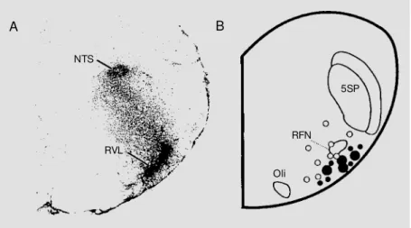
In 1988, two studies were published that demonstrated for the first time that Ang II can directly excite pressor neurons in the VLM. Allen et al. (4) reported that microin-jection of Ang II into the pressor region of the rostral VLM increased arterial pressure and heart rate, while Andreatta et al. (12) reported the same effect following applica-tion of Ang II to the nearby ventral surface. In a later and more detailed study by Sasaki and Dampney (13), it was found that the distribution of sites in the rostral VLM of the rabbit at which Ang II microinjection evoked increases in arterial pressure and renal sym-pathetic nerve activity closely matched the distribution of Ang II receptor binding sites (Figure 1), which, as discussed above, were
later demonstrated to be AT1 binding sites.
Consistent with this, Hirooka et al. (14) found that the pressor and sympathoexcitatory re-sponse evoked by microinjection of Ang II into the rostral VLM was prevented by prior
administration of the selective AT1 receptor
antagonist losartan into the same region (Fig-ure 2).
Similarly, studies in the rat have con-firmed that microinjection of Ang II into the of these receptors in cardiovascular
regula-tion. This brief review will begin with a
description of the distribution of AT1
recep-tors in the VLM, but will then discuss mainly recent studies which have provided informa-tion on the funcinforma-tional effects of activainforma-tion or blockade of these receptors, ranging from in vivo studies in whole animals to in vitro studies on single neurons. We shall then consider the fundamental question as to the
normal physiological role of VLM AT1
re-ceptors in cardiovascular function. In this review we will not consider the role of these receptors in brain regions outside the VLM, which have been discussed in detail in other recent reviews (1,2).
D istributio n o f AT1 re ce pto rs in the VLM
Studies using in vitro autoradiography have demonstrated a high density of angio-tensin (Ang) receptor binding sites in the VLM of most species studied, including
hu-Figure 1 - A, Distribution of Ang II receptor binding sites in the rostral VLM (RVL) of the rabbit, using in vitro autoradiography (adapted from Ref. 3 and reprinted w ith the kind permission of the publisher). B, Distribution of sites in the rostral VLM of the rabbit at w hich microinjections of Ang II (20 pmol in 20 nl) elicited rises in mean arterial pressure of 6-20 mmHg (small filled circles) or more than 20 mmHg (large filled circles). The open circles indicate sites at w hich no significant change in mean arterial pressure w as observed. (Adapted from Ref. 13, w ith the kind permission of the publisher). Note the close match betw een the receptor binding sites and the pressor sites. NTS, Nucleus tractus solitarius; RFN, retrofacial nucleus; 5SP, spinal trigeminal nucleus; Oli, inferior olivary nucleus.
A B
NTS
RVL
5SP
RFN
rostral VLM of this species also evoked a rise in arterial pressure (11,15). Furthermore, Chan and co-workers (16), using single-unit recording, found that many but not all rostral VLM neurons were excited by direct appli-cation of Ang II. These observations were confirmed by in vitro studies, which demon-strated using patch clamp recordings that Ang II evokes a depolarisation in some but not all spinally projecting neurons, an effect which is due to a reduction in potassium conductance (17). This effect was also blocked by administration of losartan,
dem-onstrating that it is mediated by AT1
recep-tors. A similar mechanism of action of Ang II on hypothalamic neurons has also been demonstrated (18).
With regard to the caudal and intermedi-ate VLM, Sasaki and Dampney (13) found that microinjection of Ang II into this region evoked a fall in arterial pressure and renal sympathetic nerve activity. As with the ros-tral VLM, the distribution of sites evoking cardiovascular responses closely matched
that of AT1 receptor binding sites (6). The
depressor and sympathoinhibitory response evoked by Ang II in this region can be ex-plained as a consequence of activation of inhibitory interneurons that project directly to rostral VLM sympathoexcitatory neurons (19). Such interneurons have been identified in the caudal and intermediate VLM of the rabbit and rat (20-22). Anatomical studies have shown that these neurons are not cate-cholamine neurons, although they are co-localised with catecholamine neurons (22, 23).
Allen et al. (24) found that microinjec-tion of Ang II into the caudal VLM also evoked a rise in the level of circulating vaso-pressin. This can be explained as a result of activation of caudal VLM neurons that project to and excite vasopressin-secreting neurons in the hypothalamic paraventricular and su-praoptic nuclei. Unlike caudal VLM neu-rons that project to the rostral VLM, the hypothalamus-projecting neurons in the
cau-Arterial pressure
(mmHg) 200
100
0 300 200 100
200
100
0 Heart
rate (bpm)
Integrated renal nerve activity (% control)
Ang II Ang II
1 min
Figure 2 - Chart recording show ing the effects on cardiovascular variables of microinjection of angiotensin II (Ang II, 40 pmol) into the pressor region in the rostral ventrolateral medulla before and after microinjection of losartan (1 nmol) into the same site (reprinted from Ref. 14, w ith the kind permission of the publisher).
dal VLM are mainly catecholamine neurons of the A1 group (25).
A recent study in the conscious rabbit used the method of immediate early gene expression to identify the population of neu-rons in the VLM that are activated by admin-istration of Ang II into the fourth ventricle (26). These experiments were performed in both intact and sinoaortic denervated rabbits (to eliminate possible secondary effects on immediate early gene expression arising from blood pressure changes induced by the Ang II administration). In both groups of animals, Fos, the protein product of the immediate early gene c-fos, was detected in neurons in both the NTS and VLM, with a distribution
that closely matched the distribution of AT1
receptors in this region (Figure 3). The neu-rons in the VLM that were activated by Ang II consisted of both catecholamine and non-catecholamine neurons, as shown by double-labeling for tyrosine hydroxylase immunore-activity (Figure 3). This study showed that approximately 60% of the rostral VLM
rons that expressed Fos in response to Ang II were catecholamine neurons. It is interesting to note that a similar proportion (63%) of rostral VLM neurons that express Fos in response to baroreceptor unloading in the conscious rabbit are also catecholamine neu-rons (27). Thus, the results of these two studies using different experimental ap-proaches indicate that although the majority
of rostral VLM neurons with functional AT1
receptors may be catecholamine neurons, this also appears to be the case for the gen-eral population of neurons within the rostral VLM that have a sympathoexcitatory func-tion.
Similarly, neurons within the intermedi-ate and caudal parts of the VLM that ex-pressed Fos in response to Ang II also con-sist of catecholamine and non-catecholamine neurons (26). Thus, these findings are con-sistent with the results of the physiological studies described above indicating that both catecholamine and non-catecholamine neu-rons in the intermediate and caudal VLM can be activated by exogenous Ang II. The former group are likely to be the A1 neurons that project to vasopressin-secreting neu-rons in the supraoptic and paraventricular nuclei in the hypothalamus, while the latter are presumably inhibitory interneurons that project to rostral VLM sympathoexcitatory neurons.
An important question is the specificity of action of Ang II in the VLM. Although the anatomical and functional studies
summa-rised above provide ample evidence that AT1
receptors are associated with presympathetic vasomotor neurons in the rostral VLM, it is not clear whether they are also associated with neurons subserving other functions, such as respiratory regulation or antinociception, since such neurons are also located within this region (19). To test the possibility that
AT1 receptors in the rostral VLM may be
associated with non-cardiovascular func-tions, Li et al. (28) determined the effects of microinjection of Ang II into the rostral VLM on phrenic nerve activity as well as arterial pressure. The results of these studies showed clearly that, although pressor responses evoked by microinjections of glutamate into the rostral VLM were invariably associated with changes in phrenic nerve activity, this was never the case with Ang II, even when large doses were injected. These results are consistent with the hypothesis that the pep-Figure 3 - Upper panel, Distribution of cells immunoreactive for Fos (indicated by small filled
circles) in the medulla oblongata at the level of the rostral VLM follow ing fourth ventricular infusion of Ang II in the conscious rabbit. Low er panel, Distribution of Fos-positive cells (small filled circles), cells immunoreactive for tyrosine hydroxylase (TH, a marker of cate-cholamine cells) (large open circles) and neurons immunoreactive for both Fos and TH (shaded circles enclosing small filled circles) in the rostral VLM in the region indicated by the rectangle in the upper panel. Vsp, Spinal nucleus of the trigeminal nucleus; RFN, retrofacial nucleus; TS, solitary tract. (Adapted from Ref. 26, w ith the kind permission of the publisher).
TS
Vsp
RFN
tide acts exclusively on vasomotor neurons in this region.
Effe cts o f blo ckade o f AT1 re ce pto rs in the VLM o n re sting sym pathe tic vaso mo to r activity
Sasaki and Dampney (13) first reported that unilateral microinjection of the peptidic
Ang receptor antagonist [Sar1,Thr8]Ang II
resulted in a fall in arterial pressure and renal sympathetic nerve activity, suggesting that endogenous Ang II is tonically released in the rostral VLM and contributes to the rest-ing activity of rostral VLM neurons and hence sympathetic activity. Later, Ito and Sved (29) showed that bilateral
microinjec-tions of [Sar1,Thr8]Ang II or another
pep-tidic antagonist, [Sar1,Ile8]Ang II, resulted in
a profound fall in arterial pressure, close to the level seen after complete blockade of the sympathetic vasomotor activity. On the ba-sis of these observations, Ito and Sved (29) suggested that endogenous Ang II makes a very major contribution to the maintenance of sympathetic vasomotor tone. Recently, we have confirmed this observation and ex-tended it by demonstrating that renal
sympa-thetic nerve activity is also profoundly re-duced (30). However, the sympathoinhibi-tory effects resulting from bilateral
injec-tions of [Sar1,Thr8]Ang II or [Sar1,Ile8]Ang
II into the rostral VLM were not altered by prior blockade of glutamate or GABA recep-tors in this region (Figure 4), indicating that they are not mediated by presynaptic modu-lation of tonic glutamatergic or GABAergic activity. Thus, these findings suggest that
[Sar1,Thr8]Ang II and [Sar1,Ile8]Ang II act
postsynaptically to inhibit the tonic activity of rostral sympathoexcitatory neurons.
However, [Sar1,Thr8]Ang II and [Sar1,
Ile8]Ang II are broad-spectrum Ang receptor
antagonists, and therefore block AT1, AT2
and Ang-(1-7) receptors. In contrast to the
effects of [Sar1,Thr8]Ang II or [Sar1,Ile8]Ang
II, however, selective blockade of AT1 or
AT2 receptors has little effect on resting
arte-rial pressure and renal sympathetic nerve activity (11,14), at least under normal condi-tions. On the other hand, bilateral selective blockade of Ang-(1-7) receptors in the ros-tral VLM of the rat does result in a signifi-cant fall in arterial pressure (11), but the hypotensive effect is much less than that
resulting from [Sar1,Thr8]Ang II and
Figure 4 - Example of the ef-fects on arterial pressure, heart rate and integrated renal sym-pathetic nerve activity (RSNA) evoked by bilateral microinjec-tions of [Sar1,Ile8]Ang II (Sarile) into the left (L) and right (R) ros-tral VLM , before and after bilat-eral injections of kynurenic acid (Kyn) plus bicuculline (Bic) into the same sites. Note that the depressor and sympathoinhibi-tory response evoked by [Sar1, Ile8]Ang II w as unaffected by prior injection of kynurenic acid plus bicuculline (reprinted from Ref. 30, w ith the kind permis-sion of the publisher). Arterial
pressure (mmHg)
200
100
0 500 400 300 200
100
0 Heart
rate (bpm)
Integrated RSNA (% control)
1 min
L R 20 min L R L R L R 20 min
Sarile (1 nmol)
Kyn (4.5 nmol)
Bic (200 pmol)
Sarile (1 nmol)
Arterial pressure
(mmHg) 300
100
0 500
400
300
150
100
0 Heart
rate (bpm)
Integrated RSNA (% baseline)
1 min
L R
Bic (0.2 nmol)
acsf (100 nl)
A
Figure 5 - Examples of the ef-fects on arterial pressure, heart rate and integrated renal sympa-t hesympa-t ic nerve acsympa-t ivisympa-t y (RSNA) evoked by bilateral microinjec-tions into the rostral VLM of ei-ther artificial cerebrospinal fluid (acsf, the vehicle solution) (A) or the AT1 receptor antagonist L-158,809 (B) after prior bilateral microinjections of the GABA re-cept or ant agonist bicuculline (Bic) into the same region. The paired arrow s indicate the times of the first and second bilateral injections. Note that there w as a decrease in arterial pressure, heart rate and RSNA follow ing injections of L-158,809, indicat-ing that there is a tonic excita-tory action on rostral VLM sym-pathoexcitatory neurons medi-ated by AT1 receptors under con-ditions w hen GABAergic inhibi-tory inputs are blocked.
Arterial pressure
(mmHg) 300
100
0 500 400
300
150
100
0 Heart
rate (bpm)
Integrated RSNA (% baseline)
1 min Bic
(0.2 nmol)
L-158,809 (1 nmol)
B
[Sar1,Ile8]Ang II. Thus, it seems likely that
the inhibitory effects of these compounds on rostral VLM sympathoexcitatory neurons are due to mechanisms that are independent of
AT1, AT2 or Ang-(1-7) receptors. Further
studies are required to elucidate these mechanisms.
neu-rons (19,31,32). It is clear, however, that these neurons are subject to a tonic GABAergic inhibition (33). In the presence of GABA receptor blockade, a powerful tonic excitatory effect on rostral VLM neurons is unmasked, resulting in a large increase of sympathetic activity and arterial pressure (30,34). Recently, we have obtained
evi-dence that AT1 receptors do make a small
contribution to the maintenance of the rest-ing activity of rostral VLM sympathoexcita-tory neurons, which can be revealed when the effect of tonic GABAergic inhibition of these neurons is removed. As shown in Fig-ure 5A, bilateral microinjections of bicucul-line into the rostral VLM pressor region resulted in a large and sustained rise in sym-pathetic activity and arterial pressure.
How-ever, when a selective AT1 receptor
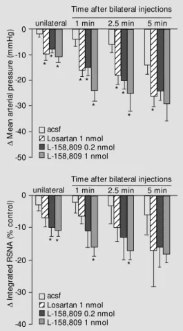
antago-nist (losartan or L-158,809, gift of Merck Co., Rahway, NJ, USA) was injected during the period when the arterial pressure and sympathetic activity were elevated follow-ing GABA receptor blockade in the rostral VLM, both arterial pressure and renal sym-pathetic nerve activity decreased substan-tially (Figures 5B, 6). Thus, tonic activation
of AT1 receptors may contribute to the
rest-ing activity of rostral VLM neurons, but this effect is only apparent under conditions where the tonic inhibition of these neurons is elimi-nated.
Effe cts o f blo ckade o f AT1 re ce pto rs in the VLM o n synaptic re gulatio n o f ro stral VLM sym patho e xcitato ry ne uro ns
Although there is abundant evidence, as
summarised above, that activation of AT1
receptors excites cardiovascular neurons in the VLM, the major question that remains is: what is the normal physiological role of these receptors in the synaptic regulation of these neurons? Synaptic excitation of rostral VLM neurons that occurs in response to stimulation of a wide variety of peripheral
receptors as well as some supramedullary regions appears to be mediated primarily by glutamate receptors, since it is blocked by kynurenic acid, a broad-spectrum ionotropic glutamate receptor antagonist (19,31,35-38). At the same time, there is evidence that some excitatory inputs to rostral VLM sympatho-excitatory neurons are mediated by non-glu-tamate receptors. In particular, Kiely and Gordon (39) found that the pressor response evoked by stimulation of certain hypotha-lamic areas (the paraventricular nucleus (PVN) and the perifornical area) is reduced by non-specific blockade of synaptic trans-mission in the rostral VLM, but not by block-ade of glutamate receptors in this region.
These observations therefore raise the possibility that excitatory inputs to the ros-tral VLM sympathoexcitatory neurons are
mediated by AT1 receptors. To test this
hy-pothesis, we have recently determined the
Figure 6 - Histogram summaris-ing the effects of unilateral and bilateral injections into the ros-tral VLM of either artificial cere-brospinal fluid (acsf, N = 6) or the AT1 receptor antagonists lo-sartan (1 nmol, N = 6) or L-158,809 (0.2 nmol, N = 6 or 1.0 nmol, N = 7) on arterial pressure and renal sympathetic nerve ac-tivity (RSNA). In all cases the GABA receptor antagonist bicu-culline had been injected bilater-ally into the rostral VLM approxi-mately 10 min before the injec-tions of these different com-pounds. * P<0.05 for the effects of losartan or L-158,809 com-pared w ith that of the vehicle solution (acsf). D M e a n a rt e ri a l p re s s u re ( m m H g ) 0 -20 -30 -40 -50 -10
Time after bilateral injections unilateral12 1 min 2.5 min 5 min
12 12 12 12 12 123 123 123 123 123 123 123 123 12 12 12 12 12 12 12 12 12 12 12 12 12 12 12 12 12 12 12 12 12 12 12 12 acsf
Losartan 1 nmol L-158,809 0.2 nmol L-158,809 1 nmol
Time after bilateral injections unilateral12 1 min 2.5 min 5 min
12 12 12 12 123 123 123 123 123 12 12 12 12 12 12 12 123 123 123 123 123 123 123 123 123 123 123 12 12 acsf
Losartan 1 nmol L-158,809 0.2 nmol L-158,809 1 nmol
effect of selective blockade of AT1 receptors in the rostral VLM on the cardiovascular response evoked by activation of the hypo-thalamic PVN (40). For this purpose, micro-injections of bicuculline were made into the PVN, since this has been shown to be a highly effective and reproducible means of activating PVN neurons (41,42).
This study demonstrated that the increase in arterial pressure, heart rate and renal sym-pathetic nerve activity that was evoked by a unilateral microinjection of bicuculline into the PVN was reduced by approximately 50% after microinjection of the potent neuroin-hibitory compound muscimol into the ipsi-lateral rostral VLM. However, the response was unaffected by muscimol injection into the contralateral PVN, which is consistent with anatomical studies showing that the descending pathway from the PVN to the rostral VLM is almost entirely ipsilateral (43). Thus, these experiments demonstrated that the pressor and sympathoexcitatory re-sponse evoked from the PVN is partly medi-ated by the ipsilateral rostral VLM, and partly via a descending pathway that is independ-ent of the rostral VLM. The most interesting observation, however, was that the response evoked from the PVN was attenuated by about 50% following microinjection of the
selective AT1 receptor antagonists losartan
or L-158,809 into the ipsilateral rostral VLM, but was unaffected by microinjection of kynurenic acid into the same region. In con-trast to the response evoked from the hypo-thalamus, the sympathoexcitatory response evoked reflexly by stimulation of the sciatic nerve (the somatosympathoexcitatory reflex) was unaffected by losartan or L-158,809 in the rostral VLM, but was abolished by kynurenic acid in this region. Thus, these findings suggest that the excitatory input to rostral VLM sympathoexcitatory neurons arising from activation of the hypothalamic PVN, but not that reflexly evoked by stimu-lation of sciatic nerve afferents, is mediated
by AT1 receptors.
Many questions remain unanswered,
however, as to the role of AT1 receptors in
the rostral VLM. For example, assuming that Ang II is the endogenous ligand for these receptors, what is the source of Ang II that is released in response to activation of the PVN? There is evidence that both the PVN and the lateral parabrachial nucleus contain neurons that are immunoreactive for Ang II (44). Both of these nuclei contain neurons that project to the rostral VLM (19), but it has not been shown whether some of these neurons are also Ang II-containing neurons. Alterna-tively, Lippoldt et al. (45) have proposed that Ang II can be formed in the extracellular fluid from angiotensinogen, which itself is produced in astrocytes. Thus, it is possible that Ang II in the rostral VLM is not formed within the neurons, but is instead taken up from the extracellular fluid into nerve termi-nals from which it is subsequently released. Another important question is whether Ang II is the sole neurotransmitter released from nerve terminals in response to PVN activation, or else is a co-transmitter together with another neurotransmitter such as gluta-mate. This possibility is consistent with the fact that Ang II and glutamate appear to be co-transmitters in the angiotensinergic path-way from the subfornical organ to the PVN (2).
A further question is whether AT1
recep-tors in the rostral VLM mediate synaptic excitatory inputs only from the PVN, or whether they have a more generalised role in synaptic transmission. Given that excitatory inputs to the rostral VLM sympathoexcitato-ry neurons that originate from peripheral receptors and/or are relayed via the NTS are, at least in every case so far examined, de-pendent upon glutamate receptors in the ros-tral VLM (e.g., Refs. 35-38 and 40), it is
possible that the AT1 receptors have a much
in-clude all supramedullary inputs, because it is established that some excitatory inputs to rostral VLM sympathoexcitatory neurons arising from or passing through the hypo-thalamus are also glutamatergic (35).
A final possibility that should be briefly
considered is whether AT1 receptors in the
rostral VLM play a functionally important role only under conditions of stress. This would be consistent with the finding that they mediate sympathoexcitatory responses originating from the PVN, since it is well known that this nucleus is activated during stress. Further, in a recent study in the con-scious rabbit it was found that administra-tion of losartan into the fourth ventricle re-sulted in an increase in renal sympathetic nerve activity under conditions of barore-ceptor unloading and hypoxia, but not under
Re fe re nce s
1. Allen AM , M acGregor DP, M cKinley M J & M endelsohn FAO (1999). Angiotensin II receptors in the human brain. Regulatory Peptides, 79: 1-7.
2. Ferguson AV & Washburn DLS (1998). Angiotensin II - a peptidergic neurotrans-mitter in central autonomic pathw ays.
Progress in Neurobiology, 54: 169-192. 3. M endelsohn FAO, Allen AM , Clevers AJ,
Denton DA, Tarjan E & M cKinley M J (1988). Localization of angiotensin II re-ceptor binding in rabbit brain by in vitro
autoradiography. Journal of Comparative Neurology,270: 372-384.
4. Allen AM , Dampney RAL & M endelsohn FAO (1988). Angiotensin receptor binding and pressor effects in cat subretrofacial nucleus. American Journal of Physiology, 255: H1011-H1017.
5. Allen AM , Chai SY, Clevers J, M cKinley M J, Paxinos G & M endelsohn FAO (1988). Localization and characterization of angio-tensin II receptor binding and angioangio-tensin converting enzyme in the human medulla oblongata. Journal of Comparative Neu-rology,269: 249-264.
6. Aldred GP, Chai SY, Song K, Zhuo J, M acGregor DP & M endelsohn FAO (1993). Distribution of angiotensin II re-ceptor subtypes in the rabbit brain. Regu-latory Peptides, 44: 119-130.
7. M acGregor DP, M urone C, Song K, Allen AM , Paxinos G & M endelsohn FAO (1995). Angiotensin II receptor subtypes in the human central nervous system.
Brain Research,675: 231-240.
8. Song K, Allen AM , Paxinos G & M endelsohn FAO (1992). M apping of an-giotensin II receptor subtype heterogene-ity in rat brain. Journal of Comparative Neurology,316: 467-484.
9. Yang SN, Lippoldt A, Jansson A, Phillips M I, Ganten D & Fuxe K (1997). Localiza-tion of angiotensin II AT1 receptor-like im-munoreactivity in catecholaminergic neu-rons of the rat medulla oblongata. Neuro-science, 81: 503-515.
10. Lin KS, Chan JY & Chan SH (1997). In-volvement of AT2 receptors at NRVL in tonic baroreflex suppression by endoge-nous angiotensins. American Journal of Physiology,272: H2204-H2210. 11. Fontes M A, Silva LC, Campagnole-Santos
M J, Khosla M C, Guertzenstein PG & Santos RA (1994). Evidence that angio-tensin-(1-7) plays a role in the central con-trol of blood pressure at the ventro-lateral medulla acting through specific receptors.
Brain Research,665: 175-180.
12. Andreatta SH, Averill DB, Santos RA & Ferrario CM (1988). The ventrolateral me-dulla. A new site of action of the
renin-angiotensin system. Hypertension, 11 (Suppl I): I-163-I-166.
13. Sasaki S & Dampney RAL (1990). Tonic cardiovascular effects of angiotensin II in the ventrolateral medulla. Hypertension, 15: 274-283.
14. Hirooka Y, Potts PD & Dampney RAL (1997). Role of angiotensin II receptor subtypes in mediating the sympathoexci-tatory effects of exogenous and endoge-nous angiotensin peptides in the rostral ventrolateral medulla of the rabbit. Brain Research,772: 107-114.
15. M uratani H, Averill DB & Ferrario CM (1991). Effects of angiotensin II in ventro-lateral medulla of spontaneously hyper-tensive rats. American Journal of Physiol-ogy,260: R977-R984.
16. Chan RK, Chan YS & Wong TM (1991). Responses of cardiovascular neurons in the rostral ventrolateral medulla of the normotensive Wistar Kyoto and sponta-neously hypertensive rats to iontophoretic application of angiotensin II. Brain Re-search, 556: 145-150.
17. Li YW & Guyenet PG (1996). Angiotensin II decreases a resting K+ conductance in rat bulbospinal neurons of the C1 area.
Circulation Research,78: 274-282. 18. Sumners C, Zhu M , Gelband CH & Posner
P (1996). Angiotensin II type 1 receptor resting conditions (46,47). Although the site
of action of losartan in these studies is un-known (it could be the NTS, caudal or rostral VLM or even A5 area in the pons), the results are at least consistent with the
hypo-thesis that medullary AT1 receptors are
func-tionally important only under conditions of stress, such as hypotension or hypoxia. At present, however, this hypothesis is specula-tive.
In conclusion, the results of studies to
date have demonstrated that AT1 receptors in
modulation of neuronal K+ and Ca2+ cur-rents: intracellular mechanisms. American Journal of Physiology, 271: C154-C163. 19. Dampney RAL (1994). Functional
organi-zation of central pathw ays regulating the cardiovascular system. Physiological Re-view s,74: 323-364.
20. Agarw al SK & Calaresu FR (1991). M ono-synaptic connection from caudal to rostral ventrolateral medulla in the baroreceptor reflex pathw ay. Brain Research,555: 70-74.
21. Gieroba ZJ, Li Y-W & Blessing WW (1992). Characteristics of caudal ventrolateral med-ullary neurons antidromically activated from rostral ventrolateral medulla in the rabbit. Brain Research, 582: 196-207. 22. Polson JW, Potts PD, Li Y-W & Dampney
RAL (1995). Fos expression in neurons projecting to the pressor region in the rostral ventrolateral medulla after sus-tained hypertension in conscious rabbits.
Neuroscience, 67: 107-123.
23. Blessing WW, Hedger SC, Joh TH & Willoughby JO (1987). Neurons in the area postrema are the only catecholamine-syn-thesizing cells in the medulla or pons w ith projections to the rostral ventrolateral medulla (C1 area) in the rabbit. Brain Re-search,419: 336-340.
24. Allen AM , M endelsohn FAO, Gieroba ZY & Blessing WW (1990). Vasopressin re-lease follow ing microinjection of angio-tensin II into the caudal ventrolateral me-dulla in the anaesthetized rabbit. Journal of Neuroendocrinology, 2: 867-874. 25. Saw chenko PE & Sw anson LW (1982).
The organization of noradrenergic path-w ays from the brainstem to the paraven-tricular and supraoptic nuclei in the rat.
Brain Research Review s,4: 275-326. 26. Hirooka Y, Head GA, Potts PD, Godw in
SJ, Bendle RD & Dampney RAL (1996). M edullary neurons activated by angio-tensin II in the conscious rabbit. Hyper-tension,27: 287-296.
27. Li Y-W & Dampney RAL (1994). Expres-sion of fos-like protein in brain follow ing sustained hypertension and hypotension in conscious rabbits. Neuroscience, 61: 613-634.
28. Li YW , Polson JW & Dam pney RAL (1992). Angiotensin II excites vasomotor neurons but not respiratory neurons in the rostral and caudal ventrolateral me-dulla. Brain Research, 577: 161-164. 29. Ito S & Sved AF (1996). Blockade of
an-giotensin receptors in rat rostral ventrolat-eral medulla removes excitatory vasomo-tor tone. American Journal of Physiology, 270: R1317-R1323.
30. Tagaw a T, Horiuchi J, Pot t s PD & Dampney RAL (1999). Sympathoinhibition after angiotensin receptor blockade in the rostral ventrolateral medulla is independ-ent of glutamate and GABA receptors.
Journal of the Autonomic Nervous Sys-tem, 77: 21-30.
31. Guyenet PG (1990). Role of the ventral medulla oblongata in blood pressure regu-lation. In: Loew y AD & Spyer KM (Edi-tors), Central Regulation of Autonomic Functions. Oxford University Press, New York, 145-167.
32. Lipski J, Kanjhan R, Kruszew ska B, Rong WF & Smith M (1996). Pre-sympathetic neurones in the rostral ventrolateral me-dulla of the rat: electrophysiology, mor-phology and relationship to adjacent neu-ronal groups. Acta Neurobiologiae Experi-mentalis,56: 373-384.
33. Sun M -K & Guyenet PG (1985). GABA-m ediat ed barorecept or inhibit ion of reticulospinal neurons. American Journal of Physiology, 249: R672-R680. 34. Dampney RAL, Blessing WW & Tan E
(1988). Origin of tonic GABAergic inputs to vasopressor neurons in the subretrofa-cial nucleus of the rabbit. Journal of the Autonomic Nervous System, 24: 227-239. 35. Sun M -K & Guyenet PG (1986). Hypotha-lamic glutamatergic input to medullary sympathoexcitatory neurons in rats. A-merican Journal of Physiology, 251: R798-R810.
36. Sun M -K & Guyenet PG (1987). Arterial baroreceptor and vagal inputs to sympa-thoexcitatory neurons in rat medulla. A-merican Journal of Physiology, 252: R699-R709.
37. Koshiya N, Huangfu D & Guyenet PG (1993). Ventrolateral medulla and
sympa-thetic chemoreflex in the rat. Brain Re-search, 609: 174-184.
38. Kiely JM & Gordon FJ (1993). Non-NM DA receptors in the rostral ventrolateral me-dulla mediate somatosympathetic pres-sor responses. Journal of the Autonomic Nervous System, 43: 231-240.
39. Kiely JM & Gordon FJ (1994). Role of rostral ventrolateral medulla in centrally mediated pressor responses. American Journal of Physiology, 267: H1549-H1556. 40. Tagaw a T & Dampney RAL (1999). AT1 receptors mediate excitatory inputs to RVLM pressor neurons from hypothala-mus. Hypertension, 34: 1301-1307. 41. M artin DS, Segura T & Hayw ood JR
(1991). Cardiovascular responses to bicu-culline in the paraventricular nucleus of the rat. Hypertension, 18: 48-55. 42. Zhang K & Patel KP (1998). Effect of nitric
oxide w ithin the paraventricular nucleus on renal sympathetic nerve discharge: role of GABA. American Journal of Physi-ology, 275: R728-R734.
43. Shafton AD, Ryan A & Badoer E (1998). Neurons in the hypothalamic paraventricu-lar nucleus send collaterals to the spinal cord and to the rostral ventrolateral me-dulla in the rat. Brain Research,801: 239-243.
44. Lind RW , Sw anson LW & Ganten D (1985). Organisation of angiotensin II im-munoreactive cells and fibres in the rat CNS. Neuroendocrinology, 40: 2-24. 45. Lippoldt A, Paul M , Fuxe K & Ganten D
(1995). The brain renin-angiotensin sys-tem: molecular mechanisms of cell to cell interactions. Clinical and Experimental Hypertension, 17: 251-266.
46. Bendle RD, M alpas SC & Head GA (1997). Role of endogenous angiotensin II on sympathetic reflexes in conscious rabbits.
American Journal of Physiology, 272: R1816-R1825.

![Figure 4 - Example of the ef- ef-fects on arterial pressure, heart rate and integrated renal sym-pathetic nerve activity (RSNA) evoked by bilateral microinjec-tions of [Sar 1 ,Ile 8 ]Ang II (Sarile) into the left (L) and right (R) ros-tral VLM , before](https://thumb-eu.123doks.com/thumbv2/123dok_br/15807753.650371/5.918.83.667.727.1080/example-arterial-pressure-integrated-pathetic-activity-bilateral-microinjec.webp)